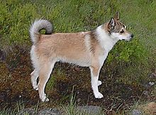
Lundehund Syndrome
The Lundehundsyndrom ( syn . Lundehund gastroenteropathy ) is a complex of diseases of the digestive tract , which in Norwegian Lundehunds occurs. It is a hereditary disease .
Pathophysiology
The Lundehundsyndrom belongs to the complex of chronic inflammatory bowel disease (IBD) and comprises gastritis , protein loss through the intestine ( protein Losing Enteropathy , PLE), lymphangiectasia and malabsorption .
clinic
Signal element
Lundehund syndrome occurs only in Norwegian Lundehounds. It is most often diagnosed in middle age (about five years), but can in principle occur at any age. There is no sexual disposition , males and bitches seem to be affected equally often. The typical changes in the gastrointestinal tract are often found in lundehounds that are not or not yet clinically ill.
Symptoms
Affected dogs can develop a variety of gastrointestinal symptoms, such as intermittent diarrhea , gastritis , vomiting , weight loss, and ascites . In addition, due to protein losses through the intestine, edema of the subcutaneous tissue ( anasarca ) can occur, which occurs particularly on the hind legs. In severe cases, hydrothorax and hydropericardium can also occur.
diagnosis
The diagnosis is made by microscopic examination of tissue samples of the gastrointestinal mucosa, which are taken during endoscopy . Macroscopically, edema and partial erosions of the gastric mucosa and a stomach wall thickened by fibrosis are found . A chronic atrophic gastritis with mononuclear infiltration and degeneration of the fundus glands can be seen under the microscope . In some dogs, there are also enlarged lymph vessels (lymphangiectasia). Near the pylorus can inflammatory nodules develop.
In contrast to chronic inflammatory bowel disease in humans, Lundehund syndrome does not appear to lead to metaplasia of the intestinal epithelial cells. Dilated lymph vessels are also often found in the small intestine. The intestinal lining is thickened and rough. The villi are stunted and partly fused together, while the crypts are hyperplastic and partly filled with cell fragments. Partly it comes to the separation of the epithelium and blistering between basement membrane and epithelium. Granulomas can also occur.
Lundehund syndrome does not cause characteristic changes in blood counts . Unlike other breeds affected by PLE, lymphocyte counts in Lundehund Syndrome are normal. Total protein, albumin , calcium, cobalamin and folic acid may be reduced.
As an additional diagnostic test, there is also the option of examining the dog's feces for faecal α 1 -proteinase inhibitor. An increased value indicates a loss of protein via the intestines, which can be considered diagnostic for Lundehund syndrome in Lundehounds.
In 2016, a causal mutation in the LEPREL1 gene was identified that is simply inherited as an autosomal recessive trait. A genetic test to identify carriers and dogs with clinical disposition is available. An influence of other genes (especially NOD1 ) is also likely.
Therapy and prognosis
The treatment of Lundehund syndrome is purely symptomatic, as the cause of the gastrointestinal changes is not yet known. There is no therapeutic protocol that can be used in all dogs, and symptom improvement with treatment varies widely from person to person.
In general, a high quality, easily digestible diet with low fat and high protein content is recommended to relieve the lymphatic system of the intestine. Occasionally, the addition of medium-length triglycerides , which can also be ingested via a non-lymphatic route, is recommended. Vitamins can also be useful, especially in cases where cobalamin and folic acid are low; Cobalamin must be administered parenterally . Anti-inflammatory drugs and immunosuppressants such as prednisolone can also improve symptoms, with prednisolone also appearing to improve the gut's ability to absorb nutrients. Antiemetics may also be given if vomiting is frequent . The treatment is lifelong, a cure is not possible.
Complications of Lundehund Syndrome are bacterial colonization of the small intestine ( SIBO ) that can be treated with antibiotics . Edema formation is treated with diuretics . The loss of antithrombin III through the intestine can lead to an increased tendency for blood to clot , which can lead to thrombosis and embolism . Chronic gastritis can degenerate into a malignant tumor of the stomach wall ( gastric carcinoma ).
The prognosis varies widely and depends on how the dog responds to treatment. However, the disease often progresses even in treated dogs and can ultimately lead to death. Some lundehounds only need treatment in acute phases and have long symptom-free intervals in between, while others never recover from the first symptoms.
Genetics and Breeding Hygiene
Today's Lundehund population goes back to five individuals who began controlled breeding in 1961. One of these dogs developed symptoms similar to those of Lundehund Syndrome by the age of three. Since then, the disease has been observed repeatedly in dogs of the breed. One can therefore assume that it is a hereditary disease that was able to spread through a genetic bottleneck within the entire breed ( founder effect ). The prevalence of the disease within the breed is very high, with about half of the Lundehounds examined, a protein loss via the intestine was detected.
Thanks to the availability of a genetic test, it is now possible to avoid mating two carrier animals and / or affected dogs with carriers and thus reduce the risk of clinically apparent Lundehund syndrome.
literature
- N. Berghoff: Prevalence and partial characterization of gastroenteropathies with protein loss in the Norwegian Lundehund in North America (PDF; 934 kB) Diss. Med. vet. Hanover 2006, accessed on February 2, 2011
- N. Berghoff et al: Gastroenteropathy in Norwegian Lundehunds. In: Compendium (Yardley, PA). Volume 29, Number 8, August 2007, pp. 456-65, 468, ISSN 1940-8307 . PMID 17849700 . (Review, online at Vetlearn.com, accessed February 28, 2016.)
- K. Flesjå, T. Yri: Protein-losing enteropathy in the Lundehund. In: The Journal of small animal practice Volume 18, Number 1, January 1977, pp. 11-23, ISSN 0022-4510 . PMID 853728 .
- T. Landsverk, H. Gamlem: Intestinal lymphangiectasia in the Lundehund. Scanning electron microscopy of intestinal mucosa. In: Acta pathologica, microbiologica, et immunologica Scandinavica. Section A, Pathology Volume 92, Number 5, September 1984, pp. 353-362, ISSN 0108-0164 . PMID 6507100 .
Individual evidence
- ↑ a b J. Metzger, p Pfahler, O. Distl: Variant detection and runs of homozygosity in next generation sequencing data elucidate the genetic background of Lundehund syndrome. In: BMC genomics. Volume 17, August 2016, p. 535, doi : 10.1186 / s12864-016-2844-6 , PMID 27485430 , PMC 4971756 (free full text).
- ↑ N. Berghoff: Prevalence and partial characterization of gastroenteropathies with protein loss in the Norwegian Lundehund in North America (PDF; 934 kB) Diss. Med. vet. Hanover 2006, accessed on February 2, 2011
