Uterine fibroid
| Classification according to ICD-10 | |
|---|---|
| D25.- | Leiomyoma of the uterus
Including: Fibromyoma of the uterus |
| D25.0 | Submucosal leiomyoma of the uterus |
| D25.1 | Intramural leiomyoma of the uterus |
| D25.2 | Subserous leiomyoma of the uterus |
| D25.9 | Leiomyoma of the uterus, unspecified |
| ICD-10 online (WHO version 2019) | |
The uterine myoma ( myoma of the uterus ; also fibroid ) is the most common benign tumor in women. So have about 25 percent of women after age 30 uterine fibroids ( English Fibroids ) that about 25 percent of them have problems. Fibroids can occur individually (solitary fibroids), but they are often distributed in large numbers in the uterus . A uterus enlarged by fibroids is called a uterus myomatosus . Myomas are usually round in shape, and histologically mostly leiomyomas (tumors of the smooth muscles ).
Emergence
Fibroids develop and grow under the influence of estrogens , progesterone and growth factors , therefore only in childbearing age (in the time between the first and last menstruation of the woman): Young girls cannot have fibroids, after menopause no new fibroids develop and existing ones Myomas can then shrink and possibly calcify. Fibroids do not disappear during menopause, but then due to the lack of bleeding symptoms (with the exception of patients on hormone replacement therapy) they rarely require treatment. The appearance of myomas can be hereditary (familial risk groups), chromosome aberrations are often found . The FH gene coding for fumarase can be affected. Fibroids are much more likely to occur in African women, around twice as often as in Caucasian women. They also occur more frequently in the West Indies and the French overseas departments ; some sources speak of an up to nine times higher risk for African, Afro-Caribbean and Afro-American women.
Classification
A distinction is made between several different types according to the position of the myoma in relation to the different layers of the uterus:
- submucosal fibroids : directly under the uterine lining, or in the myometrium with contact to the uterine lining; submucosal fibroids often cause increased menstrual bleeding; The myoma in statu nascendi is a special form ; it refers to a submucosal, pedunculated myoma which enters the cervical canal and is "born".
- intramural fibroids : located in the muscle layer (myometrium) of the uterus; Often larger than submucosal myomas, they compress the bladder and, in addition to discomfort when urinating (dysuria), often lead to painful menstrual bleeding.
- subserous myomas : lying on the outside of the uterus (under the peritoneum of the uterus), either broad-based or pedicled; Subserous myomas often become very large there and can remain symptom-free for a long time. Due to their size and the resulting pressure on the bladder and intestines, it can lead to frequent urination or pressure pain in the lower abdomen.
- myomas growing intraligamentously : less common myomas, localized in the connective tissue layers on both sides of the uterus (mostly the broad uterine ligaments ); there they can compress the ureters and then possibly lead to kidney disease due to the resulting urine build-up.
- Cervical fibroids : occur in around 8 percent of cases, grow within the muscles of the cervical area and can compress the cervical canal as well as exert pressure on nearby structures (bladder, rectum, etc.).
Symptoms
Depending on the size and location of the fibroids in the uterus, most women with a fibroid are symptom-free, but there can also be massively increased, prolonged menstrual periods (in the case of intramural fibroids because of weak contraction of the uterus or in submucous fibroids because of impaired mucosal regeneration) and / or Intermenstrual bleeding occurs, possibly leading to secondary anemia .
Other possible complaints are:
- Pain, feeling of pressure, feeling of foreign body in the abdomen, especially with large tumors or subserous myomas
- Constipation (due to pressure on the intestines)
- Discomfort when urinating (due to pressure on the bladder; possible consequences: dysuria , pollakiuria , incontinence )
- Discomfort during intercourse ( dyspareunia )
- Lower back pain and (nerve) pain in the legs (due to the pressure of the tumor on the presacral nerves) are also possible.
- Acute abdomen : by twisting the stem of a pedunculated subserous myoma with necrosis and reactive peritonitis
Fibroids and pregnancy
During pregnancy, usually between the third and sixth month, in rare cases, severe pain in the area of the myoma can occur in isolation, due to an impairment of the blood supply, which leads to infarction and necrosis of the myoma (also known as red degeneration ). Very rarely a large fibroid can be a birth obstacle so that an appropriate location for caesarean section ( cesarean section must be) carried out. The risk of miscarriage and premature birth may be slightly higher in women with large intrauterine (in the uterus) fibroids. Bleeding during pregnancy and (rarely) premature placenta detachment can occur as a consequence of a subplacental position of a myoma. Fibroids can sometimes be a cause of sterility (infertility). However, they are only considered to be the main cause of infertility in 3% of cases.
Secondary changes
Apart from the change in size, fibroids can also change structurally, mostly due to impaired blood flow:
- The most common change concerns a softening , which can take several forms. By far the most common form is hyaline degeneration (around 60%), cystic degeneration with central cavity formation and myxomatous degeneration can also be mentioned. A rare form is the painful red degeneration that occurs almost exclusively during pregnancy .
- A hardening usually occurs as a result of calcification or fibrosis (fibroleiomyoma).
- Necrosis is either hemorrhagic or ischemic.
- Infections occur through ascending germs
- Malignant degeneration : About 0.1% of myomas turn out to be malignant (leiomyosarcomas). While a malignant degeneration of benign myomas was previously assumed, later genetic investigations (Levy et al., 2000) strongly suggest that the sarcomas develop primarily as such and are initially only misunderstood due to differential diagnostic difficulties. Leiomyosarcomas can resemble a myoma both sonographically and tomographically; mostly it is the rapid progression of size that arouses the suspicion of a potential malignancy. A leiomyosarcoma can only be detected pathologically after its complete removal, either by means of enucleation of the myoma or in the context of a uterine removal.
- The rare secondary changes in the fibroids include edematous loosening as well as angiomatous (vascular), endometriotic , lipomatous (fatty) or chronic inflammatory changes.
therapy

Uterine fibroids are benign tumors that do not require any therapy. However, due to their location in the uterus and / or their size (from very small to 15 cm or larger), they can cause discomfort, which may then require therapy.
- Conservative therapy: fibroids that cause no symptoms usually do not require any therapy and can be checked clinically and with ultrasound at regular intervals. Conservative drug therapy is usually used first. The drug groups used are anti-inflammatory, non-steroidal drugs such as ibuprofen or naproxen , ovulation inhibitors, as well as hormonal therapies with GnRH analogues, which, usually carried out over six months, create a reversible endocrine environment corresponding to the postmenopause. The GnRH analogues often reduce the size of the fibroids. After discontinuing therapy, however, the growth may return to its original size. The possible side effects of the GnRH analogues are similar to those of menopausal status , such as osteoporosis , vaginal dryness and hot flashes .
- Since March 2012, ulipristal acetate from the group of selective progesterone receptor modulators (SPRM) has been approved in Germany for the treatment of symptomatic myomas for suitable patients in whom surgery is planned after 12 weeks of daily oral administration of a 5 mg tablet. The effectiveness of this treatment has been demonstrated in randomized, double-blind studies. In the case of bleeding disorders, the bleeding stops on average within 7 days and an average volume reduction of 36% occurs after three months. While the menstrual bleeding returned quickly, usually at a reduced rate, after discontinuation, the reduction in volume lasted for more than 6 months. The most common side effects of ulipristal acetate were headaches in about 20% of users. In contrast to GnRH analogues, the estrogen levels are not influenced, so that typical temporary menopausal deficits do not occur. Reversible benign changes in the endometrium (PAEC) were observed in 60% of users during therapy. The importance of therapy with ulipristal acetate (UPA) with regard to fulfilling the desire to have children is unclear: to date (December 2016) there are no comparative data UPA vs. Myoma removal for this indication.
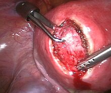
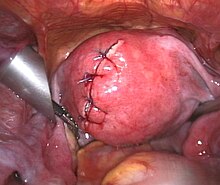
- Surgical therapy: Myomas with symptoms are usually treated surgically. A distinction is made between uterine preservation and ablative procedures. In uterine preservation procedures , the fibroids are peeled from the uterus. This can be done by means of an abdominal incision, but increasingly also by means of a laparoscopy , under certain conditions (submucosal position) also by means of hysteroscopy (uterine mirror image ). The hysterectomy (removal of the uterus) is considered an ablative procedure . This can be done vaginally, by incision in the abdomen (abdominal) or by laparoscopy ( laparoscopy ). The technique of chopping up fibroids and uteri ( morcellement ) can, regardless of the type of operation (abdominal incision, operation via the vagina or laparoscopy), in very rare cases lead to the spread of benign, but also of initially unknown malignant tissue in the abdomen. When choosing the right treatment option, the size and location of the fibroids play a key role in addition to the patient's age and her desire for treatment (e.g. still wanting to have children?).
- Myomembolisation or Uterusarterienembolisation (UAE): The uterine artery embolization is a radiological, minimally invasive treatment of uterine fibroids. To do this, a catheter is pushed into the artery supplying the uterus through a skin incision in the right groin under X-ray control. Embolization usually takes place via a microcatheter with a small outer diameter of 2.5-2.8 F (this corresponds to 0.8-0.9 mm). When the catheter is securely placed in the supplying vasculature of the myoma, it is used to embolize the supplying vasculature by injecting small gelatine or plastic particles. The small particles (500-900 micrometers) flow into the end arteries of the fibroid and remain in it (thus closing all fibroids in the uterus in one procedure). The supplying vessels are slowly blocked for a few minutes. This procedure must also be carried out in the same way in the vascular system on the opposite side. To numb the pain during and after the procedure, a PCA pump (patient-controlled analgesia) is usually required , where the patient can independently call up pain medication boluses via a perfusor syringe. Most patients describe the procedure as being well tolerated. With uterine fibroid embolization, 78-94 percent of women treated in this way can become symptom-free, depending on the type of myoma and pre-interventional symptoms. With regard to patients who wish to have children, the following consensus was reached at the 2nd and 3rd radiological-gynecological expert meeting: “UAE is not a method in the context of fertility treatment. Before considering a hysterectomy in a patient with incomplete family planning, the possibility of UAE should be assessed. The role of UAE as a treatment option has not been clarified for patients who wish to have children. So far there are no prospectively collected data, the results of which allow a statement with the necessary evidence about the influence of UAE on the fertility rate and the outcome of pregnancy. ”However, a hysterectomy is generally not indicated in patients who wish to have children from a gynecological point of view. Retrospective data indicate significantly increased complication rates in pregnancies after fibroid embolization.
- Focused ultrasound (MRgFUS, MR-HIFU): a method that has been available since around 2002 and is becoming increasingly important is the targeted ultrasound heating of the fibroids to up to 80 ° C under MRI control. Most contraindications result from those of the MRI procedure itself (e.g. metal implants, pacemakers, claustrophobia). Furthermore, the inclusion and exclusion criteria relate to the location, size, number and blood flow of the fibroids. In principle, the method is suitable for women who want to have children, since the uterus is preserved. However, due to the lack of sufficient data, German-speaking experts (radiological-gynecological consensus meeting) do not recommend such treatment for women who want to have children. The therapy is usually carried out on an outpatient basis. Only a few health insurance companies cover the costs at a flat rate; many others do this on a case-by-case basis. The method is currently (as of May 2014) available at eight locations in Germany (Berlin, Bochum, Bottrop, Dachau, Heidelberg, Lübeck, Munich and Stuttgart).
literature
- TK Helmberger, TF Jakobs, MF Reiser : Technique and methods in uterine leiomyoma embolization. In: Radiologe , 2003, 43 (8), pp. 634-640.
- TJ Kröncke, B. Hamm: Role of magnetic resonance imaging (MRI) in establishing the indication for, planning, and following up uterine artery embolization (UAE) for treating symptomatic leiomyomas of the uterus. In: Radiologe , 2003, 43 (8), pp. 624-633.
- B. Radeleff, S. Rimbach, GW Kauffmann u. a .: Risk and complication rate of uterine fibroid embolization (UFE). In: Radiologe , 2004, 43 (8), pp. 641-650.
- GM Richter, B. Radeleff, S. Rimbach u. a .: Uterine fibroid embolization with spheric micro-particles using flow guiding: safety, technical success and clinical results. In: Röfo , 2004, 176 (11), pp. 1648-1657.
- J. Pelage: Treatment of uterine fibroids. In: Lancet , 2001, 12 (357 (9267)), p. 1530.
- JH Ravina, A. Aymard, N. Ciraru-Vigneron et al. a .: Uterine fibroids embolization: results about 454 cases. In: Gynecol Obstet Fertil. , 2003, 31 (7-8), pp. 597-605.
- JB Spies, J. Bruno, F. Czeyda-Pommersheim u. a .: Long-term outcome of uterine artery embolization of leiomyomata. In: Obstet Gynecol. , 2005, 106 (5), pp. 933-939.
- DT Rein, T. Schmidt, M. Fleisch, R. Wagner, W. Janni: Multimodal treatment of the uterus myomatosus. In: Frauenarzt , 50, 2009, pp. 752–758, Frauenarzt.de (PDF; 520 kB)
- A. Taran, G. Gaffke, M. Rüsch, H. Heuer, B. Hosang, J. Ricke, S.-D. Costa: The modern multimodal therapy of the uterus myomatosus: when is a hysterectomy indicated? In: Ärzteblatt Sachsen-Anhalt , 19, 2008, pp. 38–45.
- Jacques Donnez et al. a .: Ulipristal Acetate versus Placebo for Fibroid Treatment before Surgery. In: N Engl J Med. , 2012, 366, pp. 409-420.
- Jacques Donnez et al. a .: Ulipristal Acetate versus Leuprolide Acetate for Uterine Fibroids. In: N Engl J Med. , 2012, 366, pp. 421-432.
- M. Kirschbaum, K. Münstedt: Checklist gynecology and obstetrics. Georg Thieme Verlag, Stuttgart, ISBN 3-13-126292-3 .
- Diedrich u. a .: Gynecology & Obstetrics. Springer Medizin Verlag, Heidelberg, ISBN 978-3-540-32867-4 .
- A. Strauss: Ultrasound Practice: Obstetrics and Gynecology. 2nd Edition. Springer Medizin Verlag, Heidelberg 2008, ISBN 978-3-540-78252-0 .
- C. Keck, J. Neulen, H. Behre, M. Breckwoldt: Practice of gynecology / endocrinology, reproductive medicine, andrology. Georg Thieme Verlag, Stuttgart, ISBN 978-3-13-107162-0 .
Web links
- Pictures of a fibroid operation
- Consensus paper from radiologists and gynecologists on fibroid embolization (2010) (PDF; 208 kB)
- Information on myomas from the Working Group for Gynecological Endoscopy of the German Society for Gynecology and Obstetrics
- Uterus-Myomatosus.net Non-profit patient information portal on myoma therapies
Individual evidence
- ↑ a b ulipristal acetate in symptomatic uterine myomatosus and myoma-related hypermenorrhea. Joint statement of the German Society for Gynecological Endocrinology and Reproductive Medicine (DGGEF) e. V. and the professional association of gynecologists (BVF) e. V., online
- ↑ Minimally invasive therapy of myomas of the uterus (uterine fibroid embolization) University Hospital Tübingen; Retrieved December 17, 2008.
- ↑ Chronic pain in gynecology - pelvipathies. ( Memento of the original from December 12, 2013 in the Internet Archive ) Info: The archive link was inserted automatically and has not yet been checked. Please check the original and archive link according to the instructions and then remove this notice. In: Journal for applied pain therapy. 3/2002.
- ↑ JP Pelage: Everything you should know about the embolization of uterine fibroids ( Memento of the original from February 19, 2009 in the Internet Archive ) Info: The archive link was inserted automatically and has not yet been checked. Please check the original and archive link according to the instructions and then remove this notice. (PDF; 244 kB) European Congress of Radiology, 2008.
- ↑ What are fibroids? St. Elisabeth Hospital Tilburg; Retrieved December 17, 2008.
- ↑ Classification of myomas, portal Frauenaerzte-im-netz.de
- ^ Fibroids and pregnancy
- ↑ Dissertation on the topic of uterine fibroids (PDF)
- ↑ Hysteroscopic myoma enucleation ( Memento of the original dated June 6, 2014 in the Internet Archive ) Info: The archive link was inserted automatically and has not yet been checked. Please check the original and archive link according to the instructions and then remove this notice. presented in the portal of the Marienhospital Stuttgart
- ↑ Michael A. Seidman, Titilope Oduyebo, Michael G. Muto, Christopher P. Crum, Marisa R. Nucci, Bradley J. Quade: Peritoneal Dissemination Complicating Morcellation of Uterine Mesenchymal Neoplasms. In: PLoS ONE. 7 (2012), p. E50058, PMID 23189178 , doi: 10.1371 / journal.pone.0050058
- ^ MW Beckmann, I. Juhasz-Böss, D. Denschlag, P. Gaß, T. Dimpfl, P. Harter, P. Mallmann, SP Renner, S. Rimbach, I. Runnebaum , M. Untch, SY Brucker, D. Wallwiener: Surgical Methods for the Treatment of Uterine Fibroids - Risk of Uterine Sarcoma and Problems of Morcellation: Position Paper of the DGGG. In: Obstetrics Frauenheilkd. 75 (2015), pp. 148-164, doi: 10.1055 / s-0035-1545684 , German version (PDF; 2.39 kB)
- ↑ "Patient information on fibroid embolization, Jena University Hospital"
- ↑ TJ Kröncke, M. David: Results of the 2nd radiological-gynecological expert meeting - Uterine artery embolization (UAE) for myoma treatment. (Consensus paper) In: Fortschr Röntgenstr. , 2007, 179, pp. 325-326, doi: 10.1055 / s-2007-972191
- ^ Thomas Kröncke, Matthias David: Uterine artery embolization for myoma treatment. In: Gynecologist. 51 (2010), pp. 644–646, online ( memento of the original dated December 12, 2013 in the Internet Archive ) Info: The archive link was inserted automatically and has not yet been checked. Please check the original and archive link according to the instructions and then remove this notice. (PDF; 213 kB)
- ↑ Thomas Römer, Hans-Rudolf Tinneberg: Commentary on: Thomas Kröncke, Matthias David: Uterus artery embolization for myoma treatment. In: Gynecologist. 51 (2010), pp. 647–648, online ( memento of the original dated December 12, 2013 in the Internet Archive ) Info: The archive link has been inserted automatically and has not yet been checked. Please check the original and archive link according to the instructions and then remove this notice. (PDF; 213 kB)
- ↑ M. David, T. Kröncke: Uterine Fibroid Embolization - Potential Impact on Fertility and Pregnancy Outcome. In: Obstetrics Frauenheilkd 2013; 73, pp. 247-255.
- ↑ Destroy fibroids with focused ultrasound under magnetic resonance control
- ↑ Magnetic resonance-guided (MR) therapy of uterine fibroids with focused ultrasound - a non-invasive treatment to improve quality of life. ( Memento of the original from September 6, 2009 in the Internet Archive ) Info: The archive link was automatically inserted and not yet checked. Please check the original and archive link according to the instructions and then remove this notice.
- ↑ T. Kröncke, M. David: Magnetic resonance-guided focused ultrasound for myoma treatment - results of the 2nd radiological-gynecological expert meeting. (Consensus paper) In: Fortschr Röntgenstr. , 2015, 187, pp. 480-482, doi: 10.1055 / s-0034-1399342
- ↑ Presentation of the MRgFUS procedure ( memento of the original from June 7, 2014 in the Internet Archive ) Info: The archive link was automatically inserted and not yet checked. Please check the original and archive link according to the instructions and then remove this notice. in the portal of the Dachau Clinic


