Cystic echinococcosis
| Classification according to ICD-10 | |
|---|---|
| B67.0 | Echinococcus granulosus [cystic echinococcosis] infection of the liver |
| B67.1 | Echinococcus granulosus infection of the lungs |
| B67.2 | Echinococcus granulosus infection of the bones |
| B67.3 | Echinococcus granulosus infection in multiple and other locations |
| B67.4 | Echinococcus granulosus infection, unspecified |
| ICD-10 online (WHO version 2019) | |
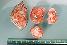
The cystic hydatid disease is the formation of cysts by the fin of some representatives of the genus Echinococcus (tapeworms) in the intermediate host . The symptom is called an echinococcal bladder , also known as a leg worm , bladder worm , metacestode , hydatide or hydatid cyst . This shows an "expansive" growth and displaces the surrounding tissue.
The most frequent metacestodal infestation in humans is caused by the three-part dog tapeworm ( Echinococcus granulosus ). For the dog tapeworm, the dog , wolf and dingo are the definitive final hosts . Sheep in particular act as intermediate hosts , but also goats , cattle , pigs , horses, but also wild ungulates and occasionally humans. According to the Infection Protection Act, notification is required in Germany.
The alveolar echinococcosis , however, is transmitted by the fox tapeworm .
distribution
Echinococcosis, which is widespread worldwide but with different regional concentrations, occurs in Europe mainly in the Mediterranean countries, but is relatively rare in Germany. The imported cases of illness observed (foreigners, German tourists) come mainly from the southern countries of the Mediterranean region.
transmission
The eggs of the dog tapeworm are usually transmitted by contact infection or smear infection from the dog's excrement, the fur or the muzzle via the hands that are then contaminated (with pathogen adhesions) and the mouth. Indirect infections are also possible, for example through food or drinking water that has been contaminated with Echinococcus eggs.
diagnosis
The disease can mainly be detected with imaging methods such as sonography , X-rays , and computed tomography (CT) as well as magnetic resonance imaging (MRI), which should be combined with serological methods ( IFT , PHA). A serological differentiation between E. granulosus and E. multilocularis ( fox tapeworm ) is possible using ELISA . In addition, cross-reactions with other tapeworms are possible and indicated to clearly identify the pathogen.
Differential diagnosis
Other space-occupying nodules such as an amoebic abscess should be excluded for a clear diagnosis . Furthermore, dysontogenetic cysts , tumors or abscesses of the liver (caused by abnormal development of certain tissues, occurring individually or in clusters) should be excluded.
Course of disease
The length of the incubation period varies widely. The period can extend over months to years. Basically anyone - adults as well as children - can be affected by this disease, but it occurs most frequently between the third and fifth decades of life.
In the dog tapeworm, the fin is a liquid-filled, single or multi-chambered bladder. The organism reacts to this foreign body by forming a layer of connective tissue around it, so that a very firm connective tissue capsule (incubator capsules) is created, especially in the liver tissue. Starting from the inner germ layer , many small vesicles form after about six months, which contain the preliminary stages of the finished tapeworms, which move freely in the liquid after hatching. These "hydatids" have a diameter of a few millimeters up to 30 cm.
The disease mainly affects the liver (50–70%), lungs (15–30%) and rarely also the spleen , kidneys , brain and other organs, although usually only one organ is affected.
Symptoms
Although the infection with the dog tapeworm usually goes hand in hand with no symptoms and therefore often goes undetected, symptoms can arise at some point in the course.
Liver chinococcosis (liver disease) often only causes clinical symptoms in a very large number of cysts by compressing blood vessels or biliary tract . If the liver capsule is stretched , more or less severe abdominal pain may occur. Sometimes, if the infection is extensive, jaundice ( jaundice ) with yellowing of the patient's eyes and skin.
In pulmonary echinococcosis (involvement of the lungs), the thin-walled lung cysts burst ( rupture ) and are accompanied by pain, coughing and breathing difficulties .
When the central nervous system is affected , the echinococcosis cysts cause neurological focus symptoms, depending on their location in the brain or spinal cord .
With all possible infestations, there is an additional risk of allergic shock if an echinococcus cyst bursts and the fins subsequently spread. In rare cases such an event can also lead to spontaneous healing .
Complications
Occasionally, tissue destruction and insatiable bleeding can occur in the liver . In addition, the increased pressure in the blood vessels leading to the liver can cause water to build up in the abdomen. When parasites die, they usually leave cavities of decay, into which bleeding can then take place.
therapy
Correct treatment of cystic echinococcosis tailored to the individual patient requires interdisciplinary collaboration between surgeons, radiologists, gastroenterologists and tropical medicine specialists, parasitologists and infectiologists.
A radical surgical treatment is sought for a completely possible cure, in which it is particularly important for the surgeon to remove the fin undamaged and completely so that there is no additional spread of the pathogen during and after the operation. In the event that the parasite cannot be completely removed or the patient is inoperable (inoperable), only long-term therapy with drugs such as mebendazole and albendazole remains . In the case of dog tapeworms, the pathogen can even be completely killed off in some cases.
Easily accessible cysts of the dog tapeworm should be carefully removed under perioperative chemotherapy (performed during the operation). If necessary, to be on the safe side , the cyst can be emptied and rinsed using the PAIR procedure (puncture - aspiration - injection - reaspiration) before surgical removal . If a surgical procedure leads to a rupture of the cyst (rupture of the cyst) and the corresponding sowing of the fins, long-term treatment with mebendazole or albendazole can prevent or slow the progression of the disease, as in an inoperable patient.
prevention
Regular examinations and, if necessary, deworming of dogs and cats are recommended to prevent such a disease in good time . Such preventive measures are particularly indicated if the said pets have been taken on trips to or from Mediterranean countries.
See also
Individual evidence
- ^ Robert Koch Institute: Echinococcosis
- ^ Carlos Thomas: Atlas of infectious diseases (= pathology. ). Schattauer, Stuttgart 2010, ISBN 978-3-7945-2762-5 , p. 222 → Section 2.4.1 Large cystic echinococcosis. ( at Google books ).
- ↑ Gerd Poeggel: Short textbook biology. 3rd, revised edition, Thieme, Stuttgart a. a. 2013, ISBN 978-3-13-150813-3 , p. 177 → dog tapeworm. ( at Google books ).
Web links
- Information from the Robert Koch Institute
- Information from the Echinococcosis Working Group of the Paul Ehrlich Society for Chemotherapy ( Memento from January 6, 2015 in the Internet Archive )
- Information from the consulting laboratory for echinococcosis at the University of Würzburg
- Video of a liver hydatidectomy

