Biosensor
Biosensors are measuring sensors that are equipped with biological components. These are used in biotechnological measurement technology. The term was coined in 1977 by Karl Cammann; IUPAC has had a definition for it since 1997.
Structure and principle
Biosensors are based on the direct spatial coupling of an immobilized biologically active system with a signal converter ( transducer ) and an electronic amplifier . Biosensors use biological systems at different levels of integration to identify the substances to be determined. Such recognition elements can either be natural (e.g. antibodies , enzymes , nucleic acids , organelles or cells ) or synthetic (e.g. aptamers , molecularly imprinted polymers , macrocycles or synthetic peptides ) systems. The immobilized biological system of the biosensor interacts with the analyte . This leads to physicochemical changes, such as B. Changes in the layer thickness, the refractive indices , the light absorption or the electrical charge. These changes can by means of the transductor, such as. B. optoelectric sensors, amperometric and potentiometric electrodes or special field effect transistors ( chemically sensitive field effect transistor ) can be determined. The initial state of the system must be restored after the measurement process. A problem in the development of biosensors is the corrosion of the biosensor due to a coating with cells ( biocorrosion ) or due to the culture medium . For example, typical cell culture media for eukaryotic cell cultures corrode silicon at a rate of about 2 nm / h.
The measurement of an analyte by means of a biosensor therefore takes place in three steps. First, the specific detection of the analyte is carried out by the biological system of the biosensor. The physicochemical changes that result from the interactions between the analyte and the receptor are then converted into an electrical signal. This signal is then processed and amplified. Signal conversion and electronics can be combined, e.g. B. in CMOS-based microsensor systems. A biosensor derives its selectivity and sensitivity from the biological system used.
Types of biosensors
- Piezoelectric sensors
- The oscillation frequency of a quartz is inversely proportional to the square root of its mass. A quartz crystal coated with enzymes , antibodies or other binders can thus be used as a microbalance . A particularly sensitive (sensitive) special case are the surface wave sensors (SAW sensors, Surface Acoustic Waves ). Here, two coatings are applied to a piezoelectric quartz, which serve as a transmitter or receiver and emit surface acoustic waves after electrical excitation. By binding an antigen to an antibody, immune reactions cause a change in the surface and thus a change in the resonance frequency of the wave.
- Optical sensors
- In practice, these sensors are primarily used to track the oxygen content in liquids. The measurement principle here is based on fluorescence quenching . An optical waveguide , at the end of which an indicator is attached, serves as the measuring device . The luminescence or absorption properties of this indicator are dependent on chemical parameters such as the oxygen concentration. Another method that can be used is based on evanescence , which occurs during total reflection at the transition from the optically denser to an optically thinner medium. Here, fluorescent light from a fluorescence- labeled analyte can be coupled into the light guide and a statement can be made about the concentration. This method is used to determine antigens via a reaction with a specific antibody on the surface of a light guide. The method can be made more sensitive by adding a thin metal film to the surface of the light guide. Density fluctuations of free charge carriers ( plasmons ) occur in the metal film . With such a sensor based on the principle of surface plasmon resonance , the metal film is additionally coated with dextrans to which analyte-specific antibodies can be bound.
- Electrochemical detection
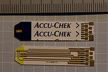
- by amperometry : In amperometry, the current flow is measured in a measuring chamber on two electrodes with the voltage kept constant. It is suitable for metabolic products that can easily be oxidized or reduced. Often, mediators are also used, i.e. redox pairs that intervene indirectly during the oxidation of the actual substrate and serve for electron transfer . Is z. If , for example, a substrate to be determined is oxidized by FAD , which is a coenzyme of most oxidases , FAD is reduced to FADH, then FADH is then oxidized again to FAD by the oxidized form of the mediator. The resulting reduced form of the mediator is anodically oxidized again. Using recordings of the current-voltage curves, statements can be made about the redox behavior and the concentration of the actual substrate. As mediators z. B. hydroquinone or derivatives of ferrocene are used. The advantage of mediators is that you can specify a much lower voltage and thus avoid undesirable side reactions. Amperometric biosensors are used e.g. B. used to determine glucose , cholesterol , fatty acids and L- amino acids with the corresponding enzymes as oxidases.
- by potentiometry : Potentiometry is used for ionic reaction products. The quantitative determination of these ions is carried out on the basis of their electrical potential on a measuring electrode that is coated with a suitable enzyme to determine a substrate. In hydrolases , e.g. B. urease , the change in pH or the change in ammonium ions or hydrogen carbonate ions is determined. Ion -sensitive field effect transistors (ISFET) or metal-oxide-coated acid electrodes ( MOSFET ) are often used as measuring electrodes . An electrode of the same type, but without an enzyme coating, is used as the reference electrode. The potentiometric method is used to determine z. B. urea , creatinine or amino acids .
- with ion-selective electrodes : If these are coated with an enzyme, they work according to the same principle as described for potentiometry.
- Interferometric detection
- Here, the biomolecules interact with a polymer layer, the change in thickness of which is monitored with reflectometric interference spectroscopy.
Applications
The first measuring system, which can be called a biosensor according to the definition given above, was developed by Clark and Lyons in 1962. A measuring system has been described which enables the determination of glucose in the blood during and after operations. This biosensor consisted of either a Clark oxygen electrode or a pH electrode as a transducer, in front of which the enzyme glucose oxidase was applied between two membranes . The glucose concentration could be determined as a change in the pH value or as a change in the oxygen concentration as a result of the oxidation of glucose under the catalytic effect of the enzyme glucose oxidase.
In this construction, the biological material is enclosed between two membranes, or the biological system is applied to a membrane and is connected directly to the surface of the transducer. The areas of application for biosensors in the analysis of water and wastewater can be divided into biosensors for the determination of individual components, biosensors for the determination of toxicity and mutagenicity and biosensors for the determination of the biochemical oxygen demand (BOD).
Biosensors for the determination of proteins were realized with silicon field effect sensors (so-called ChemFETs ). They allow the marker-free analysis of proteins in the field of protein analysis by in-situ process, since they detect the protein binding on the intrinsic charge amount of the protein by means of field effect.
The bacterial content of bathing water or sewage can be determined using a biosensor. Antibodies against certain types of bacteria are attached to a vibrating membrane . If the corresponding bacteria swim past the sensor, they attach themselves to the antibodies and thereby slow down the vibrations of the membrane. If the vibrations fall below a certain value, an alarm is triggered.
The penicillin concentration in a bioreactor in which fungal strains are cultivated can be determined with a biosensor. The biological component of the sensor used here is the enzyme acylase . This penicillin-splitting enzyme is placed on a membrane that rests on a pH electrode. If the penicillin concentration in the medium increases, the enzyme splits off ever larger amounts of an acid , phenylacetic acid . This changes the pH value on the electrode. So one can now deduce the concentration of penicillin from the pH value.
The biosensors also include surface plasmon resonance spectroscopy . The binding of substances is measured by means of plasmon detection.
A new development for monitoring food is based on nanosensors. The fluorescence of nanoparticles that are in an agarose nutrient medium changes significantly when the pH changes due to bacterial metabolism in the food. Two fluorescent dyes are embedded in the nanoparticles. The first is a fluorescein water-repellent dye . It lights up green when excited by a light-emitting diode and reacts sensitively to a change in the pH value. The second, a dye with pH-independent red fluorescence, serves as an internal reference.
With a new type of pH sensor, changes in the pH value in living cells can be tracked over longer periods of time. The principle is based on a combination of fluorescent nanocrystals with flexible oligonucleotides that fold or stretch depending on the surrounding pH value. This changes the distance between the nanocrystal energy donor with a green fluorescent dye and a FRET acceptor, which consists of a red fluorescent dye, in a pH-dependent manner. A FRET energy transfer and thus the glow of the red fluorescent dye occurs when the distance is small. The relationship between green and red fluorescence is observed with a fluorescence microscope .
swell
- RD Schmid , U. Bilitewski: Biosensors. In: Chemistry in Our Time . 26th year, No. 4, 1992, pp. 163-173, ISSN 0009-2851
- Brian R. Eggins: Chemical Sensors and Biosensors. Analytical Techniques in the Sciences. 2nd Edition. Wiley, 2002, ISBN 0-471-89914-3 .
- M. Perpeet, S. Glass, T. Gronewold, A. Kiwitz, A. Malavé, I. Stoyanov, M. Tewes, E. Quandt: SAW sensor system for marker-free molecular interaction analysis. In: Analytical Letters . Volume 39, No. 8, 2006, pp. 1747-1757.
literature
- Reinhard Renneberg, Dorothea Pfeiffer, Fred Lisdat, George Wilson, Ulla Wollenberger, Frances Ligler, Anthony PF Turner: Frieder Scheller and the short history of biosensors. In: Advances in Biochemical Engineering / Biotechnology. Springer-Verlag, Berlin / Heidelberg 2008, ISBN 978-3-540-75200-4 , pp. 1–18 (brief outline of the history of biosensors)
Web links
- Homepage of Eugenii Katz: Biosensors & Bioelectronics
- Helmholtz Center for Environmental Research UFZ: Biosensors
Individual evidence
- ^ Reinhard Renneberg, Dorothea Pfeiffer, Fred Lisdat, George Wilson, Ulla Wollenberger, Frances Ligler, Anthony PF Turner: Frieder Scheller and the short history of biosensors. In: Advances in Biochemical Engineering / Biotechnology. Springer-Verlag, Berlin / Heidelberg 2008, ISBN 978-3-540-75200-4 , p. 3, there the name is given as "Karl Camman".
- ↑ Can Dincer, Richard Bruch, Estefanía Costa-Rama, Maria Teresa Fernández-Abedul, Arben Merkoçi: Disposable Sensors in Diagnostics, Food, and Environmental Monitoring . In: Advanced Materials . May 15, 2019, ISSN 0935-9648 , p. 1806739 , doi : 10.1002 / adma.201806739 .
- ↑ Florinel-Gabriel Bănică: Chemical Sensors and Biosensors: Fundamentals and Applications . John Wiley & Sons, Chichester, UK 2012, ISBN 978-1-118-35423-0 .
- ^ Graham J. Triggs, Gareth JO Evans, Thomas F. Krauss: Degradation of silicon photonic biosensors in cell culture media: analysis and prevention. In: Biomedical Optics Express . Volume 8, No. 6, 2017, p. 2924, doi: 10.1364 / BOE.8.002924 .
- ↑ A. Hierlemann , O. Brand, C. Hagleitner, H. Baltes: Microfabrication techniques for chemical / biosensors. In: Proceedings of the IEEE. Volume 91, No. 6, 2003, pp. 839-863. ISSN 0018-9219 .
- ^ A. Hierlemann , H. Baltes: CMOS-based chemical microsensors. In: The Analyst . Volume 128, No. 1, 2003, pp. 15-28.
- ^ LC Clark, C. Lyons: Electrode systems for continuous monitoring in cardiovascular surgery. In: Ann. NY Acad. Sci. Volume 31, No. 102, 1962, pp. 29-45. PMID 14021529
- ^ SQ Lud, MG Nikolaides, I. Haase, M. Fischer, AR Bausch: Field Effect of Screened Charges: Electrical Detection of Peptides and Proteins by a Thin Film Resistor. In: ChemPhysChem. Volume 7, No. 2, 2006, pp. 379-384.
- ↑ Xu-dong Wang, Robert J. Meier, Otto S. Wolfbeis: Fluorescent pH-Sensitive Nanoparticles in an Agarose Matrix for Imaging of Bacterial Growth and Metabolism . In: Angewandte Chemie . tape 124 , no. 45 , 2012, doi : 10.1002 / anie.201205715 .
- ↑ Euan R. Kay, Jungmin Lee, Daniel G. Nocera, Moungi G. Bawendi : Conformational Control of Energy Transfer: A Mechanism for Biocompatible Nanocrystal-Based Sensors . In: Angewandte Chemie . tape 124 , no. 52 , 2012, ISSN 1521-3757 , doi : 10.1002 / anie.201207181 .
