Canine Hip Dysplasia
The hip dysplasia or canine hip dysplasia ( HD ) is a failure of the hip joint . All dog breeds are affected , with large breeds developing the disease particularly frequently. It was first diagnosed in the German Shepherd Dog and is therefore mistakenly mainly associated with this breed, although other breeds are now more affected. The frequency of occurrence ( prevalence ) can be over 50 percent depending on the breed. Even with domestic cats may hip dysplasia occur, especially among the Maine Coon cats .
HD is largely genetic ( heritability is between 20 and 40 percent), which is why many breeding associations demand freedom from HD for breeding approval . Since improper nutrition and posture can promote the development and progression of the disease, it is a multifactorial (dependent on many factors) event. Clinically , HD manifests itself in increasing restricted mobility and pain, which arise as a result of the pathological remodeling processes at the hip joint ( hip joint arthrosis ). In the advanced stage, only the removal of the hip joint with or without the insertion of an artificial hip joint can bring about a significant improvement. If this is not possible, permanent pain therapy can often maintain an adequate quality of life for a long time.
Symptoms and Diagnosis

The severity of clinical symptoms of HD varies depending on the age or stage of the disease. At relatively young animals, aged between one-half to one year, it comes to pain, because the head of the femur in the acetabulum (acetabulum) finds only insufficient support and its abnormal mobility pain registering nerve fibers of the periosteum of the glenoid rim irritated. Older animals tend to develop pain as a result of progressive degenerative changes ( arthrosis ) of the hip joint.
The onset of HD manifests itself in increasing pain during walks, the dog no longer wants to walk far, sits down more often, occasionally cries out when playing and shows an unstable gait. When presenting the hind limbs, the pelvis is moved sideways in the direction of the presented limb ( LSÜ twist ). When the joint moves, a cracking, clicking, or crunching sound can be heard. If you notice any of the symptoms, it is advisable to go to the vet immediately .
Palpation
Even unclear lameness of the hind limbs in the presence of HD can often quickly be assigned to the hip joint by stressing individual joints . The evaluation of severe hip dysplasia often requires special tests in order to be able to make a statement about joint stability. The Ortolani test is used most often: the thigh is positioned at right angles to the spine when the animal is lying on its healthy side. A hand placed on the knee joint pushes the thigh bone in the direction of the spine under strong pressure . If the joint is extremely unstable, this leads to dislocation or subluxation of the hip joint. If the thigh is now moved away from the body axis , the head of the thigh slides back into the pan with a clicking sound (Ortolani click) .
If possible, this test should only be carried out by a veterinarian, possibly under anesthesia.
roentgen
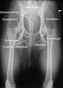
An X-ray examination is a reliable way to determine the severity of the disease . The joints have to be overstretched, which causes severe pain in the presence of HD. Therefore, it is performed under short anesthesia . Prerequisite for a meaningful diagnosis is the exact positioning of the animal in the supine position with straight, parallel mounted thighs and perpendicular to the beam path screwed kneecaps . Proper positioning can be judged by the shape of the obturator foramina , the shape of the iliac blades , the width of the iliac columns, and the position of the kneecaps. Additional images can be taken in the “frog position” of the thighs or in the lateral (latero-lateral) beam path; in Germany this is only carried out in the case of senior expert opinions.
In the assessment, a distinction is made between primary criteria such as the shape of the hip joint and determination of an incongruence , the degree of looseness of the joint and the shape of the joint socket and the femoral head, as well as secondary criteria , which indicate osteoarthritis.
The breeding evaluation of HD recordings is only possible in the VDH by experts approved by the breed associations, to whom the practicing veterinarian sends the x-ray images.
Primary criteria
The pelvic socket should be deep and form an even, narrow and parallel joint space with the head of the thigh bone. The anterior contour of the acetabulum should be rounded. The compression (sclerosis) at the front edge of the pan should be drawn evenly and finely. If it is emphasized towards the lateral edge of the socket and reduced towards the inside, this indicates greater stress on the outer joint area and thus increased instability. The healthy femoral head is spherical, without deposits and its center lies on the inside of the upper edge of the socket.
An essential evaluation criterion is the Norberg angle . It is defined as the angle that is drawn between the line connecting the centers of the two femoral heads and the respective anterior edge of the socket (see illustration). In an HD-free animal, it should be more than 105 ° (yellow lines).
The formation of the hip joints and the Norberg angles also show some breed-typical variations, which is taken into account in the evaluation by the experts appointed by the respective breed association.
Secondary criteria
Secondary criteria are indications of arthritic processes as a result of improper loading. These include deformations and "lip formation" on the femoral head, cylindrical thickening of the femoral neck, marginal bulges on the joint socket, compression of the bone substance located under the cartilage in the socket area and the accumulation of bone material ( osteophytes ) at the base of the joint capsule ( Morgan line , caudodorsal curvilinear osteophyte , CCO). The Morgan line is a sensitive early marker for instability in the hip joint, but not all animals with a Morgan line also have dysplasia or osteoarthritis.
Degrees of severity
A distinction is usually made between five different degrees of severity. The percentages relate to a study of 3749 dogs in Switzerland between 1991 and 1994 and indicate the distribution of dogs among the various HD grades.
| A. | HD-free | Joints inconspicuous in every respect, Norberg angle 105 ° or more. Sometimes A1 if the edge of the socket encompasses the femur even further. | 25% |
| B. | HD suspicion | The thigh head or the socket roof are slightly uneven and the Norberg angle is 105 ° (or more), or the Norberg angle is less than 105 ° but the thigh head and the socket roof are uniform. | 33% |
| C. | Light HD | The head of the femur and the socket are uneven, the Norberg angle is 100 ° or less. Possibly slight arthritic changes. | 27% |
| D. | Medium HD | The head of the femur and socket are clearly uneven with partial dislocations. Norberg angle greater than 90 °. Arthritic changes and / or changes in the edge of the socket occur. | 11% |
| E. | Heavy HD | Noticeable changes in the hip joints (e.g. partial dislocations), Norberg angle less than 90 °, the edge of the socket is clearly flattened. Various arthritic changes occur. | 4% |
Sometimes the grades AD are divided into A1 and A2, B1 and B2, C1 and C2 as well as D1 and D2.
Differential diagnoses
Hip dysplasia has to be distinguished from other disorders of the skeletal system . In addition to bone fractures and dislocations , these are mainly tumors of the bones in large dog breeds , which occur relatively frequently in the area of the thigh bone. In small animals, aseptic femoral head necrosis ( Legg-Calvé-Perthes disease ) must be differentiated. In addition, fast-growing dogs often detach the joint cartilage ( osteochondrosis dissecans ), which is also painful. Furthermore, diseases of the knee joint (e.g. torn cruciate ligament ), pelvic hernias and diseases of the spine ( herniated disc , especially in small breeds of dogs) and instability at the lumbar-sacrum junction of the spine ( cauda equina syndrome , more common in German Shepherds) must be excluded.
treatment
There is no cure for HD; it can only delay the onset of clinical symptoms and the progression of the disease, or reduce the pain. The more frequently the dog performs certain movements, the faster the hip wears out . These movements include those that particularly compress the joints, such as walking upstairs, jumping on hard surfaces and the like. One can enable the dog to lead a normal life with early detection and correct handling of the disease.
There are the following treatment options:
- Drug therapy with anti-inflammatory and pain reliever drugs ( anti-inflammatory drugs )
- PIN operation : cutting or removal of the pectineus muscle and cutting around the edge of the joint capsule to ligate the nerve fibers that conduct pain . This is a very effective pain therapy that lasts for several years.
- Capsule ruffling: Here the joint capsule is surgically tightened. The operation is only useful in young animals if no significant signs of wear have yet occurred and prevents the subluxations and thus a progression of the disease.
- Osteotomy of the pelvis : To do this, all three pelvic bones ( iliac , ischium and pubic bone ) are severed, the pelvis is tilted a little to one side and the bones are then connected again using osteosynthesis . The aim is that the head of the thigh is better positioned against the acetabulum again. This operation is time-consuming and is only advisable for young dogs in which there are no visible changes in the shape of the femoral head in the sense of the onset of osteoarthritis .
- The insertion of an artificial hip joint is a very expensive treatment. As a rule, the operation leads to freedom from symptoms into old age. It is important to support the dog with plenty of exercise during the subsequent muscle building. Cycling and swimming are ideal. Good results are also achieved with the additional medication of muscle building preparations.
- Resection of the femoral head : This involves removing the joint head of the thigh bone (Caput ossis femoris) , whereupon a connective tissue connection develops between the pelvis and thigh bone. Combined with intensive physiotherapy , this method offers good chances of leading a pain-free life. However, this treatment method often leaves a permanent dysfunction.
- Stem cell therapy : With stem cell therapy it is possible to rebuild cartilage and bring about a reduction in pain.
- Gold implantation : This treatment method is classified in the field of alternative medicine , its effectiveness has not been proven.
- Physiotherapy: for pain relief and muscle building.
- Orthopedic dog bed: for pain relief, but an effect has not been proven.
prevention
Prevention of progression can by eating right and not too much sports are achieved - - especially by little stress and avoiding upsetting and overstretching of the hip joint. A physical therapy can relieve through the specific structure of the pelvis and thigh muscles, the hip joint. The addition of cartilage-building supplementary feed is also possible.
To avoid the inheritance of the malformation, most dog breeding associations require a certificate of freedom from HD for breeding approval. But mating from HD-free parent animals does not guarantee that the offspring will be HD-free either. Many breeding associations also use a breeding value estimate for their selection .
Literature and evidence
- Leo Brunnberg: lameness diagnostics in dogs. Examination, diagnosis, therapeutic advice. Parey-Verlag, Berlin 1999, ISBN 3-8263-3275-X .
- ↑ HD statistics by breed on the Orthopedic Foundation for Animals website , accessed on January 31, 2013.
- ↑ Bernd Tellhelm, Ottmar Distl, Antje Wigger: Hip dysplasia (HD) - development, detection, control . In: Small Animal Practice . tape 53 , no. 4 , 2008, p. 246-260 .
- ↑ a b c d e Kerstin Amort ao: X-ray diagnostics of hip dysplasia - preparation of X-rays, findings and special features. In: Small Animal Medicine. 5-2013, pp. 212-226.
- ↑ Combating hip dysplasia (HD). In: Implementation provisions for the breeding regulations of the VDH. Status: January 1, 2011 ( online ; PDF; 617 kB)
- ↑ SM Linnmann: The hip dysplasia of the dog. 2nd Edition. Veterinärspiegel-Verlag, 2012.
- ↑ Ute Klimt et al: The importance of the “Morgan line” for the examination of HD in dogs. In: Small Animal Practice. 37 (1992), pp. 211-217.
- ↑ Anette Quandt, Andreas Zohmann: capsulorrhaphy at the hip joint. In: Small animal specifically. 10 (2007), volume 3, pp. 4-7 and volume 4, pp. 5-9.
- ↑ LL Black et al .: Effect of adipose-derived mesenchymal stem and regenerative cells on lameness in dogs with chronic osteoarthritis of the coxofemoral joints: a randomized, double-blinded, multicenter, controlled trial. In: Veterinary therapeutics: research in applied veterinary medicine. Volume 8, Number 4, 2007, pp. 272-284, ISSN 1528-3593 . PMID 18183546 .

