Pituitary gland


The pituitary gland (also Latin hypophysis , from ancient Greek ὑπόφυσις hypóphysis "the plant attached below") or pituitary gland , Latin glandula pituitaria , is a pea-sized hormonal gland that is "hanging" at the base of the brain , about the size of a pea and controlled by the hypothalamus Plays a role in regulating the hormonal system in the body. It is a kind of interface with which the brain regulates processes such as growth , reproduction and metabolism by releasing hormones . The pituitary gland sits on the Turkish saddle ( Sella turcica ), a bony depression in the middle cranial fossa at the level of the nose.
Structure and Physiology
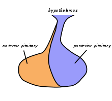
The pituitary gland is connected to the hypothalamus by the pituitary stalk (infundibulum) and is in
-
Anterior pituitary lobe (anterior pituitary lobe, adenohypophysis), consisting of
- Pars distalis (the anterior, largest section of the anterior lobe),
- Pars infundibularis (Pars tuberalis), covering the pituitary stalk, and
- Pars intermedia (intermediate lobe of the pituitary gland , a narrow median strip), and
- Posterior pituitary lobe (lobe posterior hypophysis, neurohypophysis)
assigned. The pituitary lobes differ from each other historically and functionally. While the adenohypophysis emerges from a protuberance in the roof of the pharynx, the so-called Rathke pocket , and attaches to the neurohypophysis, the neurohypophysis is an protuberance of the diencephalon . This difference can be recognized histologically, because while the adenohypophysis has various endocrine gland cells arranged in balls, the neurohypophysis is dominated by nerve cell processes, so-called axons , whose cell bodies are located in the hypothalamus. Thus, the adenohypophysis is able to produce hormones under the control of the hypothalamus and the neurohypophysis, on the other hand, is responsible as a storage and secretion organ for the hormones produced in the hypothalamus.
- Blood supply
The pituitary gland is supplied with blood through four arteries. From the pars cavernosa of the internal carotid artery arise two inferior hypophysial arteries, which form a capillary network, especially in the area of the neurohypophysis, into which the corresponding hormones are released. From the pars cerebral arteries of the internal carotid artery two spring hypophysiales the superior, in the region of the median eminence and the pituitary stalk Primärplexus form, in which some areas of the hypothalamus their hormones, Liberine and statins , secrete . They reach the secondary plexus , which is located on the adenohypophysis, via the venae portal hypophysiales . In this secondary plexus, the hormones of the hypothalamus reach their place of action and the hormones of the adenohypophysis are released from where they flow into the cavernous sinus and thus into the body's circulation in order to develop their effects.
Anterior pituitary hormones (adenohypophysis)
A distinction is made between hormones that act directly on their target organs (non- glandotropic hormones) and those that stimulate the hormone production of downstream endocrine glands (glandotropic hormones). The glandotropic hormones are also called control hormones , as they regulate the function of other endocrine glands. The growth hormone somatotropin (STH for somatotropic hormone or GH for growth hormone ) and prolactin have a direct effect on their target organs . In the case of glandotropic hormones, the gonadotropic hormones which act on the gonads ( gonads ) are follicle-stimulating hormone (FSH) and luteinizing hormone (LH) as well as the non-gonadotropic hormones, namely the adrenocorticotrophic hormone, which stimulates the adrenal gland (TSH ) and the thyroid gland stimulating hormone (TSH ) and the thyroid hormone (TSH) ) differentiated. By processing of a larger precursor peptide, the Proopiomelanocortins arise next to ACTH also melanotropin (MSH), β- endorphin and met-enkephalin . The hormone production of the pituitary gland is regulated by the hypothalamus by means of liberins and statins .
Pituitary lobe hormones
The intermediate pituitary lobe is among other things the place where melanocyte-stimulating hormones (MSH, melanotropins) are produced.
Posterior pituitary (neurohypophysis) hormones
The hormones that are stored and released in the posterior lobe of the pituitary gland are oxytocin and antidiuretic hormone (ADH), which is also known as adiuretin or vasopressin. ADH is formed in the nucleus supraopticus (core area that is above the optic nerve), oxytocin in the nucleus paraventricularis (core area in the hypothalamus) of the hypothalamus.
Diseases and diagnostics
In 1905, the Italian pathologist Gaetano Fichera (1880–1935) discovered a strong growth inhibition in chickens from which the pituitary gland had been removed, as was later also demonstrated in mammals. Herbert M. Evans then achieved a kind of giant stature in California by administering pituitary extract.
Underactive pituitary gland ( pituitary insufficiency , panhypopituitarism ) can have a variety of causes.
Tumors of the adenohypophysis are called pituitary adenomas . They often cause excessive hormone production. Large tumors can press on the optic nerves, causing significant visual disturbances. If left untreated, blindness is the result. Such tumors are often surgically removed through the nose, and the patient can usually see normally again immediately after the operation.
The physical examination is followed by hormone measurements and function tests. If there is clinical suspicion, hormone examinations should be carried out by an endocrinologist prior to imaging procedures, as imaging procedures often give false positive results (" incidentomas "). X-rays of the sella turcica in the lateral image of the bony skull, computer tomography , magnetic resonance tomography and somatostatin receptor scintigraphy are used as imaging methods .
Other tumors that can occur in the area of the pituitary- diencephalon (in addition to eosinophilic, basophilic and chromophobic tumors of the anterior pituitary) include craniopharyngiomas , pituitary duct cysts , gliomas , teratomas and Schüller-Christian granulomas .
literature
- Helga Fritsch, Wolfgang Kühnel: Pocket Atlas Anatomy. Vol. 2: Internal organs. Thieme, Stuttgart 2005, ISBN 3-13-492109-X .
- Lois Jovanovic, Genell J. Subak-Sharpe: Hormones. The medical manual for women. (Original edition: Hormones. The Woman's Answerbook. Atheneum, New York 1987) From the American by Margaret Auer, Kabel, Hamburg 1989, ISBN 3-8225-0100-X , pp. 11 f., 55 f., 74-77, 181 f., 290-292 and 376 f.
- Ulrich Welsch: Sobotta textbook histology. Elsevier, Munich 2006, ISBN 3-437-42421-1 .
Web links
Individual evidence
- ↑ TH Schiebler (Ed.): Textbook of the entire human anatomy. Cytology, histology, history of development, macroscopic and microscopic anatomy. 3. Edition. Springer-Verlag, Berlin / Heidelberg / New York / Tokyo 1983, ISBN 3-540-12400-4 , p. 684.
- ^ Otto Westphal , Theodor Wieland , Heinrich Huebschmann: life regulator. Of hormones, vitamins, ferments and other active ingredients. Societäts-Verlag, Frankfurt am Main 1941 (= Frankfurter Bücher. Research and Life. Volume 1), p. 28 f.
- ↑ Ludwig Weissbecker: Diseases of the pituitary-diencephalon system. In: Ludwig Heilmeyer (ed.): Textbook of internal medicine. Springer-Verlag, Berlin / Göttingen / Heidelberg 1955; 2nd edition, ibid. 1961, pp. 1008-1013, here: p. 1010.


