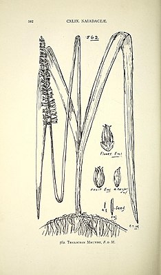
Maundia triglochinoides
| Maundia triglochinoides | ||||||||||||
|---|---|---|---|---|---|---|---|---|---|---|---|---|

Maundia triglochinoides , illustration |
||||||||||||
| Systematics | ||||||||||||
|
||||||||||||
| Scientific name of the family | ||||||||||||
| Maundiaceae | ||||||||||||
| Nakai | ||||||||||||
| Scientific name of the genus | ||||||||||||
| Maundia | ||||||||||||
| F. Garbage. | ||||||||||||
| Scientific name of the species | ||||||||||||
| Maundia triglochinoides | ||||||||||||
| F. Garbage. |
Maundia triglochinoides is arepresentative of the monocot plants that occurs only in Australia . It is the only representative of the genus Maundia and family Maundiaceae .
features
Vegetative characteristics
Maundia triglochinoides is an herbaceous, perennial plant . It forms rhizomes up to 5 mm thick . The leaves are tufted along the rhizome. The leaves are spongy and inflated, they are triangular in cross section. They are up to 80 cm long and 5 to 10 mm wide. The leaves are not differentiated into stalk and blade, a ligula is missing.
Inflorescence stalk
The peduncle is circular in cross section. It lifts the inflorescence above the surface of the water. It has neither leaves nor scales. The epidermis of the stem is a layer of cells thick. A layer of cortex is missing or consists of one to eight layers of thin-walled cells. The stele consists of numerous vascular bundles that are distributed in cross-section without a pattern. The central bundles are larger than the outer ones. They are collateral vascular bundles. In the larger vascular bundles there is a lacuna at the site of the protoxylem. The tracheids form a horseshoe-shaped row around the phloem . The tracheids are stiffened in a spiral shape with up to five parallel spirals per cell. Trachea were not observed. The peripheral vascular bundles are mostly inverted, ie the xylem is on the outside. The larger bundles are almost completely surrounded by a bundle sheath of thin-walled, lignified fibers, smaller bundles only have fibers on the phloem side, medium-sized bundles have two fiber strands on the phloem and xylem side. The space between the bundles is filled with an aerenchyme . Large air ducts are separated by unified rows of thin-walled cells. In the longitudinal axis, the air channels are divided into chambers by transverse septa. The septum cells are perforated and thus provide a connection between the individual chambers. Apart from the fiber cells in the vascular bundles, there are no specialized mechanical elements in the stem.
inflorescence
The inflorescence is an unbranched spike . It is up to 10 cm long and 2.5 cm wide. The flowers are in regularly alternating threefold whorls along the inflorescence . Before the anthesis , the internodes are very short, the flowers are very dense, so that the axis is not visible. Six orthostitches from flowers can be seen. After the anthesis, the internodes are stretched, the axis can be seen between the flowers or fruits. No flower-bearing bracts or rudiments can be seen. The flowers are completely sessile, sometimes with the exception of the uppermost flowers in the spike. The fruits are therefore also seated and stand vertically from the axis.
blossom
With the exception of the top flowers of an ear, all have a stable basic plan. The inflorescence consists of two tepals . These are transverse-abaxial. They are green and protrude 1.5 mm above the stamens . They have a narrow base and are attached with the receptacle to the radii of the two transverasl-abaxial stamens of the inner stamen whorl. You have a short nail that gradually widens into an almost circular plate . This is noticeably thick, there are numerous stomata on the abaxial side . Their guard cells are bean-shaped.
There are six stamens in two alternating, trimeric whorls: in the outer whorl there is a median abaxial stamen and two transverse-adaxial stamens, in the inner one a median adaxial and two transverse-abaxial stamens. The stamens are yellow, tetrasporangiate and bithekish. Stamens are missing. The connectors are wide and very short, only around a third of the length of the counter. The counters continue freely over the connective and just below. The anthers open extrors. The endothecium cells have fibrous thickenings. The pollen grains are spherical, inaperturate, the pollen surface is reticulate.
The gynoeceum consists of four carpels , only at the tip of the inflorescence the number can vary (3–5). The carpels are 6 to 8 mm long. They are distinctly tubular (ascidian). At the base they are congenitally connected to each other via the flower center. The connected part is about as long as the free one. Each carpel contains a single ovule that attaches ventrally. The ovule is pendulous, bitegmic and orthotropic. The micropyle is formed by the inner integument. The outer integument consists of three to four layers of cells and contains conspicuous air channels.
Alternative interpretations of the flower organs
Before the detailed study by Sokoloff et al. In 2013 there were different interpretations of the identity of the flower organs. Either the flower has two perianth segments or these are interpreted as two bracts . The stamens are either counted as 12 per bilocular stamens, or the six outer ones are interpreted as perianth segments, while the inner six are interpreted as stamens; or there are 12 unilocular stamens that have grown together in pairs. All 12 make pollen and open extrors. The structure, which is usually interpreted as a flower (euanthia interpretation), has also been interpreted on various occasions as an inflorescence consisting of several unisexual flowers (pseudanthia thesis).
fruit
After fertilization, the fused part of the carpels in particular develops, so that it is much more developed than the free part. Stamens and partly also the tepals remain on the fruit when the fruit ripens.
In the developing ovule , an unusual process occurs in the area of the chalaza (in the nucellus ) that is very rare among angiosperms: the cell walls of the nucellus cells closest to the chalaza dissolve, and a very large, multicellular nucleus is formed Coenocyte . This contains a large vacuole with numerous cytoplasmic strands. The cell nuclei should all be functional. The function of this structure is unknown. Between this coenocyte and the endosperm there are several layers of single-cell nucellus cells.
The information on whether the fruits open to ripeness vary. According to one interpretation, the fruits open to ripeness but remain connected to the central axis. However, according to several other authors, there is no morphological evidence that the fruit is opening.
The exocarp is a layer of cells thick and consists of short, thin-walled epidermal cells. They have stomata with bean-shaped guard cells; the number of cells surrounding the guard cells does not seem to be fixed. The mesocarp is multi-layered and consists mainly of medium-sized, thin-walled cells. In the middle and outer area of the mesocarp there are large spherical or elongated intercellular spaces . In the middle part of the fruit, each fruit compartment is surrounded by several layers of fibers that have rather thin woody cell walls. The endocarp consists only of the innermost layer of the pericarp.
The seeds fill the compartment almost completely. The Mesotesta and Exotesta are no longer visible in the ripe seed.
distribution
Maundia triglochinoides is only found in Australia. Here it is limited to the states of New South Wales and Queensland . To the south the area extends to southern Sydney. It grows in swamps and shallow fresh water on heavy clay soil. In both states, Maundia triglochinoides is classified as endangered (vulnerable). The cause of the threat is habitat loss and fragmentation.
Flowering occurs in the warm months. Maundia triglochinoides is probably wind pollinated ( anemophilia ).
Systematics
The Maundiaceae are a family of the Alismatales . Within this order they are the sister group of ( Potamogetonaceae + Zosteraceae ) within the "petaloid clade" .
The species Maundia triglochinoides and the genus Maundia were first described by Ferdinand von Mueller in 1858 . Traditionally, Maundia was placed in the Juncaginaceae family . Nakai Takenoshin established his own Maundiaceae family in 1943, but this was essentially only taken over by Armen Takhtajan in 1997. Only molecular genetic work by von Mering and Kadereit 2010 and Les and Tippery 2013 showed that Maundia does not belong to the Juncaginaceae. Based on this work, the Angiosperm Phylogeny Group recognized the genus as an independent family in 2016.
Individual evidence
- ↑ a b c d e f g Genus and species on Flora of New South Wales Online , accessed May 19, 2016.
- ↑ a b c d e f g h i j k l m n o p q Dmitry D. Sokoloff, Sabine von Mering, Surrey WL Jacobs, & Margarita V. Remizowa: Morphology of Maundia supports its isolated phylogenetic position in the early-divergent monocot or Alismatales . Botanical Journal of the Linnean Society 2013, Volume 173, pp. 12-45 doi : 10.1111 / boj.12068
- ^ New South Wales government, Environment & Heritage, Maundia triglochinoides (a herb) - vulnerable species listing , accessed May 24, 2016.
- ↑ a b The Angiosperm Phylogeny Group : An update of the Angiosperm Phylogeny Group classification for the orders and families of flowering plants: APG IV . Botanical Journal of the Linnean Society, 2016, Volume 181, pp. 1-20. doi : 10.1111 / boj.12385
- ↑ Donald H. Les, Nicholas P. Tippery: In time and with water ... the systematics of alismatid monocotyledons . In: P. Wilkin, SJ Mayo: Early Events in Monocot Evolution . Cambridge University Press 2013, pp. 118-164.
- ^ Sabine von Mering, Joachim W. Kadereit: Phylogeny, systematics and recircumscription of Juncaginaceae - a cosmopolitan wetland family . In: Seberg, Petersen, Barfod and Davis (eds.): Diversity, Phylogeny, and Evolution in the Monocotyledons . Aarhus University Press, Aarhus, Denmark, pp. 55-79. (PDF)
- ^ Ferdinand von Mueller: Fragmenta phytographiae Australiae . Vol. 1. Melbourne: Auctoritate Gubern. 1858.
- ↑ Nakai Tanegoshin: Maundiaceae. Ordines, Familiae, Tribi, Genera, Sectiones ... novis edita. Appendix , 1943, p. 213, Tokyo, Imperial University.
Web links
- Maundiaceae in: P. Stevens: APWebsite