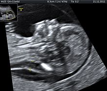
Neck transparency
The term neck transparency refers to a subcutaneous, i.e. , an accumulation of fluid ( edema ) located under the skin between the skin and the soft tissue above the cervical spine in the neck area of an unborn child.
The edema occurs between the 11th and 14th week of pregnancy , when the lymphatic system and the functions of the kidneys develop. As a result, the fluid cannot yet be drained away and lymph accumulates in the neck area, where the skin is very flexible. As a result of this fluid build-up, the neck transparency is created. Subsequently, in the course of further development, this lymphatic accumulation recedes again. The term has gained importance in the context of prenatal diagnostics in recent years and is derived from the fact that liquids appear black as an echo-free space on conventional ultrasound monitors and thus appear transparent. If there is a noticeable increase in neck transparency, the probability of various malformations is considered to be increased.
Neck transparency measurement
The terms neck density measurement , NT screening and, colloquially , neck fold measurement are used synonymously for the term neck transparency measurement . NT means nuchal translucency , so N Acken t ransluzenz or N Acken t ransparency. The NT measurement is a search test and not a diagnostic examination. This means that some of the children with a corresponding peculiarity are not recognized ( false negative results) and regular children are often attested to having “conspicuous” prognoses ( false positive results).
NT screening is often combined with an examination of two biochemical laboratory values ( PAPP-A and free β-hCG ) in the pregnant woman's blood and then referred to as first trimester screening .
The measurement is used to identify pregnant women who, from a statistical point of view, have a particularly increased probability of expecting a child with a particular chromosomal feature and / or a heart defect , so that further diagnostics (e.g. fine ultrasound , chorionic villus sampling , amniocentesis ) are particularly recommended can be.
Particularities in which an unusually increased nuchal transparency can be found with remarkable frequency are:
- Trisomy 10
- Patau syndrome (trisomy 13)
- Trisomy 15
- Trisomy 16
- Edwards syndrome (trisomy 18)
- Down syndrome (trisomy 21)
- Trisomy 22
- Triplo-X Syndrome (Trisomy X)
- Tetrasomy 12 p
- Cornelia de Lange Syndrome
- Noonan's Syndrome
- Turner syndrome (monosomy X / sometimes very pronounced accumulation of fluid, which in severe forms, which usually have a heart defect - typically coarctation of the aorta - can also have spread to the forehead, back, chest, abdomen and back of the baby's feet / cystic neck hygroma , isolated hydrothorax , ascites )
- Smith-Lemli-Opitz syndrome
- Joubert syndrome
- Ectrodactyly-ectodermal dysplasia syndrome
- Multiple pterygia syndrome
- Fryns Syndrome
Other causes, some of which are independent, some of which may be in connection with one of the chromosome peculiarities mentioned in the baby and some of which cause a particular degree of neck transparency, include:
- Heart defects or functional disorders of the heart (cardiovascular changes), especially in the case of coarctation of the aorta , i.e. a narrowing of the transition between the aortic arch and the thoracic aorta ( thoracic artery )
- Malformations of the lungs (pulmonary changes)
- Skeletal malformations (e.g. achondrogenesis / thanatophoric dysplasia )
- delayed or unusual development of the lymph vessels (lymphovascular changes)
- Congestion of vessels on the neck or head
- Sedentary lifestyle of the child
- Early fetofetal transfusion syndrome : In pregnancies with monochorionic diamniotic (and therefore identical and always same-sex) twins with early onset fetofetal transfusion syndrome, the neck transparency is often greater in the acceptor (larger twin) than in the donor (smaller twin)
- Diaphragmatic hernia (diaphragmatic hernia)
- Umbilical hernia (umbilical hernia)
- Malformation (s) of the kidneys
- Malformation (s) of the abdominal wall
An unusually large nuchal transparency is a so-called soft marker for the special features mentioned.
Qualification of the examiner
Doctors who carry out a neck transparency measurement require special training and a sufficiently resolving ultrasound device. In principle, therefore, internal quality assurance is to be aimed for. External quality control is offered by the Fetal Medicine Foundation , which receives three ultrasound images and the values measured in the year from the doctors involved in this control and then checks these according to a catalog of criteria. After the images have been checked, the doctors involved are given a certificate.
The attending physician is obliged to advise the pregnant woman in detail in advance of the examination and to discuss the advantages and disadvantages of measuring the neck transparency in an understandable manner. This also includes the indication that no diagnoses are possible through the examination alone, but invasive and comparatively high-risk examinations if chromosomal peculiarities are suspected, e.g. B. an amniocentesis or chorionic villus sampling would have to be followed to obtain a diagnosis.
In addition, their doctor must inform pregnant women in advance of the examination that certain physical characteristics such as B. Heart defects are meanwhile usually (operative) therapy or treatment methods, but there is no therapy for causal healing for chromosomally-related peculiarities , and thus ultimately only the acceptance of the child with his peculiarity, the postnatal release of the child for adoption or the postnatal transfer of the child to a foster family , a home or the termination of pregnancy exist as alternatives.
Examination time and course
The measurement of the neck transparency can be carried out between 11 + 0 and 13 + 6 weeks of pregnancy by ultrasound (or up to a crown-rump length of the child of max. 84 mm); the best results are obtained when the examination is carried out at 12 weeks of pregnancy. It does not matter whether the ultrasound is done vaginally (= via the vagina) or transabdominally (= via the abdominal wall), because the results do not differ significantly. The examination procedure for pregnant women does not differ from other ultrasound examinations and, according to the current state of knowledge, is neither harmful to the pregnant woman nor to the unborn child.
To calculate the degree of nuchal transparency, the unborn child is shown by ultrasound in the sagittal section , ie in the side view with the measurement attachment parallel to the central axis . The baby should fill the entire ultrasound monitor if possible, his spine should be down and his head should be neither in the flexion position nor in the hyperextension position .
The special accumulation of fluid in the neck area regresses in most babies after the 14th week of pregnancy, so that a measurement after this point in time can no longer produce a useful result.
Several measurements should always be made, from which the average value is then determined as the result. It is interesting that different doctors often get different readings from one and the same patient at the same gestational age. In the so-called FASTER study in the USA, in which more than 38,000 pregnant women took part, the neck transparency could not be shown in 4.5% of the cases.
Based on the measured nuchal transparency of the baby, the age of the pregnant woman, the age of pregnancy (pregnancy week), the length of the crown of the head of the child (it must be at least 45 mm and no more than 84 mm in size for this test) and any previous pregnancies with a baby with chromosome peculiarities Using a computer program or a table of values, a statistics-oriented individual probability statement for a child with a chromosome peculiarity is created.
Readings
Due to the usual physical developments already mentioned, it is by no means unusual, but completely normal for babies between the 11th and 14th week of pregnancy to have an accumulation of fluid in the neck area, the value of which averages 1 to 2.5 mm. An NT value from approx. 3.0 mm is considered to be significantly increased , a value of approx. 6.0 mm is considered to be greatly increased . The likelihood of a chromosomal aberration such as B. Trisomy 21 , Trisomy 18 , Trisomy 13 or Turner syndrome with an NT value of 3 mm is very dependent on the age of the pregnant woman. All NT values that are lower are within the normal range, although there are doctors who recommend a chromosome analysis from an NT value of 2.5 mm .
There is a tendency to observe a steady downward shift in the limit values. This results from the fact that the neck transparency measurement generally has a comparatively low informative value, which means that even if the values are basically inconspicuous, the child e.g. B. may have a trisomy 21. Conversely, children with increased neck transparency values can also be chromosomally and organically normal.
In some babies with one of the above-mentioned peculiarities, however, the neck area is noticeable to the extent that the values are viewed as an indication (as a so-called soft marker ) of a peculiarity. The statistics say that many babies with z. B. a form of trisomy an unusually high neck transparency is only found because they also have a heart defect or other physical developmental features that are associated with the neck edema.
So it happens that, statistically speaking, 5% of all neck transparency measurements have a particularly high neck transparency value (exceeding the 95th percentile ) in the unborn child, but then only in about 10 out of 100 cases special features with their own disease value can be found in further examinations.
Obviously, the high rate of false positive prognoses is often due to the fact that the measurements are often set too far down in the neck area and, as a result, larger values are measured than are actually available. It is also more common for the child's neck skin to be confused with the amnion (= the amniotic sac surrounding the child) and, as a result, an unusually large neck edema is found, although there is actually no particular expression.
Forecast and further action
The value of the measurement can be used as an opportunity to have further prenatal diagnostic examinations carried out, through which certain chromosome peculiarities and physical malformations can be diagnosed or excluded with a high degree of certainty.
Information on the exact prognostic correlation between the absolute NT values (in millimeters) and the various chromosome aberrations, genetic syndromes and malformations is not possible. The results known so far from the various studies show too great a spread for this.
For the assessment of a possible heart defect and other physical characteristics, ultrasound diagnostics are required, such as B. fine ultrasound , Doppler sonography or recordings in 3D ultrasound . However, these can or should only be carried out in later weeks of pregnancy, as they have little informative value beforehand or do not allow precise assessments of the respective malformation . Even in later stages, many heart defects cannot be precisely assessed in terms of their severity and prenatal and postnatal treatability.
For the clarification of chromosomal peculiarities the use of invasive examination methods is necessary and thus "the ultrasound examination (...) also becomes the rail for prenatal-invasive measures such as amniotic fluid puncture , chorionic villus sampling and chordocentesis " (Maier, 2000, p. 128).
New aspects
In the last few years, the working group headed by Professor Nicolaides in London has not only tried to advance the quality assurance of the neck transparency measurement, but also tried to find signs (markers) in the same cutting plane in which the measurement is taken –13 weeks of pregnancy not or only with difficulty recognizing malformations. Initial pilot studies show promising results for spina bifida , Dandy Walker malformation and corpus callosum agenesis . However, here too, such pilot studies must be confirmed by large follow-up studies. The first such results are available for spina bifida and dandy-walker malformations. In addition, the ultrasound examination, which originally only included screening for chromosomal disorders, has been significantly further developed so that screening for the risk of premature birth and preeclampsia is also possible. This enables early detection of the high-risk population to prevent both of these dreaded pregnancy complications. Compared to chromosomal disorders, both preeclampsia (simplified: high blood pressure disorders during pregnancy, "pregnancy poisoning") and premature birth occur significantly more frequently, and their risk can be reduced with relatively simple means (drug application) through an early diagnosis compared with the classification as high risk with 20-22 Pregnancy weeks (Chaoui, 2009; Scheier, 2011; Brückmann, 2013; Lachmann, 2010; Lachmann, 2011; Lachmann, 2012a; Lachmann 2012b; Lachmann, 2013; Lachmann and Schleussner, 2013; Lachmann and Schlembach, 2013; Lachmann R, 2013; Lachmann R, 2014).
Situation in individual countries
Germany
A neck transparency measurement is not part of the usual preventive examinations during pregnancy in Germany. It may only be carried out if the pregnant woman or the parents expressly request it, because according to the maternity guidelines it is not part of the maternity provision and is therefore a private benefit that must be paid for yourself. However, it is under discussion to make the measurement of the nuchal transparency an integral part of one of the three ultrasound examinations anchored in the guidelines.
In its September 2005 edition , for example, the Lebenshilfe newspaper , for example, spoke out against the assumption of neck transparency measurement by the health insurance companies . It is "not the task of the statutory health insurance to offer examinations that only look for a disability without a therapeutic approach" , especially since subsequent examinations such as amniocentesis, for example, “bring clarity about the possible diagnosis of a disability weeks later” . Health insurance companies are also rather skeptical of a corresponding innovation with a view to follow-up costs, since diagnostic certainty is only possible through follow-up examinations.
Switzerland
As part of the first trimester test (ETT), which is covered by the compulsory health insurance (basic insurance), a neck transparency measurement is carried out if the pregnant woman agrees. If the calculated risk as part of the first trimester test for trisomy 21, 18 or 13 is higher than 1: 1000, a non-invasive trisomy blood test (NIPT) will also be covered by the basic insurance.
See also
Hygroma colli - Dorsonuchal edema - Hydrops fetalis - Haemolyticus neonatorum disease
literature
- N. Bock: Analysis of two sonographic first-trimester screening concepts at the Women's Clinic of the MHH: a prospective follow-up study. Dissertation . Medical University, Hanover 2003. (pdf)
- JW Dudenhausen (ed.): Early diagnosis and advice BEFORE pregnancy. Pregravid risks.
- T. Degener and others: The main thing is that it is healthy? Female self-determination under human genetic control.
- M. Willenbring: Prenatal diagnosis and the fear of a disabled child. A psychosocial conflict between women from a systemic point of view.
- H. Friedrich, K.-H. Henze, S. Stemann-Acheampong: An Impossible Decision: Prenatal Diagnostics - Its Psychosocial Requirements and Consequences. Berlin 1998, ISBN 3-86135-274-5 .
- K. Griese: But a Mongi z. B. would be nice. How women deal with the offer of prenatal diagnostics.
- C. Swientek: What are the benefits of prenatal diagnostics? Information and experience.
- I. Schmid-Tannwald et al.: Prenatal medicine between healing mandate and selection. 2001.
- I. Dietschi: Test case child - The dilemma of prenatal diagnostics.
- E. Kirchner-Asbrock et al.: Being pregnant - a risk? Information and decision support for prenatal diagnostics.
- A. Brückmann et al.: Intima-media thickness of the carotid artery in the first trimester is a predictive parameter for preeclampsia. In: obstetric women's health. 2013; 73 - V02.
- R. Lachmann et al .: Posterior brain in fetuses with Dandy-Walker malformation with complete agenesis of the cerebellar vermis at 11-13 weeks: a pilot study. In: Prenat Diagn. 32 (8), Aug 2012, pp. 765-769.
- R. Lachmann et al: Midbrain and falx in fetuses with absent corpus callosum at 11-13 weeks. In: Fetal Diagn Ther. 33 (1), 2013, pp. 41-46.
- R. Lachmann: Correspondence regarding research letter published by Arigita et al. In: Prenat Diagn. 32 (2), Feb 2012, p. 201; author reply 202-3.
- M. Scheier et al .: Three-dimensional sonography of the posterior fossa in fetuses with open spina bifida at 11-13 weeks' gestation. In: Ultrasound Obstet Gynecol. 38 (6), Dec 2011, pp. 625-629.
- R. Lachmann et al .: Posterior brain in fetuses with open spina bifida at 11 to 13 weeks. In: Prenat Diagn. 31 (1), Jan 2011, pp. 103-106.
- R. Lachmann et al: Frontomaxillary facial angle in fetuses with spina bifida at 11-13 weeks' gestation. In: Ultrasound Obstet Gynecol. 36 (3), Sept. 2010, pp. 268-271.
- R. Chaoui et al .: Assessment of intracranial translucency (IT) in the detection of spina bifida at the 11-13-week scan. In: Ultrasound Obstet Gynecol. 34 (3), Sept. 2009, pp. 249-252.
- R. Lachmann, D. Schlembach: Preeclampsia screening. Prediction and prevention in the 1st, 2nd and 3rd trimester. In: The gynecologist. 4, 2013, pp. 326-331.
- R. Lachmann, E. Schleussner: Premature birth, prediction, prevention and diagnostics. In: Gynakol Gebh. 18 (4), 2013, pp. 32-38.
- R. Lachmann: Premature birth screening, prevention and obstetric risks of conization. In: Gyn. (18) 2013, pp. 1–8.
- R. Lachmann: The concept "Turning The Pyramid Of Care". New, extended first trimester screening. In: Gynakol Gebh. 19 (5), 2014, pp. 29–32.
Individual evidence
- ↑ Nina Bock: Analysis of two sonographic first-trimester screening concepts at the gynecological clinic of the MHH: a prospective follow-up study. 2003, p. 23.
- ^ LH newspaper. No. 3, 26th year, 09/2005, p. 11.
- ↑ Swissmom: The first trimester test

