Pinworm
| Pinworm | ||||||||||||
|---|---|---|---|---|---|---|---|---|---|---|---|---|

Head of a pinworm ( Enterobius vermicularis ), artificially colored |
||||||||||||
| Systematics | ||||||||||||
|
||||||||||||
| Scientific name | ||||||||||||
| Enterobius vermicularis | ||||||||||||
| Linnaeus |
The pinworm , Spring worm , Pfriemenschwanz or after Made ( Enterobius vermicularis , synonym : Oxyuris vermicularis of Greek ὀξύς oxys , tip 'οὐρά, Tail' and Latin vermiculus , worms') is a parasitic living in human intestines Fadenwurmart of up to 13 millimeters in length, . The pinworm is the most common intestinal worm in humans in Europe and one of the most common parasites in humans .
The species specializes in human hosts and is generally heavily committed to this host species. Monkeys are also more rarely attacked (in zoos) and possibly also cockroaches. Around 500 million infections are recorded worldwide every year, regardless of the age and social status of the infected person. The term `` child pinworm '', which can also be found, is a misleading worm, which is mainly observed in children with nocturnal itching of the anus and thus impaired performance, whose eggs can get under the fingernails and from there into the mouth by scratching the anus. About 50% of all people are attacked at least once in a lifetime. The pathological worm infestation itself is referred to as enterobiasis , enterobiosis , oxyuriasis or oxyuriosis .
Its occurrence accumulates in areas with a temperate climate, mainly in Europe and Asia.
features
The white worms are elongated and about half a millimeter thick. The females are 8 to 13 millimeters long and have a tapering tail and a characteristic bubble-like swelling of the cuticle at the front end. The males measure 2 to 6 millimeters, are trimmed at the end and have their tails curled up. The female can be distinguished from the male due to its size and pointed rear end. There are up to three larval stages. Under the microscope, the eggs appear typically elongated-oval and conspicuously asymmetrical on the longitudinal axis with one-sided flattening and measure around 20 to 30 by 50 to 60 micrometers (0.001 mm) .
Life cycle
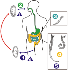
Without intermediate host stages , the egg ingested by the host - possibly inhaled - enters the stomach, where the egg shell is softened. The first larvae develop from the egg after just six hours. In the duodenum , the larvae hatch from the egg shell and moult. These migrate from the small intestine, where they shed their skin up to three times, to their preferred location on the intestinal wall around the appendix . There they suck on the intestinal wall and form a commensal (eating community) with their host . Here the animals grow out for about two to three weeks and reach sexual maturity.
After mating, the males die. After mating and the eggs have matured for about two weeks, the females migrate towards the anus to lay eggs. Mostly at night they crawl out of the anus, lay their eggs (5,000 to 17,000) on the anus folds within 10 to 30 minutes, and then die. The eggs already contain a tadpole-shaped embryo which, after being laid, develops into the second larval stage. If oxygen is exposed outside, the eggs become infectious within a few (4 to 8) hours. This comparatively very rapid development draws its energy mainly from glycogen stores , which are rapidly degradable compared to fat , which are concentrated in the middle and rear body area of the worms, where most of the differentiation and movement takes place in this development phase.
When scratching in the perianal area, eggs can stick to the fingers, especially under the nails, which are often picked up by the host itself through anus-finger-mouth contact, which leads to renewed infection ( self- infection ). Otherwise, the main route of spread is by airborne dusting of the tiny eggs, with eventual ingestion through contaminated food or inhalation of the eggs. Some authors claim that larvae that have hatched in the anus can also crawl directly into the intestine. The eggs of the pinworm are viable for up to three weeks.
history
Eggs of the pinworm have been found in human coprolites around 10,000 years old (approx. 7800 BC, found in Utah ). With settling down and the emergence of agriculture, the spread of infections with Enterobius vermicularis increased . It is assumed that humans have long had knowledge of the intestinal worms due to the fact that excreted worms can be easily recognized with the naked eye and their independent movement. Early mentions in literature can be found in the ancient Egyptian Ebers papyrus . Hippocrates of Kos (approx. 460-370 BC) the pinworm was known under the name "Ascaris" as a pathogen and treatment with strong laxatives. Physicians in the Roman Empire and later Arab physicians were also familiar with the species. The Roman physician Paulus Aegineta (625 to 690 AD) gave a good description of the clinical symptoms of the infection.
After the publication of the germ theory in the 19th century, a systematic investigation of its harmful effects began. Carl von Linné described it as Ascaris vermicularis , as a roundworm. Johann Gottfried Bremser (1767 to 1827) separated it from the genus Ascaris for the first time in 1819, still against the opposition of important scientific colleagues, and assigned it to the genus Oxyuris introduced by Karl Asmund Rudolphi , after he found the similarity to some in the intestines of rabbits and from had discovered species assigned to him here. Leach postulated the genus Enterobius in a paper published in 1853 and assigned the species to it.
In 1947 piperazine was introduced to treat enterobiasis. In 1958 Pyrvinium was introduced, which, among other things, is easier to use. In 1962 the broadband benzimidazole thiabendazole was introduced. In doses that are effective against worms, it is also quite toxic to the human organism. Since it is largely absorbed in the body as the hydrochloride, it often develops undesirable systemic side effects . It was the drug of choice until ivermectin was introduced . In 1968 pyrantel and 1969 levamisole were introduced, which are much better tolerated than thiabendazole. The thiabendazole derivatives mebendazole (1973), flubendazole (1976) and albendazole (1981), which hardly pass into the bloodstream, come from a further development of anthelmintics in veterinary medicine .
Enterobiasis
Symptoms, clinical picture
Pinworm infections often go unnoticed by the host. The main symptom of worm disease caused by pinworms is itching (especially at night) in the anal area , which occurs when the female pinworms lay their eggs around the anus. The itching, in turn, can lead to sleep disorders and their consequences, such as irritability, nervousness, difficulty concentrating, paleness or dark circles under the eyes. It also induces intensive scratching, which in turn can lead to skin abrasions ; these can become infected with bacteria. A massive worm infestation can lead to abdominal pain and weight loss, chronic diarrhea, rectal bleeding, or symptoms of chronic appendicitis ( appendicitis ).
Unlike many other intestinal parasites, the pinworm does not enter the bloodstream or organs other than the intestine. In rare cases, however, girls' genital tracts are affected, which can lead to vulvovaginitis . In extreme cases, adult worms can migrate through the vagina into the retroperitoneum, where they can lead to eosinophilic inflammation with accompanying ascites . The urethra and bladder can also be affected.
Diagnosis
The diagnosis takes place in several steps:
- Suspicion in the case of typical symptoms or infestation in the immediate vicinity
- In the morning before going to the toilet, you apply an adhesive strip to the region around the anus, remove it and stick it on a slide. Then you can microscope the eggs with the 5 to 10 lens. The double contour of the eggs is typical and movements of the larvae can usually be observed at higher magnifications. The examination should be repeated several times if the result is negative but the suspicion persists.
- Adult worms are occasionally found in the perianal region.
- The worms can also be seen in the stool. However, a parasitological examination of stool samples for the detection of oxyuriasis is less suitable than the adhesive tape technique.
- Dead female worms may be found in bed or in nightwear .
treatment
The worm treatment is medicated with anthelmintics . These are pyrvinium embonate from the 4th month of life (see Pyrvinium ) (one-time 5 milligrams per kilogram of body weight, leads to stool discoloration), from the 7th month of life pyrantel (one-time 10 milligrams per kilogram of body weight, maximum 1 gram) and also for children from 2 years of age 100 milligrams of mebendazole (Vermox) once. In the event of renewed or persistent infestation, one of the medications should be given three times in the dosage given above on days 1, 14 and 28 in order to prevent recurrences of autoinfection. If the infestation persists, the family members should be treated at the same interval with 3 doses as indicated above. In the case of vulvovaginitis due to oxyurides, which can be the cause of persistent infestation, therapy with albendazole (Eskazole) is recommended, as only this can be absorbed enterally in sufficient quantities . The single dose for children over 2 years of age and over 10 kilograms is 400 milligrams, children in the 2nd year of life and under 10 kilograms receive half the dose. Therapy should be carried out on days 1, 14 and 28 for successful rehabilitation. The day after the treatment, bedclothes, underwear and pajamas should be washed at at least 60 ° C.
In addition, it is advisable to adhere to the following hygiene rules during the duration of the infestation (hygiene measures alone, however, do not replace medication):
- In the morning after getting up, the anus region should be cleaned thoroughly with a wet washcloth. Hands and fingernails should be freed of any eggs immediately afterwards with a nail brush.
- Hands must be washed before eating.
- Avoid touching the anus region if possible; Wash your hands thoroughly after touching, especially your fingernails.
- The fingernails should be cut as short as possible.
- After every bowel movement, hands must be washed thoroughly and the space between the fingernail and finger must be thoroughly cleaned with a hand brush.
- Avoid raising dust, for example when making beds.
The eggs seem to be insensitive to disinfectants.
literature
- J. Dönges: Parasitology. With special consideration of human pathogenic forms. Thieme, Stuttgart 1988.
- H. Mehlhorn, G. Piekarski: Outline of parasite science. 6th edition. Heidelberg 2002.
Web links
- Pinworms - kindergesundheit-info.de: independent information service of the Federal Center for Health Education (BZgA)
Individual evidence
- ↑ OT Chan, EK Lee, JM Hardman, JJ Navin: The cockroach as a host for Trichinella and Enterobius vermicularis: implications for public health. In: Hawaii medical journal. Volume 63, Number 3, March 2004, pp. 74-77. PMID 15124739 .
- ↑ Hans Adolf Kühn: intestinal parasites. In: Ludwig Heilmeyer (ed.): Textbook of internal medicine. Springer-Verlag, Berlin / Göttingen / Heidelberg 1955; 2nd edition ibid. 1961, pp. 834-841, here: pp. 839 f. ( Oxyuris vermicularis ).
- ↑ a b c d Urania animal kingdom . Invertebrates 1. Urania, Leipzig 1993, ISBN 3-332-00501-4 , p. 348 f .
- ↑ a b c Anthony Fauci, Eugene Braunwald, Dennis L. Kasper, Stephen L. Hauser, Dan L. Longo, JL Jameson, Joseph Loscalzo: Harrison's internal medicine . Ed .: Manfred Dietel, Norbert Suttorp, Martin Zeitz. 17th edition. tape 1 . ABW Wissenschaftsverlag, 2008, ISBN 978-3-936072-82-2 , p. 1637 .
- ↑ a b c Hans-Eckhard Gruner (Ed.): Textbook of Special Zoology . tape 1 : invertebrates . Spektrum Akademischer Verlag, 1999, ISBN 3-334-60474-8 , pp. 515 f .
- ↑ Peter O'Donoghue: Enterobius. In: Para-Site. Faculty of Science, The University of Queensland, May 2010, accessed December 13, 2012 .
- ↑ a b Lexicon of Biology . Spectrum Academic Publishing House, Heidelberg 2002.
- ↑ Wilfried Westheide, Reinhard Rieger (Ed.): Special Zoology . 2nd Edition. Part 1: Protozoa and invertebrates . Spektrum Akademischer Verlag, 2006, ISBN 3-8274-1575-6 , p. 751 .
- ↑ Hans Engelbrecht: About Enterobius vermicularis (Linné 1758, Leach 1853) . I. Glycogen and Fat in the Ovary. In: Journal of Parasitic Studies . tape 23 , no. 4 , 1963, pp. 384-389 , doi : 10.1007 / BF00331237 .
- ^ Gary F. Fry, Jennifer G. Moore: Enterobius vermicularis: 10,000-year-old human infection . tape 166 , no. 3913 , 1969, p. 1620 , doi : 10.1126 / science.166.3913.1620 , PMID 4900959 (English).
- ↑ Alena Mayo Iñiguez, Karl Reinhard, Marcelo Luiz Carvalho Gonçalves, Luiz Fernando Ferreira, Adauto Araújo, Ana Carolina Paulo Vicente: SL1 RNA gene recovery from Enterobius vermicularis ancient DNA in pre-Columbian human coprolites . In: Australian Society for Parasitology (Ed.): International Journal for Parasitology . tape 36 , no. 13 November 2006, pp. 1419–1425 , doi : 10.1016 / j.ijpara.2006.07.005 (English).
- ↑ Constantinos Trompoukis, Vasilios German, Matthew E. Falagas: From the Roots of Parasitology: Hippocrates' First Scientific Observations in Helminthology . In: Journal of Parasitology . tape 93 , no. 4 , August 2007, p. 970-972 , doi : 10.1645 / GE-1178R1.1 (English).
- ^ Francis EG Cox: History of Human Parasitology . In: American Society for Microbiology (Ed.): Clinical Microbiology Reviews . tape 15 , no. 4 , October 2002, p. 595–612 , doi : 10.1128 / CMR.15.4.595-612.2002 (English, cmr.asm.org ).
- ^ Maurice Ernst: Oxyuris vermicularis (the threadworm) . A treatise on the parasite and the disease in children and adults, together with the particulars of a rapid, harmless and reliable cure. Ernst Homeopathic Consulting Rooms and Dispensary, London 1910 (English, archive.org ).
- ^ J. Horton (WHO), GlaxoSmithKline: The efficacy of anthelminthics: past, present, and future . In: World Health Organization (Ed.): Controlling disease due to helminth infections . Geneva 2004, ISBN 92-4156239-0 , p. 143 ff . (English, who.int [PDF]).
- ↑ a b c d German Society for Pediatric Infectious Diseases [DGPI] (Ed.): DGPI Handbook. Infections in children and adolescents . 6th, completely revised edition. Thieme Verlag, Stuttgart / New York 2013.
- ↑ Gerald Hellstern, Martin Bald, Claudia Blattmann, Hans Martin Bosse: Short textbook pediatrics . 1st edition. Thieme, 2012, ISBN 978-3-13-149941-7 , pp. 202-203 .
- ↑ H.-A. Oelkers et al .: Investigations on oxyure eggs . In: Journal of Parasitic Studies . tape 14 , no. 6 , November 2, 1950, p. 574-581 ( cabdirect.org ).


