Armpit
In human anatomy, the armpit ( lat .: Axilla ) describes the area between the shoulder and the chest wall. In the narrower sense, however, the armpit means the armpit ( axillary fossa ), whereby it should be noted that the anatomical definition of the armpit does not correspond to the colloquial term. In anatomy, the armpit is the space under the shoulder that is bounded in front by the anterior axillary fold, formed by the large pectoral muscle , behind by the posterior axillary fold, formed by the large back muscle , and inwardly by the rib cage , but lies under the skin. The colloquial term armpit, on the other hand, only means the skin fold between the chest wall and the upper arm, i.e. the skin of the axillary fossa.
As a rule, the armpits are hairy from puberty . In various cultures, however, armpit hair is considered unaesthetic and is therefore removed by the wearer (see also the section “Cultural Aspects” in the “Body Hair” article ).
In the armpit there are many sweat and sebum glands ( axillary glands ), such as the apocrine sweat glands (Glandulae sudoriferae apocrinae), which enable the armpit hair to send more sexual attractants ( pheromones ) (see also vomeronasal organ ). By means of a deodorant (short Deo , also deodorant ) that is mainly applied in the armpits can body odor suppressed and / or the activity of the eccrine sweat glands are inhibited (see antiperspirant ). The skin glands can be responsible for a variety of diseases , including underarm wetness and carbuncle . The axillary lymph nodes lie under the skin .
In the armpit, the body temperature is often measured with a clinical thermometer (axillary temperature measurement) in order to diagnose any fever , even if this method is particularly inaccurate compared to rectal or oral measurements.
The axillary blockade is a regional anesthetic procedure that enables surgical interventions on the arm.
Armpit anatomy
Axillary fossa
When the upper arm is abducted (spread apart), the axillary fossa forms a four-sided pyramid, the spatial extent of which is approximately the following:
| Base: | Axillary fascia |
| ventral (front) wall: | Fascia clavipectoralis , musculus pectoralis major , musculus pectoralis minor |
| dorsal (rear) wall: | Subscapularis , latissimus dorsi , teres major |
| medial (towards the middle of the body) wall: | Serratus anterior muscle |
| lateral (outer) margins: | anterior and posterior axillary folds (formed by the pectoralis major and latissimus dorsi muscles ) |
| cranial (upper) wall: | Shoulder joint ( articulatio humeri ), proximal end of the humerus, coracobrachialis and short head of biceps |
| Top: | Middle of the collarbone |
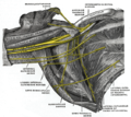
Superficial muscle layer of the rib cage and upper arm, viewed from the dorsal right side
The branches of the brachial artery
The right brachial plexus (the infraclavicular part), viewed from the front and below
Axillary gaps
Since the space between the teres major muscle and the teres minor muscle is divided by the long triceps head , two gaps arise, which are known as the lateral axillary foramen (lateral axillary gap ) and the medial axillary foramen (central axillary gap). By the square foramen lateral axillary the contact axillary nerve , of the deltoid muscle innervated and the above preferred skin, and the posterior humeral circumflex artery ; through the triangular foramen axillary mediale the arteria circumflexa scapulae .
Physiological microbiome of the armpit
The microbiome generally designates - here in the area of the human armpit - in a broader sense the entirety of all microorganisms colonizing humans. In the narrower sense, this denotes the entirety of all microbial genes or genomes (DNA) in the human organism and distinguishes it from the term microbiota, which denotes all microorganisms.
In a rough overview, the human skin can be divided into three zones: into oily, moist and dry regions. Oily skin, d. H. rich in sebum glands can be found between the eyebrows, next to the nose, on the back of the head, on the chest and on the upper back. Moist regions can be found at the nasal entrance, under the armpits, in the crook of the elbows, in the hollow of the knee, on the sole of the foot, in the navel or in the buttock folds. The dry regions include the skin on the buttocks, palms and forearms.
One of the earliest studies of the armpit microbiota was carried out by James Leyden et al. (1981) published, it was possible to identify members of the bacterial family Micrococcaceae and representatives of the bacterial genera Corynebacterium and Propionibacterium in bacterial cultures in over 200 women and men examined .
Jackman and Noble (1983) examined the bacterial composition of the armpits of 163 male and 122 female test persons and were able to show that the aerobic bacteria of the genus Corynebacterium spp. prevailed, while in both women the axillary bacterial flora was dominated by Micrococcaceae.
Patrick Zeeuwen et al. (2012) were able to show in their experimental set-up (injured vs. regenerating skin) that the actual microbiome of the skin is not located on the surface of the horny layer ( stratum corneum ), but in the deeper layers of the cornea underneath and that there is not just one There are different microbial compositions of the uppermost horny layer of normal healthy skin with that in the deeper skin layers of the skin in the individual individuals, but that there were also clear differences in the composition of the microbiome between women and men . Strikingly, they found bacteria from the genital area , even if only in small numbers, in the deeper layers of the skin . In women, bacteria from the vulva and vagina and in men, bacteria from the penis .
literature
- G.-H. Schumacher: Topographical Human Anatomy. Georg Thieme Verlag, 5th edition, 1988
- Franz-Viktor Salomon et al. (Ed.): Anatomy for veterinary medicine. Enke-Verlag Stuttgart, 2nd ext. 2008 edition, ISBN 978-3-8304-1075-1
Web links
Individual evidence
- ↑ axilla . Roche Medical Lexicon, 5th edition. Urban & Fischer, 2003.
- ^ Gerhard Aumüller, Gabriela Aust, Andreas Doll, Jürgen Engele, Joachim Kirsch, Siegfried Mense, Dieter Reissig, Jürgen Salvetter, Wolfgang Schmidt, Frank Schmitz, Erik Schulte, Katharina Spanel-Borowski, Werner Wolff, Laurenz J. Wurzinger and Hans-Gerhard Zilch: Dual Series Anatomy . 2nd Edition. Georg Thieme, 2010, ISBN 978-3-13-136042-7 , p. 424 .
- ↑ a b Michael Schünke, Erik Schulte, Udo Schumacher: PROMETHEUS - learning atlas of anatomy . 4th edition. General anatomy and musculoskeletal system. Thieme, Stuttgart New York 2015, ISBN 978-3-13-139544-3 , pp. 382 .
- ↑ Urs Jenal - Biozentrum Universität Basel: Humans and their microorganisms: Interactions between illness and well-being. (How many people is a person?) Biozentrum.unibas.ch ( Memento of the original from March 4, 2016 in the Internet Archive ) Info: The archive link was inserted automatically and has not yet been checked. Please check the original and archive link according to the instructions and then remove this notice. (PDF).
- ^ EA Grice: The skin microbiome: potential for novel diagnostic and therapeutic approaches to cutaneous disease. In: Seminars in cutaneous medicine and surgery. Volume 33, Number 2, June 2014, pp. 98-103, PMID 25085669 , PMC 4425451 (free full text) (review).
- ^ Eugenie Fredrich-Vahle: Metatranscriptome analyzes of the microbial communities of the human ear and the armpit. Dissertation, Bielefeld University, Bielefeld 2016 [1]
- ↑ Jackman PJH, Noble Toilet .: Normal axillary skinmicroflora in various populations. Clin Exp Dermatolog 1983; 8: 259-68.
- ↑ PL Zeeuwen, J. Boekhorst, EH van den Bogaard, HD de Koning, PM van de Kerkhof, DM Saulnier, II van Swam, SA van Hijum, M. Kleerebezem, J. Schalkwijk, HM Timmerman: Microbiome dynamics of human epidermis following skin barrier disruption. In: Genome biology. Volume 13, number 11, November 2012, p. R101, doi : 10.1186 / gb-2012-13-11-r101 , PMID 23153041 , PMC 3580493 (free full text).


