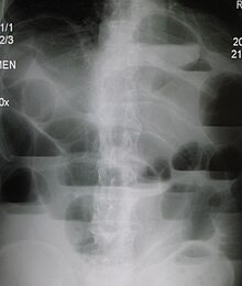
Intestinal obstruction
| Classification according to ICD-10 | |
|---|---|
| K56 | Paralytic ileus and mechanical ileus without a hernia |
| K56.0 | Paralytic ileus |
| K56.1 | Intussusception |
| K56.2 | Volvulus |
| K56.3 | Gallstone ileus |
| K56.4 | Other obturation of the intestine |
| K56.5 | Intestinal adhesions with ileus |
| K56.6 | Other and unspecified mechanical ileus |
| K56.7 | Ileus, unspecified |
| ICD-10 online (WHO version 2019) | |
The ileus ( Latinized form of the Greek εἰλεός ileós , from ancient Greek εἰλεῖν eilein , German ' to include, to push together' ), German for intestinal obstruction , is an interruption of the intestinal passage. As a life-threatening condition , it generally requires immediate hospitalization and possibly surgical intervention. In intestinal obstruction (ileus), a distinction is made between a mechanical ileus ( intestinal stenosis ) and a paralytic ileus (due to paralysis of the intestinal muscles, intestinal paralysis ).
Classification
Classification according to the cause
One differentiates here
- the mechanical ileus with obstruction of the intestinal passage from the outside or from the inside (seen from the intestinal lumen ). In addition, insufficient blood supply to the intestinal wall can occur, especially in the case of a pinched fracture ( incarceration ), a strangulation ileus or a volvulus .
- the functional ileus with paralysis of the smooth muscles that are responsible for the transport of intestinal contents. The peristalsis comes to a standstill . In this regard, a distinction is again made between
- the comparatively common paralytic ileus, e.g. B. with peritonitis ,
- the vascular ileus caused by the occlusion of the blood vessels supplying the intestine (due to embolism or thrombosis with structural vasoconstriction ) and
- the very rare spastic ileus (in lead poisoning ).
Classification according to the intestinal section
According to the section of intestine in which the obstruction occurs, one can subdivide into small intestine ileus and large intestine ileus. Strangulations with vascular involvement occur more frequently in the small intestine, while obstructions predominate in the large intestine.
Classification according to age
Different types of ileus are different in frequency depending on age:
- in newborns & infants
- Atresia of the pylorus , duodenum , jejunum , ileum , colon , anal atresia
- severe stenoses as a result of u. a. Annular pancreas , duodenal membrane
- Defects such as upside-down stomach, Ledd's ligaments
- acquired with intussusception , meconium ileus , typical symptom of cystic fibrosis
- with the child
- Volvulus , incarcerated hernia , toxic megacolon , Meckel's diverticulum
- in all age groups
- postoperative adhesions
- perforation-related
- in the elderly
- Colon ileus due to colon carcinoma
Classification according to the symptoms
The distinction between ileus and subileus (preliminary stage of ileus) depends on the severity of the build-up and the symptoms. The boundary between the two forms is blurred and not clearly defined, but for pragmatic reasons it separates the group with an indication for an operation from a group with a conservative treatment attempt in mechanical ileus .
causes
Mechanical ileus
Causes of mechanical ileus can include: a. be:
-
mechanical blockage (obturation) due to:
- Meconium ( Mekoniumileus , see above), ball of feces or bezoar
- a foreign body in the intestines
- a large gallstone . Such a stone only ever reaches the intestinal lumen through an inflammatory fistula between the gallbladder and the intestinal wall and can cause the rare gallstone ileus.
-
a grown or inflammatory narrowing (obstruction)
- an intestinal tumor
- Crohn's disease with bowel stenosis
-
Laying, disconnection (strangulation)
- an adhesion cord ( bride ) clamps the intestine, leads to the ileus of the bridle
- sticky kink of the intestine (adhesion ileus) in ileitis
- an incarceration , i.e. a pinched intestinal hernia (hernia)
- a volvulus , ie a twisting of the intestine, constricts the lumen and the blood supply
- an intussusception , a di invagination of an intestinal part to another, narrowing leads both for the installation as well as for-clamping of the blood supply to the intestine
Paralytic ileus (intestinal atony)
Causes for the cessation of the peristalsis and the slackening of the intestinal wall (paralytic ileus, syn. Intestinal atony ) are
- Inflammation in the abdominal cavity ( peritonitis ) or in spaces or organs adjacent to the abdominal cavity
- with intestinal perforation
- as a complication after surgical interventions in the abdominal cavity (postoperative paralytic ileus)
- Immigration peritonitis: Any untreated mechanical ileus becomes a paralytic ileus (mixed ileus) after a certain period of time
- in pancreatitis
- in pneumonia with septic spread of the pathogen
- Poisoning
- Circulatory disorders / supply disorders of the intestine
- in acute circulatory disorders of the intestine
- in mesenteric infarction
- in severe hypokalaemia
- reflexively
- Colic of an abdominal organ outside the abdominal cavity, renal colic , biliary colic , ureteral colic
Symptoms
The symptoms of an intestinal obstruction are cramp-like abdominal pain, a bloated stomach ( meteorism ), vomiting of stomach and intestinal contents up to vomiting ( miserere ), wind and stool retention with increased peristalsis ("mechanical bowel noises" during auscultation). Typically, the pain can be clearly localized at the beginning, but is more and more diffused over the entire abdomen as it progresses. In the late stage, the symptoms of peritonitis appear because germs migrate through the intestinal wall.
diagnosis
The physical examination , including rectal examination results with local pain, guarding , hernias ( hernias ) and hyperinflation important information. In addition, blood is taken to determine laboratory values as well as imaging with sonography and possibly x-ray of the abdomen .
A distinction between mechanical and paralytic ileus can in many cases be made very quickly by listening to the abdomen. While a mechanical ileus manifests itself through hyperperistalsis (increased bowel activity and metallic-sounding noises), the paralytic ileus manifests itself as a "dead silence" in the abdomen.
If ileus is suspected, sonography provides quick information: one sees a typical pendulum peristalsis , i. H. the intestinal contents oscillate back and forth. There are also widened loops of intestine that are excessively filled with air or liquid. One can often narrow down the location of the closure. Sections of the intestine behind a mechanical obstacle have collapsed (starvation bowel) .

The blank x-ray of the abdomen while standing or lying on the left side shows in typical ileus overextended intestinal loops with fluid levels as an indication of the location of an obstruction. Depending on the patient's condition, the location and cause can be narrowed down by an enema and / or a passage with contrast medium .
In addition, a CT of the abdomen can be performed. This can often be used to find the cause of the bowel obstruction.
Differential diagnosis
Other diseases such as pseudo-obstruction Ogilvie's syndrome , spastic colon and metabolic disorders such as porphyria , toxic megacolon or Crohn's disease must be distinguished from the long list of causes of ileus .
Sometimes the cause of an ileus cannot be identified and a laparoscopy is required.
pathology
Due to the occlusion or the lack of peristalsis, the intestinal lumen distension and stool retention occurs. The lack of movement of the chyme and its build-up lead to increased bacterial growth, which can lead to translocation into the blood vessels and sepsis. At the same time, there is a massive shift in electrolytes and water caused by the local inflammatory reaction in and around the intestine and into the retroperitoneum . This can lead to severe dehydration, similar to pancreatitis . Bacterial toxins and the fluid displacement can lead to circulatory and multiple organ failure . In its effects on the whole body, one speaks of ileus disease .
If the ileus remains unknown, the intestinal wall may perforate. This causes fecal peritonitis .
treatment
Therapy depends on the underlying cause. This also applies to the procedure to be used in the event of an operation. In the case of a paralytic ileus, prokinetics can be used to stimulate bowel activity. If strangulation or a strap is present, surgical intervention, preferably using a minimally invasive technique (laparoscopy), is indicated.
Accompanying measures serve to relieve the intestines such as gastric tube , possibly parenteral nutrition or enema . A central venous catheter should be considered for rapid and adequate fluid therapy. A urinary catheter should be used for balancing.
See also
literature
- Marcel Bettex, Noel Genton, Margrit Stockmann (eds.): Pediatric surgery. Diagnostics, indication, therapy, prognosis. 2nd, revised edition. Thieme, Stuttgart et al. 1982, ISBN 3-13-338102-4 .
- Friedrich Carl Sitzmann: Pediatrics. Diagnostics - therapy - prophylaxis. 6th, enlarged and completely revised edition. Hippokrates, Stuttgart 1987, ISBN 3-7773-0827-7 .
- Martin Allgöwer , Jörg R. Siewert , Hubert J. Stein: Surgery. With integrated case quiz - 40 cases according to the new AO. 9th, revised edition. Springer, Berlin et al. 2012, ISBN 978-3-642-11330-7 .
- Hans Adolf Kühn: Organic disorders of the intestinal mobility and motor skills (intestinal stenosis and ileus). In Ludwig Heilmeyer (Hrsg.): Textbook of internal medicine. Springer-Verlag, Berlin / Göttingen / Heidelberg 1955; 2nd edition, ibid. 1961, pp. 828-820.
Web links
Individual evidence
- ↑ Klaus-Dietrich Ebel, Eberhard Willich , Ernst Richter (eds.): Differential diagnosis in pediatric radiology. Volume 2: Thorax, mediastinum, heart, large vessels, abdomen, urogenital tract. Thieme, Stuttgart / New York 1995, ISBN 3-13-128101-4 , p. 337 f.
- ↑ Wolfgang Leps, Matthias Lohr: Internal medicine. 14th edition. Georg Thieme, Stuttgart et al. 2003, ISBN 3-13-112874-7 , p. 419.
- ↑ Volker Hofmann, Karl-Heinz Deeg, Peter F. Hoyer: Ultrasound diagnostics in paediatrics and pediatric surgery. Textbook and atlas. 3rd, completely revised and expanded edition. Thieme, Stuttgart / New York 2005, ISBN 3-13-100953-5 .


