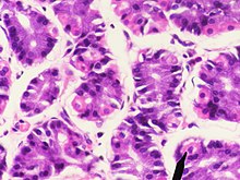
Gastric mucosa
The gastric mucosa ( Latin: tunica mucosa gastrica ) is the inner lining ( mucous membrane ) of the stomach . It consists of an epithelium ( lamina epithelialis mucosae ) and a layer of its own ( lamina propria mucosae ). The connecting layer to the smooth muscles of the stomach wall, which is located further out, is called the submucosa ( Tela submucosa ).
The gastric mucosa forms the stomach acid , pepsinogen (the inactive precursor of the protein-splitting enzyme pepsin), the intrinsic factor required for the absorption of vitamin B12 and various hormones . It also causes the stomach to be lined with a thick layer of mucus that protects the stomach wall from stomach acid.
The lining of the stomach is wrinkled depending on how full it is. The mucous membrane shows a field, these "fields" are called areas gastricae . Crater- like depressions extend from the mucous membrane to form the self- layer , which are referred to as "( Donders ) gastric dimples" ( Foveolae gastricae ).
Lamina epithelialis mucosae
The epithelium of the gastric mucosa consists of only one cell layer of highly prismatic (higher than wide) cells (single-layer, highly prismatic epithelium). The cells ( Epitheliocyti superficiales gastris ) are firmly connected to one another by so-called tight junctions . Numerous mucus-producing secondary cells are scattered in the epithelium. The mucus of these cells and that of the gastric glands (see below) protects the epithelium from the hydrochloric acid produced in the stomach .
In humans, the mucus is essentially formed by the surface epithelia or the foveolar cylindrical cells and secreted holocrine, thus by the cell molt. The so-called Adjacent cells are called mucous neck cells and are usually only found in the neck of the gland; they are not scattered between the foveolar cylindrical cells.
Lamina propria mucosae
The own layer consists of connective tissue , blood vessels , lymph vessels , cells of the immune system (partly as lymph follicles ) and glands . The gastric glands open into the pits in the stomach. These are tubular (tubular) glands. Depending on the stomach region, these are designed differently and also fulfill different functions. One differentiates between cardia, fundus and pylorus glands. In the epithelium of the gastric glands, not only exocrine cells but also endocrine active cells are integrated, which belong to the diffuse neuroendocrine system (DNES) .
Towards the submucosa, the intrinsic layer has a layer of smooth muscles, the lamina muscularis mucosae . At the transition to the submucosa there are ganglia of the Meissner plexus ( plexus submucosus ).
Cardiac glands
The cardiac glands ( glandulae cardiacae ) extend to the area of the stomach entrance ( cardia ), only in pigs they take up almost half of the stomach. In horses , they are formed on an edge area at the transition from the cutaneous to the actual mucous membrane.
The gland tubes are branched and twisted. Their wall consists of the actual gland cells ( epitheliocyti cardiaci ), which secrete mucins , an alkaline , slimy secretion.
Fundic glands
The fundus or autologous glands ( Glandulae gastricae propriae ) form the actual gastric juice. They extend to the stomach floor ( fundus ) and body ( corpus ventriculi ). They are elongated tube glands with different cell types. These hoses are divided into three sections:
- Isthmus : the narrow point at the transition from the stomach pit (Foveola gastrica ) to the glandular tube
- Neck ( cervix ) of the gland tube
- Main part ( pars principalis ): lower section and glandular base
The cells of the isthmus partly consist of epithelial cells. They form, as the rest of the epithelium, mucus ( mucus ) and bicarbonate ions, which bases a buffering effect have opposite the free protons. This alkaline mucus protects the epithelium from the acidic environment of the stomach. Further down there are stem cells that continuously divide and replace the dying epithelial cells.
In the neck of the gland, there are mainly secondary and parietal cells. In the main part are the main cells and also some parietal cells.
The secondary cells ( Mucocyti cervicales ) are iso- to highly prismatic and have a basal cell nucleus. They also secrete alkaline mucus to protect the epithelium.
The parietal cells ( Exocrinocyti parietales , also parietal cells ) lie between the other cells or on the outside of them. They secrete protons, which combine with chloride ions to form hydrochloric acid extracellularly , and the intrinsic factor necessary for cobalamin absorption (vitamin B12) . The cells form intracellular secretion canals with microvilli . In the active state of the cells, these channels are built into the luminal plasma membrane (facing the gastric cavity) and thus enlarge the contact and release surface. Here there are proton-potassium pumps that transport protons out of the cell in exchange for potassium ions. Parietal cells are relatively large and eosinophilic ( can be stained with eosin and are therefore reddish).
The enterochromaffin-like cells (also ECL cells or H cells ) are endocrine cells that are often located near the parietal cells. They produce histamine , which has a stimulating effect on the acid production of the parietal cells.
The main cells ( Exocrinocyti principales ) in the main part of the gastric glands are highly prismatic with a basal cell nucleus. They form Pepsinogens , the precursors of different enzymes that collectively Pepsine be called (in ruminants the Lab ). They are temporarily stored in the cells in so-called zymogen granules . Main cells are basophilic due to the large proportion of rough endoplasmic reticulum (rER) ( can be stained with basophilic dyes, therefore bluish).
Pyloric glands
The pyloric glands ( glandulae pyloricae ) are located at the gastric outlet ( pylorus ). Similar to the cardiac glands, they secrete an alkaline, slimy secretion. They are isoprismatic, the nucleus is partly very flattened. In contrast to the gastric glands located further orally (i.e. towards the esophagus), the pyloric glands have no main and hardly any parietal cells. In addition to the exocrine gland cells ( Exocrinocyti pylorici ) that produce mucus, endocrine cells that form hormones and release them to the surrounding blood vessels are integrated in the epithelium of the pyloric glands :
The G cells produce the hormone gastrin , which stimulates acid production in the parietal cells. This happens directly (by stimulating the acid-producing parietal cells) and indirectly, by stimulating the release of histamine in ECL cells.
The D cells make somatostatin . They are stimulated by stomach acid in the stomach lumen. Somatostatin inhibits gastrin release in G cells and histamine release in ECL cells (which has a negative effect on acid production) as well as directly acid production in parietal cells. D cells are also found in the fundus and body of the stomach, as well as in the duodenum (and in other organs of the body such as the pancreas ).
Submucosa
The tela submucosa represents a shifting layer.
Individual evidence
- ↑ a b c d e Renate Lüllmann-Rauch: Pocket textbook histology . 3. Edition. Thieme Verlag, Stuttgart 2009, ISBN 978-3-13-129243-8 , p. 373 ff .
- ^ Reinhard Hildebrand: Rudolf Albert Koelliker and his scientific contacts abroad. In: Würzburger medical historical reports 2, 1984, pp. 101–115; here: p. 107.
- ↑ Michael Gekle: Nutrition, Energy Balance and Digestion . In: Michael Gekle et al. (Ed.): Pocket textbook Physiology . Thieme Verlag, Stuttgart 2010, ISBN 978-3-13-144981-8 , p. 448 f .
literature
- Franz-Viktor Salomon: stomach, ventriculus (gaster) . In: Franz-Viktor Salomon et al. (Hrsg.): Anatomie für die Tiermedizin . 2nd ext. Edition. Enke Verlag, Stuttgart 2008, ISBN 978-3-8304-1075-1 , p. 272-293 .

