NO synthases
| Nitric oxide synthase | ||
|---|---|---|

|
||
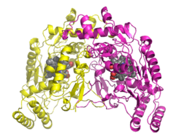
| Crystal structure of human nitric oxide synthase according to PDB 1nsi | ||
| Properties of human protein | ||
| Mass / length primary structure | 1200-1400 amino acids | |
| Secondary to quaternary structure | Homodimer | |
| Cofactor | Heme, FAD, FMN, tetrahydrobiopterin | |
| Isoforms | NOS1, NOS2, NOS3 | |
| Identifier | ||
| Gene names | 1nsi , NOS1, NOS2, NOS3 | |
| External IDs | ||
| Enzyme classification | ||
| EC, category | 1.14.13.39 , monooxygenase | |
| Response type | Dihydroxylation | |
| Substrate | L- arginine + n NADPH + m O 2 | |
| Products | L- citrulline + NO + n NADP + + m H 2 O | |
| Occurrence | ||
| Homology family | NO synthase | |
| Parent taxon | Creature | |
| Exceptions | Archaea | |
The enzyme nitrogen monoxide synthase , or NO synthase ( NOS ) for short , catalyzes the formation of nitrogen monoxide (NO) from the amino acid L- arginine . It is found in most eukaryotes , but also in some bacteria . Depending on the target structure and concentration, NO has a multitude of physiological tasks in the organism, but only has a half-life of five seconds, which is why it has to be constantly regenerated. In humans, three isoforms of NO synthase are known, which are encoded by different genes . Mutations in one of these genes ( NOS1 ) can result in type 1 pyloric stenosis (IHPS1).
Isoforms
A distinction is made between three or four isoforms of NOS , depending on the classification :
1. eNOS in the cells on the inside of blood vessels ( endothelial cells ): NO indirectly causes, by increasing the cGMP ( cyclic guanosine monophosphate ) level, the relaxation of the smooth vascular muscles , which leads to vasodilation and thus to a reduction in the afterload of the heart and of blood pressure . This reaction made it possible to understand how a whole group of drugs worked, including amyl nitrite , nitroprusside, and nitroglycerin . These drugs release NO in the body. The same mechanism underlies the dietary treatment of arteriosclerosis patients with arginine itself. The gaseous NO is administered as part of special cardiac catheter examinations to test the reaction of the pulmonary vessels to it.
2. iNOS in macrophages / microglial cells: Another effect of NO is the protection of the body from intruders. Macrophages produce large amounts of NO that kill bacteria and cells. Excessive production of NO by the macrophages can also have side effects. This explains the dangerous drop in blood pressure in septic shock .
3. nNOS in neurons: NO is also detectable in the brain (head part of the central nervous system , or CNS for short). There it takes on the function of a neurotransmitter , among other things increasing the synthesis of cGMP. The small molecule can easily diffuse in and out of cells because it is membrane permeable. It is also assumed that NO, due to its rapid diffusion, can modulate relatively large areas of the CNS.
4. mtNOS in mitochondria : mtNOS is a metabolic modulator for synthesis, proliferation , apoptosis and regulation of oxygen consumption. The classification as a separate isoform is questionable, since the mtNOS is viewed more as a splicing variant of the nNOS.
Expression
eNOS and nNOS are constitutive in the human body expressed (constantly present), which is why they sometimes (obsolete) also, jointly the cNOS ( constitutive NOS are called) - in contrast only iNOS that even constitutive, but especially after being activated by transcription factors is increasingly expressed .
The iNOS is induced by bacterial toxins ( endotoxins ) or proinflammatory cytokines . The best-known inducers of iNOS in macrophages are IFN-γ , tumor necrosis factor-α , interleukin-1β and bacterial lipopolysaccharides .
Catalyzed reaction
It is the five-electron oxidation (see reaction scheme) of one of the guanidino nitrogen atoms of L-arginine, which takes place in two steps with the intermediate NOHLA.
L- citrulline is split off during the reaction , so that this reaction can also be viewed as a short circuit in the urea cycle - bypassing the intermediates ornithine and argininosuccinate .
Regulation of activity
eNOS and nNOS
Since both mixed-functional oxidases are constantly present in the organism, their activity must be subject to a strict regulatory mechanism. However, it could be shown in the corpus pineale of the rat that the nNOS can also be induced. In rats whose circadian rhythm was disturbed by continuous light, an almost complete disappearance of the nNOS in the cells of the corpus pineale was observed. It remains to be assumed that the rat organism switches off the production of a molecule that is no longer necessary for regulation, instead of reducing its activity.
eNOS
The activity of eNOS in the vascular endothelium is also dependent on mechanical forces (shear stress), but regulation via the intracellular calcium concentration is of paramount importance. The active form of eNOS consists of a heterogeneous tetramer made up of two monomeric eNOS molecules and two Ca 2+ / calmodulin complexes. This active form does not develop at low intracellular calcium ion concentrations, but can alternatively be activated by phosphorylation even with resting calcium ion concentrations.
iNOS
In contrast, the activity of the iNOS is hardly regulated, so that after expression there is a fast, strong and long-lasting NO synthesis. The amount of NO produced by the iNOS can be 1000 times higher than by the constitutive eNOS. In this high concentration, NO has a cytotoxic effect and is therefore used, for. B. the macrophages for immune defense. In the case of sepsis, however, this can have problematic consequences, since NO also plays a role in regulating the vessel size.
Competition with arginase
Inhibition of the NO synthesis mediated by iNOS can be achieved through the availability of the substrate L- arginine , for example by increasing the expression of the extrahepatic form of the competitive enzyme arginase , which splits L-arginine into L- ornithine and urea and through the interleukins 4,10 and 13 as well as bacterial lipopolysaccharides is induced.
Neutrophil granulocytes modulate the immune response by secreting arginase. Arginase is overexpressed in psoriatic lesions. This leads to a reduced availability of nitric oxide in the tissue, since arginase competes with NO synthase for arginine . The same competition within macrophages is exploited by intracellular pathogens such as Mycobacterium tuberculosis , Toxoplasma gondii, and others to evade the immune response.
Individual evidence
- ↑ UniProt P29475
- ↑ Swiss Institute of Bioinformatics (SIB): PROSITE documentation PDOC60001. Retrieved September 20, 2011 .
- ↑ Jacobsen LC, Theilgaard-Mönch K, Christensen EI, Borregaard N: Arginase 1 is expressed in myelocytes / metamyelocytes and localized in gelatinase granules of human neutrophils . In: Blood . 109, No. 7, April 2007, pp. 3084-7. doi : 10.1182 / blood-2006-06-032599 . PMID 17119118 .
- ↑ Bruch-Gerharz D, Schnorr O, Suschek C, et al. : Arginase 1 overexpression in psoriasis: limitation of inducible nitric oxide synthase activity as a molecular mechanism for keratinocyte hyperproliferation . In: Am. J. Pathol . 162, No. 1, January 2003, pp. 203-11. PMID 12507903 . PMC 1851107 (free full text).
- ↑ El Kasmi KC, Qualls JE, Pesce JT, et al. : Toll-like receptor-induced arginase 1 in macrophages thwarts effective immunity against intracellular pathogens . In: Nat. Immunol. . 9, No. 12, December 2008, pp. 1399-406. doi : 10.1038 / ni.1671 . PMID 18978793 .
literature
- Robert F. Schmidt, Florian Lang: Human physiology with pathophysiology . 30th edition, Springer Medizin Verlag, Heidelberg 2007.
- Werner Müller-Esterl: Biochemistry - An introduction for physicians and natural scientists . Elsevier GmbH, Munich 2004.
