Optic nerve
The paired optic nerve or optic nerve ( Latinized from ancient Greek ὀπτικός optikos "for seeing properly"), also the second cranial nerve , N. II called, is the first of the retina ( retina subsequent) section of Sehleitung is He performs as. Sensory nerve fibers (especially somatoafferent) the axons of the retinal ganglion cells of the eyeball are bundled on one side and emanate from the optic nerve papilla ( papilla nervi optici ).


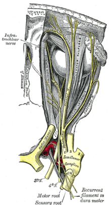
As the second cranial nerves , they are assigned to the interbrain ( diencephalon ) via the subsequent sections of the visual pathway ( tractus optici ) . Drawing by Andreas Vesalius , 1543.
Human optic nerve
The optic nerve of an adult is about 4 mm thick and, depending on the position of the eye, runs slightly meandering or in an arch, from the eye through the eye socket and through the optic nerve canal ( canalis opticus ) of the sphenoid bone into the skull. Its course - from the exit through the sieve-like lamina cribrosa sclerae of the sclera to the meeting with the optic nerve on the other side in the optic chiasm and here the partial crossing of fibers on the opposite side - is about 4.5 cm long and can be divided into
- intrabulbar part ( pars intraocularis ), still lying in the eyeball ( bulbus oculi ),
- intraorbital part ( pars intraorbitalis ), running in the eye socket ( orbit ),
- intracranial part ( pars intracanalicularis and pars intracranialis ), located in the bony optic nerve canal or in the cavity of the skull ( cranium ).
An optic nerve contains around a million nerve fibers, each of which is the individually enveloped neurite of a specific ganglion cell in the retina. It is the nerve cell extensions of the 3rd afferent neurons of the visual line - their 1st neuron are the photoreceptors in the outer, their 2nd the bipolar nerve cells in the inner granular layer of the retina. The retinal ganglion cells collect their signals and use their neurites to convey their own signal from the eye to 4th neurons in the diencephalon , most of which are located in the lateral knee cusps ( corpus geniculatum laterale , CGL).
On the way there, the nasal halves of the optic nerve fibers, which conduct signals from the nasal retinal half, cross in the optic nerve junction ( optic chiasm ) to the optic tract on the other side, so that the signals from one half of the visual field reach the other half of the brain . After the optic chiasm will train tract called.
Normally, the neurites of the retinal ganglion cells are surrounded by myelin sheaths of the oligodendrocytes when they leave the eyeball at the optic nerve papilla , which enables an increased conduction velocity in the optic nerve, but leads to functional disorders in the event of demyelination . There are also other glial cells such as astrocytes near the axons. The histologic similarity to tissues of the central nervous system lies in the fact that the retina, from the optic nerve in the course of embryonic development aussprießt, from the optic vesicle is formed which is composed of portions of the front brain bubble emerges (forebrain). The optic nerve is also how the nervous tissue of the brain surrounded by a tough shell comprising a continuous continuation of the hard meninges represents (Dura mater) and forward smoothly into the dermis merges the eye (sclera). Occur at the papilla also extending within the fiber bundle of optic nerve retinal vessels, the central retinal artery and the central vein , on or off and ensure the blood flow in the inner retinal layers. Since the photoreceptors of the retina are missing in the area of the passage , the blind spot appears as a physiological scotoma in the field of vision .
Damage to the optic nerve is often noticeable through pathological visual field defects in the affected eye. Common diseases involving the optic nerve include glaucoma ( glaucoma ), optic atrophy and neuritis nervi optici . The optic nerve and retina can be understood as advancing parts of the diencephalon ; they are to be regarded as parts of the brain and their regenerative capacity is limited.
Ophthalmic review

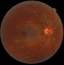
The clinical examination of the functions of the optic nerve includes the assessment of the pupils (symmetry, shape, light reaction) and the fundus by means of direct reflection. The papilla as the point where the unmarked nerve fibers of the retina converge and leave the globe as the optic nerve can be viewed directly with the ophthalmoscope and appears in front of the reddish fundus ( fundus oculi ) about 4 mm nasally from the yellow spot ( macula lutea ) as a circular shape light yellow disc ( Discus nervi optici ). Their contour, pale, bulge and vascular markings can indicate, among other things, an increase in pressure in the eye (glaucoma papilla), in the skull ( congestive papilla ) or in venous ( central vein thrombosis ) and arterial vessels ( fundus hypertonicus ).
To delimit optic nerve disease to disease elsewhere in the visual pathway frequently serving perimetry , the study of the visual field . The conduction of excitation of the optic nerve and subsequent sections of the visual conduction can be checked with the help of visual evoked potentials (VEP). Ultrasound , computed tomography and magnetic resonance tomography are available as imaging methods , with which, in addition to the conditions in the eye, those in the eye socket and within the skull can also be displayed.
Optic nerve fiber measurement
Optic nerve fiber measurement is offered or advertised by many German ophthalmologists as an individual health service (IGeL service). The measured layer thickness is compared with age-dependent standard values.
See also
literature
- Th. Axenfeld (conception), H. Pau (ed.): Textbook and atlas of ophthalmology. With the collaboration of R. Sachsenweger et al., Stuttgart: Gustav Fischer Verlag, 1980, ISBN 3-437-00255-4
- Albert J. Augustin: Ophthalmology . Berlin: Springer Verlag, 2007, ISBN 978-3-540-30454-8
- Franz Grehn: Ophthalmology. Berlin: Springer Verlag, 30th edition, 2008, ISBN 978-3-540-75264-6
Individual evidence
- ↑ Martin Trepel: Neuroanatomy - Structure and Function . 4th edition. Elsevier, Munich 2008, ISBN 978-3-437-41298-1 , pp. 60 f .
- ↑ Martin Trepel: Neuroanatomy - Structure and Function . 4th edition. Elsevier, Munich 2008, ISBN 978-3-437-41298-1 , pp. 20th f .
- ↑ Jörg Braun: Tips for station work. In: Jörg Braun, Roland Preuss (Ed.): Clinic Guide Intensive Care Medicine. 9th edition. Elsevier, Urban & Fischer, Munich 2016, ISBN 978-3-437-23763-8 , pp. 1–28, here: pp. 7 f. ( Neurological examination: head and cranial nerves ).
