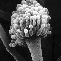
Sinunasal aspergillosis
The sinonasal aspergillosis (SNA) is a by molds caused inflammation of the nasal cavity and the paranasal sinuses. It is the second leading cause of chronic nasal discharge in dogs . Trigger is especially the Aspergillus -type Aspergillus fumigatus . Infection occurs through inhalation of mold spores. Mold toxins and the inflammatory reaction lead to meltdown in the area of the nasal cavity, the paranasal sinuses and the adjacent skull bones. Typical for the disease is a slimy, purulent or bloody-purulent nasal discharge that lasts for months. Endoscopy is particularly suitable for diagnosis . The treatment is carried out with fungal agents , whereby the local treatment is most promising.
Cause and occurrence
The main trigger of the SNA is Aspergillus fumigatus , a globally occurring, ubiquitous mold that grows on many organic materials. Other watering can mold species such as A. niger or A. flavus are less involved , and brush molds are even more rarely involved. The molds spread with their conidia via air currents and can cover long distances.
Middle-aged dogs with long or medium-sized skulls are particularly affected . In some studies, males were more often affected than bitches.
Disease emergence
Infection occurs through inhalation of fungal spores. In contrast to earlier views, favorable factors do not seem to play a role. Even if individual diseases occur in connection with injuries, foreign bodies or tumors , sinunasal aspergillosis also occur in otherwise healthy, immunocompetent animals. It is unclear what ultimately leads to SNA in some of the dogs while others are not affected. Humans breathe in about 15 watering can mold spores every day, a dog probably even more. May inhibit mold toxins , the self-cleaning and phagocytosis . The increased formation of interleukin-10 , which inhibits the elimination of fungal infections through a Th1 immune response, probably also plays a role . Perhaps an excessive inflammatory reaction from Th17 cells is also a decisive factor causing the disease.
The disease affects the nasal cavity as well as the maxillary and frontal sinuses . The inflammatory reaction and mold toxins destroy the turbinates . In severe illnesses, the surrounding skull bones are also destroyed, so that the infection can penetrate into the eye socket and through the sieve plate into the olfactory brain . The mold itself does not penetrate the nasal mucosa or underlying tissue.
The inflammation is mainly dominated by lymphocytes and plasma cells . Sometimes neutrophils are also involved , and very rarely mast cells or eosinophils are involved.
Clinical picture
Sinunasal aspergillosis is mainly characterized by chronic nasal discharge, which can last for weeks and usually begins on one side. The discharge can be slimy, purulent, or bloody and purulent. In addition, the skull is painful and the nasal surface can be ulcerated and depigmented .
If the internal structures in the nose are more severely destroyed, nosebleeds can occur, which under certain circumstances can assume life-threatening proportions. If the infection penetrates the eye socket, tearing may increase , and if the infection penetrates the cranial cavity , neurological symptoms such as seizures may occur.
Diagnosis
Chronic, on antibiotics not attractive nose river is already an important indication. Above all, tumors and foreign bodies of the nasal cavity, oronasal fistulas and dental diseases are to be excluded .
Serological methods such as ELISA or agar-gel immunodiffusion tests have a specificity of almost 100% and a sensitivity of 77 or 88%. Since false-positive results are possible, a verification of the result by imaging or endoscopy is definitely recommended.
Fungal hyphae can be detected cytologically in nasal secretions or nasal swabs, but only in about a fifth of the blind samples. Therefore, samples should be taken from visibly changed areas under endoscopic control. The same applies to the collection of samples for a mushroom culture . Endoscopic sampling increases the sensitivity to 75 to 100%. False-positive results can arise from breeding harmless commensals .
X-ray , computed tomography and magnetic resonance tomography are used as imaging methods . Typical findings are destruction of the turbinates and, more rarely, the nasal septum , thickened mucous membrane, soft tissue shading in the nose and paranasal sinuses, and changes in the frontal bone .
The endoscopy is considered the drug of choice for the diagnosis of sinonasal aspergillosis. Here white, yellow, black or greenish fungal lawns (plaques) with a flaky or velvety surface can be visualized. In addition, the destruction of the turbinates and the nasal septum can be demonstrated. The frontal sinus is not accessible for direct endoscopic examination; a trepanation must be performed here. This is of practical importance because there are patients in whom there are no fungal lawns in the nasal cavity, only in the frontal sinus. Further advantages of endoscopy are the possibility of obtaining samples and the possibility of removing altered tissue , which is usually essential for successful treatment. The endoscopy must be performed under deep anesthesia .
treatment
Antifungal drugs are used for treatment .
Treatment with orally administered antifungals such as ketoconazole or thiabendazole alone leads to a complete cure in only half of the cases. With newer antimycotics such as itraconazole or fluconazole over a period of six weeks, the chance of recovery increases to 70%. Overall, the whole-body treatment is inferior to the local administration due to the lack of mucosal penetration of the mold . Clotrimazole or enilconazole are used locally , preferably after the altered tissue has been removed . As a rule, multiple topical treatments are necessary.
Rinsing twice a day via surgically placed indwelling catheters is usually only tolerated by dogs under sedation and is therefore rarely performed. In addition, the catheters can slip and there is a risk of pneumonia if swallowed . Trephination of the frontal sinus for the temporary placement of a catheter with irrigation and the introduction of a depot of clotrimazole cream requires shorter hospital stays and achieves good therapeutic results. However, there are no comparative studies that allow a comparison of individual methods or combinations (topical and full-body treatment).
Radical clearing out of the nasal cavity is not routinely necessary, but may be indicated in severe or therapy-resistant aspergillosis.
In about half of the patients, a slight nasal discharge may persist despite successful treatment. This is attributed to irreversibly damaged turbinate parts, persistence of foci of inflammation despite the elimination of pathogens and an increased tendency to bacterial infections of the nose.
literature
- Katharina Imholt among others: Sinunasal Aspergillosis of the Dog - Symptoms, Diagnosis and Therapy. In: Small Animal Practice. 59, 2014, pp. 565-584.
- Katrin Hartmann: Aspergillosis. In: Peter S. Suter, Barbara Kohn: Internship at the dog clinic . 10th edition. 2006, ISBN 3-8304-4141-X , p. 324.
Individual evidence
- ↑ Katrin Hartmann: Aspergillosis. In: Peter S. Suter, Barbara Kohn: Internship at the dog clinic . 10th edition. 2006, ISBN 3-8304-4141-X , p. 324.
- ↑ D. Peeters et al.: Quantification of mRNA encoding cytokines and chemokines in nasal biopsies from dogs with sino-nasal aspergillosis. In: Vet. Microbiol. 114, 2006, pp. 318-326.
- ↑ Katharina Imholt et al: Sinunasal Aspergillosis of the Dog - Symptoms, Diagnosis and Therapy. In: Small Animal Practice . 59, 2014, p. 566.
