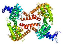
Vitamin D binding protein
| Vitamin D binding protein | ||
|---|---|---|

|
||
| PDB 1j78 | ||
|
Existing structural data: s. UniProt |
||
| Properties of human protein | ||
| Mass / length primary structure | 458 amino acids | |
| Identifier | ||
| Gene name | GC | |
| External IDs | ||
| Occurrence | ||
| Homology family | VDBP | |
| Parent taxon | Euteleostomi | |
The vitamin D-binding protein (DBP) (also: group -specific component , gene name: GC ) is a protein that binds vitamin D metabolites in vertebrates and thus mainly mediates their transport in the bloodstream. It is also found in humans in the cerebrospinal fluid , in ascites fluid and on the surface of some cell types. In addition to its vitamin D binding properties, it also binds actin and plays a role in chemotaxis . DBP belongs to the albumins family and is a glycoprotein .
General function
The most important carrier protein for vitamin D 3 and its intermediates is the vitamin D binding protein (DBP), which binds vitamin D metabolites with high affinity in the following order: 25 (OH) D 3 = 24.25 (OH) 2 D 3 > 1.25 (OH) 2 D 3 > D 3 . The plasma levels of vitamin D-binding protein are 20 times higher than the total amount of vitamin D metabolites, less than 1% of which are free, i.e. unbound, in the blood. In contrast, around 85–90% are bound to VDBP and 10–15% to albumin and other lipoproteins . Under normal conditions, the vitamin D-binding protein has a maximum binding capacity for vitamin D metabolites of approx. 1900 ng / ml. Since presumably only the small free portion of the vitamin D metabolites can be metabolized or has hormonal effects, the DBP essentially serves as a buffer function against fluctuations in tissue levels and as a reservoir. In addition, it aids in the reabsorption of free vitamin D by megalin in the kidney.
Clinical studies and animal models have shown that DBP is an important factor in removing actin from blood plasma that is released during necrosis or injury. It is also involved in the body's response to injury as a macrophage -activating factor, through chemotactic activity of neutrophils, and by amplifying C5a -mediated signals.
Variants of DBP are associated with an increased need for vitamin D in dialysis patients. The presence of the GC2 isoform of the protein may be a risk factor for familial amyotrophic lateral sclerosis (FALS). The same variant reduced the risk of developing breast cancer in another study .
Regulation of the VDBP plasma level
The blood level of vitamin D binding protein is not regulated by vitamin D metabolites themselves. Rather, it decreases with liver diseases, malnutrition (reduced education) and nephrotic syndrome (increased excretion in the kidneys); it increases with pregnancy and estrogen therapy . It is also dependent on two common polymorphisms that explain 80% of the variation between African Americans and White Americans, and shows a clear seasonal variation.
However, the concentration of free 1,25 (OH) 2 D 3 (calcitriol) remains the same when the DBP level changes (one of many examples of the complex and strict self-regulation in vitamin D metabolism).
VDBP in the kidney
In the kidney , VDBP is filtered glomerularly. In the brush border of the proximal tubular cells there is a complex of proteins (part of which is the DBP-binding receptor megalin ), which enables the reabsorbent endocytosis of VDBP from the primary urine together with the vitamin D metabolites bound to it. A VDBP-independent transport of 25 (OH) D 3 into the kidney cells must also be possible, since mice that cannot form VDBP do not get rickets , in contrast to mice that do not have megalin and their vitamin D Metabolites are therefore lost in the urine. VDBP is broken down in the tubular cell of the kidney and the vitamin D metabolites bound to it are released for further - variously regulated - metabolism.
genetics
The precursor protein is on chromosome 4q12-q13 section codes , contains 474 amino acids and has a molecular mass of approximately 55 kDa . There are over 80 variants of the human VDBP.
There are essentially two important polymorphisms that influence the blood concentration of the VDBP and, moreover, the concentration of free vitamin D in the blood. Both are single nucleotide polymorphisms . One polymorphism concerns the nucleotide rs7041 and normally encodes a guanosine (G), alternatively a thymine (T), from which in the VDBP instead of glutamine is then asparagine . The second mutation affects rs4588 with substitution of cytosine to adenine , resulting instead of threonine then lysine is incorporated. This results in three different phenotypes (which are typically abbreviated to GC because of the alternative designation of the VDBP as a group-specific component):
- GC-1F with rs7041-T and rs4588-C is found in 93% of homozygous African Americans and 6% of homozygous white Americans and has the highest binding affinity for vitamin D and its metabolites.
- GC-1S with rs7041-G and also rs4588-C is found in 5% of homozygous African Americans and 76% of white Americans and has a lower binding affinity for vitamin D and its metabolites than GC-1F.
- GC-2 with rs7041-T and rs4588-A is found in 2% of homozygous African Americans and 18% of homozygous white Americans and has a lower binding affinity for vitamin D and its metabolites than both GC-1 phenotypes.
- The variant with rs7041-G and rs4588-A practically does not exist.
In an American cohort study with over 2000 participants , the blood level for VDBP is significantly lower in African Americans with a mean 168 µg / ml versus 337 µg / ml in white Americans (and likewise the 25-OH vitamin D level with a mean 15.6 ng / ml versus 25.8 ng / ml). In a statistical model, the two polymorphisms can explain almost 80% of the variation in the blood level of the VDBP, while ethnicity explains only 0.1% of the variation.
Individual evidence
- ↑ Vieth R: Simple method for deterministic mining specific binding capacity of vitamin D-binding protein and its use to calculate the concentration of "free" 1,25-dihydroxyvitamin D . In: Clin. Chem. . 40, No. 3, March 1994, pp. 435-41. PMID 7510592 .
- ↑ a b c Dusso AS, Brown AJ, Slatopolsky E: Vitamin D . In: Am. J. Physiol. Renal Physiol. . 289, No. 1, July 2005, pp. F8-28. doi : 10.1152 / ajprenal.00336.2004 . PMID 15951480 .
- ↑ Gressner O, Meier U, Hillebrandt S, among others: Gc-globulin concentrations and C5 haplotype-tagging polymorphisms contribute to variations in serum activity of complement factor C5 . In: Clin. Biochem. . 40, No. 11, July 2007, pp. 771-5. doi : 10.1016 / j.clinbiochem.2007.02.001 . PMID 17428459 .
- ↑ Meier U, Gressner O, Lammert F, Gressner AM: Gc-globulin: roles in response to injury . In: Clin. Chem. . 52, No. 7, July 2006, pp. 1247-53. doi : 10.1373 / clinchem.2005.065680 . PMID 16709624 .
- ↑ Speeckaert MM, Glorieux GL, Vanholder R, among others: Vitamin D binding protein and the need for vitamin D in hemodialysis patients . In: J Ren Nutr . 18, No. 5, September 2008, pp. 400-7. doi : 10.1053 / j.jrn.2008.04.013 . PMID 18721734 .
- ↑ Palma AS, De Carvalho M, Grammel N, among others: Proteomic analysis of plasma from Portuguese patients with familial amyotrophic lateral sclerosis . In: Amyotroph Lateral Scler . 9, No. 6, December 2008, pp. 339-49. doi : 10.1080 / 17482960801934239 . PMID 18608108 .
- ↑ Abbas S, Linseisen J, Slanger T, among others: The Gc2 allele of the vitamin D binding protein is associated with a decreased postmenopausal breast cancer risk, independent of the vitamin D status . In: Cancer Epidemiol. Biomarkers Prev. . 17, No. 6, June 2008, pp. 1339-43. doi : 10.1158 / 1055-9965.EPI-08-0162 . PMID 18559548 .
- ↑ Zella LA, Shevde NK, Hollis BW, Cooke NE, Pike JW: Vitamin D-binding protein influences total circulating levels of 1,25-dihydroxyvitamin D3 but does not directly modulate the bioactive levels of the hormone in vivo . In: Endocrinology . 149, No. 7, July 2008, pp. 3656-67. doi : 10.1210 / en.2008-0042 . PMID 18372326 .
- ↑ Camille E. Powe, Michele K. Evans, Julia Wenger, Alan B. Zonderman, Anders H. Berg, Michael Nalls, Hector Tamez, Dongsheng Zhang, Ishir Bhan, S. Ananth Karumanchi, Neil R. Powe, Ravi Thadhani: Vitamin D – Binding Protein and Vitamin D Status of Black Americans and White Americans . New England Journal of Medicine 2013, Volume 369, Issue 21 Nov. 21, 2013, pp. 1991-2000; doi : 10.1056 / NEJMoa1306357 .
Web links
- Jassal / D'Eustachio / reactome.org: Calcidiol binds to DBP in the bloodstream and is delivered to the kidney
- Metabolic pathway between calcitriol and vitamin D binding protein