
Basalioma
| Classification according to ICD-10 | |
|---|---|
| C44 | Other malignant neoplasms of the skin |
| ICD-10 online (WHO version 2019) | |
The basal cell carcinoma ( BCC ; basal cell carcinoma ; basocellular ; engl .: basal cell carcinoma , basalioma , basal cell epithelioma ) is a malignant cancer of the skin resulting from, stem cells in the hair follicles and interfollikulär in the basal layers of the epidermis developed. Preferred locations are areas of the face exposed to the sun, such as the forehead, nose or ears. Sometimes basal cell cancer is also called "light skin cancer" or "white skin cancer" to differentiate it from "black skin cancer" ( malignant melanoma ). However, this misleading name does not take into account that pigmented (colored) growth forms of the basalioma as well as unpigmented (amelanotic) melanomas can occur and is therefore not used by medical professionals. Basal cell carcinoma, like a malignant tumor, can damage the surrounding tissue and even infiltrate bone , but it is extremely rare (0.03% of cases) to metastasize . It is therefore sometimes referred to as semi -malignant - sometimes malignant - especially in older people.
Epidemiology
Basaliomas occur mainly in people between the ages of 60 and 70. At 65%, they make up the largest proportion of malignant skin tumors and are about ten times more common than spinaliomas (squamous cell carcinomas of the skin). The prevalence varies between 20–50 (Central Europe) and 250 (Australia) diseases per 100,000 inhabitants, depending on exposure to the sun. In Germany there are around 171,000 new cases per year.
In the case of immunocompromised (e.g. HIV - infected or transplant patients ), however, the spinalioma predominates .
In the context of some syndromes , basaliomas are particularly common, such as Gorlin-Goltz syndrome , Rombo syndrome or Bazex-Dupre-Christol syndrome .
Risk Factors and Pathogenesis
The greatest risk factor is long-term exposure to sun rays. This is why basaliomas tend to develop in the face - 80% develop in the areas around the eyes, i.e. forehead, cheeks, nose, upper lip - as well as in the neck and on the thinly haired or bald head. In contrast to malignant melanoma , the tumors are not caused by frequent sunburns, but by the constant exposure of the skin to UV rays.
The long-wave portion of UV light , UV-A (320–400 nm wavelength), is crucial for the development of a basalioma due to its greater penetration depth. The short-wave UV-B radiation is for the most part already absorbed in the upper layers of the skin and primarily causes damage in the superficial epithelial cells of the skin, see squamous cell carcinoma of the skin .
In addition to light exposure, genetic predisposition and environmental factors are also responsible. Light skin ( skin type I or II) is an important risk factor. In addition, genetic disorders such as xeroderma pigmentosum and the basal cell nevus syndrome are involved in the development in rare cases. Arsenic is an important chemical noxious agent . Exposure can lead to basal cell carcinoma even years later.
Forms and clinical course
There are eight different types of basalioma:
-
Nodular solid basalioma: A mostly yellowish-reddish or brownish-gray, coarse, slowly growing lump or nodule with a glassy surface and telangiectasia and a tendency to bleeding and ulceration. (Half) spherical appearance.
- In the case of an ulcerating process (ulcerating growing basalioma, also called ulcus rodens - from Latin rodere = 'gnaw') the characteristic, coarse, pearl-like edge wall forms.
- In deeper ulcerative and destructive acting processes is called a destructive growing basal cell carcinoma, also ulcer Terebrans called .
- Superficial-multicentric basal cell carcinoma: often localized to the fuselage skin, hence Rumpfhautbasaliom called, often associated with psoriasis and in elderly patients requiring exposure to arsenic in the history exhibit. Round-oval, macular to plaque-shaped foci of reddish color with fine scaling and sharp borders. The surface is slightly folded, the lesion is prone to erosion and ulceration. In ulcerating processes, the pearl-like edge wall can be seen.
- Pigmented basal cell carcinoma: This can easily be mistaken for malignant melanoma .
- Infiltrative scleroderma basalioma: This variant has a whitish-yellowish, porcelain-white aspect, which is caused by the varying degrees of fibrosis and accordingly determines the rather coarse consistency. It is therefore often indistinguishable from normal skin with the naked eye and difficult to remove. Flat shape with horizontal extension, whereby clinically unaffected skin areas can also be included, which makes microscopic control of the incision margin absolutely necessary in the case of an excision. Particularly high risk of recurrence.
- Infiltrative non-sclerodermic basal cell carcinoma: The clinical differentiation consists in the slight to no fibrosis. The consistency is softer, there is no change in the color of the skin.
- Keratotic basal cell carcinoma
- Adenoid basal cell carcinoma
- Basal cell carcinoma with seboglandular differentiation

Nodular solid (nodular) basalioma
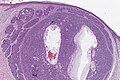
Histology of a nodular basalioma

Pigmented basal cell carcinoma

Scleroderma basalioma (morphea basalioma)

Histology of a scleroderma basal cell carcinoma


Special forms
- Wild basal cell carcinoma (syn: basosquamous (metatypical or intermediate) basal cell carcinoma): Clinically identical to a nodular basal cell carcinoma. Histological cell differentiation with regard to keratotic, bowenoid or squamous type, which grow aggressively destructively (destructively) and metastasize in the immediate vicinity (locally). The prognosis for this form of disease is correspondingly worse because this type can metastasize .
- Infundibulocystic basal cell carcinoma: Infundibulocyst-like structures with an accumulation of basaloid cells without a mature follicular papilla. Clinically, the lesion is usually a nodule with a smooth surface on the face of older people.
- Genodermatoses with basal cell carcinoma
The vast majority of basaliomas are found in the so-called centrofacial area, a strip from the hairline to the upper lip. About 15% of basaliomas are found on the auricle, on the hairy scalp and in the lower third of the face. Only about 5% of basaliomas are on the trunk or on the extremities .
Basal cell carcinomas grow very slowly over a number of years and make it differential diagnosis distinguishable to the emerging within weeks and unattractive-healing keratoacanthoma . It usually starts with a small, hard knot ("basal pearls") or a circumscribed induration. Often at the edge of the lesion there are very fine, newly formed blood vessels that shimmer delicately through the skin, the so-called telangiectasias as well pearl-like thickenings on the edge of the tumor. The epithelial layer covering the basalioma has a mother-of-pearl shimmer and is another important differential diagnostic aspect.
For a long time there is growth in the horizontal and vertical directions. The formation of ulcers (ulcerations) and destructive growth (destruction) occur in later stages and are differently pronounced depending on the clinical form.
forecast
The prognosis is generally good , since in most cases there is no tendency to metastasize . Patients with aggressive forms ( Basalioma terebrans and Basalioma exulcerans ) may have a worse prognosis, depending on the organ involvement .
therapy
Numerous forms of treatment are available:
The form of treatment with the lowest relapse rate for basaliomas, especially in the face, is still surgical treatment with histological ( microscopically controlled) cutting edge control (" Tübinger Torte "). Other forms of treatment are usually only used as the sole therapy if the operation is not possible, for example due to the patient's age or previous illnesses, the location of the tumor or the like. Because of the long-term destructive (destructive) growth, basaliomas should be surgically removed at an early stage in order to avoid damage to deeper tissue areas. However, a recurrence of the tumor ( relapse ) is always possible.
Large or inoperable basaliomas in older people are successfully treated with soft X- rays. Further treatment methods are curettage with local chemosurgical follow-up treatment, freezing treatment ( cryotherapy ), especially for superficial basaliomas, as well as local drug immunotherapy (see below) or topical chemotherapy with the application of a 5-fluorouracil- containing cream.
A new treatment method for superficial basalioma has been approved since July 2004: the active ingredient imiquimod is applied by the patient himself with a cream over several weeks. Imiquimod locally activates the body's own immune system of the skin, which then attacks the tumor cells in a targeted manner. This effect manifests itself in the form of inflammation , which can be more or less pronounced. The advantage is that this means that tumor areas that are not yet visible can also be shown and treated at the same time. Common side effects of the inflammatory reaction are itching, burning or slight pain, crusting and oozing. On the other hand, there is very rarely a flu-like feeling, in which case it is recommended to pause the therapy. If the patient allows the crusts to heal without further manipulation, scarring is not to be expected.
Since August 2013, a new drug vismodegib (trade name Erivedge , manufacturer Roche ) has been approved for the treatment of basal cell carcinoma in Germany if the measures that have been customary (surgery, radiation) are not possible or are not effective. In various publications (JEJM, PZ-Online) the substance is referred to as a "leap innovation". Another drug from the class of active ingredients of the Hedgehog signaling pathway - inhibitors is Sonidegib (trade name: Odomzo ) from Novartis . It was approved in the US in July 2015; in Europe it was approved in August 2015.
Another method for treating superficial forms of basalioma is photodynamic therapy with a methyl 5-amino-4-oxopentanoate cream (MAOP). This is mainly transformed into protoporphyrin IX in the neoplastic cells. After such a sensitization , the cells are selectively destroyed by a subsequent treatment with special red light, since protoporphyrin IX forms oxygen radicals when exposed to light, which lead to the death of these cells. This treatment option is characterized by a combination of good medical and cosmetic results, as is particularly desirable in the facial area. The risk of recurrence is very high in the case of flat-scarred (sclerodermiform) basaliomas; for nodular basaliomas, the option is not recommended due to the only shallow penetration depth.

Scleroderma basalioma

Nodular basalioma

Nodular basalioma

Terebrans ulcer

Cornu cutaneum


Aftercare
Because most localized basal cell carcinoma recurs within two years, patients who have developed basal cell carcinoma should see a doctor at least once a year for a period of three years. Pathological changes in the skin can also be detected early on through self-examination. In addition, strong exposure to the sun and visits to the solarium should be avoided, as (even artificial) UV light is harmful to the skin cells.
See also
literature
- Ingrid Moll: Dermatology (= dual series). 6th edition. Thieme, Stuttgart 2005, ISBN 3-13-126686-4 .
- H. Domarus, PJ Stevens: Metastatic basal cell carcinoma. Report of five cases and review of 170 cases in the literature. In: Journal of the American Academy of Dermatology . 10, 1984, pp. 1043-1060.
- Site of the Dermatological Information System (DermIS)
- Dirk Hasselmann: Light skin cancer - And how you put it in the shade. 2nd edition 2018. ISBN 978-3-7407-4964-4
Web links
Individual evidence
- ^ Shelby C. Peterson, Markus Eberl, Alicia N. Vagnozzi, Abdelmadjid Belkadi, Natalia A. Veniaminova: Basal Cell Carcinoma Preferentially Arises from Stem Cells within Hair Follicle and Mechanosensory Niches . In: Cell Stem Cell . tape 16 , no. 4 , April 2015, p. 400-412 , doi : 10.1016 / j.stem.2015.02.006 , PMID 25842978 , PMC 4387376 (free full text) - ( elsevier.com [accessed October 26, 2019]).
- ↑ Melanoma malignant amelanotic - P. Altmeyer - Encyclopedia of Dermatology, Venereology, Allergology, Environmental Medicine. Retrieved July 11, 2017 .
- ↑ skin cancer. Rethink! Actively preventing cancer. In: krebshilfe.de. German Cancer Aid, archived from the original on April 14, 2011 ; Retrieved April 14, 2011 .
- ^ I. Moll: Dual Series - Dermatology. 2005, p. 319 ff.
- ^ Konrad Bork, Walter Burgdorf, Nikolaus Hoede: Oral mucous membrane and lip diseases: Clinic, diagnostics and therapy. Atlas and manual. Schattauer Verlag, 2008, ISBN 978-3-7945-2486-0 , pp. 361-362.
- ↑ Carlos Thomas: Special Pathology. Schattauer Verlag, 1996, ISBN 3-7945-2110-2 , pp. 85-86.
- ↑ H. Kerl, Claus Garbe, L. Cerroni: Histopathology of the skin . Ed .: H. Wolff. 1st edition. Springer, Berlin Heidelberg 2003, ISBN 3-540-41901-2 .
- ↑ a b c M. Sand, D. Sand, C. Thrandorf, V. Paech, P. Altmeyer, FG Bechara: Cutaneous lesions of the nose. In: Head & face medicine. Vol. 6, 2010, p. 7, ISSN 1746-160X . doi: 10.1186 / 1746-160X-6-7 . PMID 20525327 . PMC 290354 (free full text). (Review).
- ↑ C. Thomas: Special Pathology. Schattauer Verlag, 1996, ISBN 3-7945-2110-2 , p. 87, (online)
- ↑ Pharmazeutische Zeitung Online: http://www.pharmazeutische-zeitung.de/?id=48488 NEJM: http://www.nejm.org/doi/full/10.1056/NEJMoa1113713 .
- ↑ FDA approves new treatment for most common form of advanced skin cancer ( Memento of July 27, 2015 in the Internet Archive ), PM FDA of July 24, 2015, accessed on July 27, 2015.
- ↑ [1] , European Public Assessment Report Odomzo, accessed on October 7, 2015.