Hippocampus
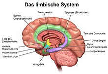
The hippocampus ( plural hippocampi ) is part of the brain , more precisely: the archicortex, which appears for the first time in reptiles . It is located on the inner edge of the temporal lobe and is a central switching station of the limbic system . There is one hippocampus per hemisphere .
Word origin


From 1706 a part of the brain was named after the seahorse ( Latin hippocampus ). The seahorse, for its part, has been named in Latinized form since the 1570s after the sea monster Hippocampus from Greek mythology , the front half of which is a horse, the rear part a fish. The name of this mythical creature ( ancient Greek ἱππόκαμπος hippokampos ) is composed of hippos 'horse' and kampos 'sea monster' .
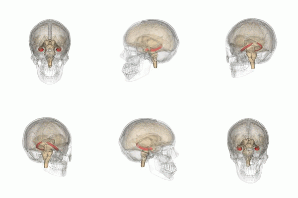
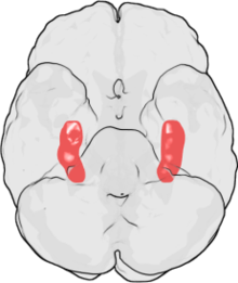
Overall, the hippocampus resembles a headless seahorse (see illustration).
anatomy
The hippocampus is located in the medial part of the telencephalon (endbrain) and forms the so-called archicortex.
Several structures belong to the hippocampus, which is why one speaks of a hippocampus formation or formatio hippocampi:
- Dentate gyrus
- Cornu ammonis (Ammon's horn, Hippocampus proprius )
- Subiculum
Cytoarchitectonics
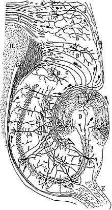
The dentate gyrus represents the entrance station of the hippocampus. Its nerve cell bodies are located in the granular cell band (stratum granulare). The main cells are excitatory, glutamatergic granule cells , which send their dendrites to the outside in the molecular layer (stratum molecularulare). In addition, there are a number of different inhibitory, GABAergic interneurons , which are differentiated by their morphology, thus also by their inputs and outputs and also by characteristic proteins. The molecular layer can be divided into the inner and outer molecular layer. In both parts there are hardly any nerve cell bodies, only nerve fibers. The entrances to the two layers differ, however, and can be visualized with different colors (see below). Within the arch of the granular cell layer is the hilus , also called the lamina multiformis. It contains some interneurons , but above all the axons of the granule cells, the so-called moss fibers (note: apart from their name, these axons have nothing in common with the moss fibers in the cerebellum ).
The actual hippocampus ( Hippocampus proprius ), as an archicortical structure, also has three layers. Similar to the dentate gyrus, the nerve cell bodies lie in one layer, the pyramidal cell layer ( stratum pyramidale ). The main cells here are also glutamatergic pyramidal cells, which send dendrites radially both inward and outward, and there is a similar diversity of interneurons . As input layers, the broad stratum radiatum and the narrower stratum lacunosum -olekulare are attached to the outside of the pyramidal cell layer , to the inside the stratum oriens , which contains the cell bodies of the inhibiting basket cells. In its tangential direction, the hippocampus proprius is subdivided into the CA1 to CA4 regions (from C ornu A mmonis), with only the CA1 and CA3 regions actually being anatomically and functionally important.
The subiculum is the transition area from the three-layer archicortical hippocampus to the six-layer neocortex . It lies between the CA1 region and the entorhinal cortex .
links
In the formation of the hippocampus, excitatory neurons are connected in a chain. The main entry point for cortical afferents is the outer molecular layer of the dentate gyrus. This is where the axons from the entorhinal cortex arrive as the so-called perforated tract . They connect to the granular cells that are also exciting; but also on some of the different classes of inhibiting interneurons (forward inhibition). Within the dentate gyrus, other interneurons, the basket and candlestick cells, cause reverse inhibition, i. That is, they receive inputs from granule cells and switch back to their somata or axon hillock. In the inner molecular layer, the granule cells receive back-coupled afferents from other granule cells and from the CA4 region of the hippocampus proprius (associative fibers), as well as commissural afferents from the dentate gyrus of the other hemisphere.
The granule cells project via the moss fibers into the inner stratum radiatum of the CA3 region of the hippocampus proprius onto the pyramidal cells there , which in turn are exciting. Here, too, there are forward and backward inhibitions due to interneurons. Further afferents are exchanged between the CA3 regions of both hemispheres; they end in the outer stratum radiatum . The pyramidal cells send axons into the fornix , which leave the hippocampal formation and reach various nuclei of the brain stem (including corpora mamillaria , substantia nigra , locus caeruleus ).
Branches of the same axons, the Schaffer collaterals, link the chain further into the CA1 region, where they in turn end at pyramidal cells. Axons of the entorhinal cortex also reach the stratum lacunosum -olekulare of the CA1 region. The axons of the CA1 pyramidal cells form another part of the fornix, and they also project into the subiculum. This projection is the main entrance to the subiculum. It also receives afferents from the cortex perirhinalis and cortex entorhinalis. In turn, it projects back into the cerebral cortex on both sides, as well as into the nucleus accumbens , the amygdala , the prefrontal cortex and the hypothalamus .
Via the projection of the fornix into the corpora mamillaria, the hippocampus is integrated into the Papez circle , which was described by James W. Papez in 1937 and postulated as the neural basis of emotions . The Papez circle closes back to the hippocampus formation via the thalamus , the cingulate gyrus and the entorhinal cortex.
The entire hippocampal formation also receives afferents of neuromodulatory pathways. Cholinergic neurons in the septum pellucidum send their axons here, serotoninergic neurons from the medial raphe and noradrenaline- containing neurons from the locus caeruleus. A very weak dopaminergic projection from the ventral tegmentum is difficult to detect histochemically.
Electrical activity
Activity in the hippocampus is low during locomotion and REM sleep . In wakefulness without locomotion and non-REM sleep , on the other hand, there is often synchronous activity of large groups of neurons that shape the extracellular electrical potential ( field potential ) in the hippocampus. Characteristic are sawtooth-shaped curves ( English sharp wave ) with periods of a few 100 ms on which short wave packets ( ripple ) are superimposed at or shortly before the maximum of the polarization . Synonymous abbreviations for this combination are SWR and SPW-R. The frequency of the ripples, 100 to over 200 Hz, varies from species to species. A ripple event almost always occurs in CA1 and CA3 and mostly in both hemispheres at the same time, whereby the ripples themselves are not synchronous.
Functional aspects
Information from various sensory systems converges in the hippocampus, is processed and sent back to the cortex from there . It is therefore extremely important for memory consolidation, i.e. the transfer of memory contents from short-term to long-term memory . People who have both hippocampi removed or destroyed cannot form new memories and thus have anterograde amnesia . However, old memories are mostly preserved. The hippocampus is thus seen as a structure that generates memories, while the memory contents are stored in various other places in the cerebral cortex .
It has been proven that in the adult brain in the hippocampus new connections between existing nerve cells are formed ( synaptic plasticity ) and that this new formation is related to the acquisition of new memory contents. The Schaffer collateral, the connection between the CA3 and CA1 region, is predestined for research into molecular learning processes. Here are special glutamate receptors ( NMDA ) that are involved in long-term potentiation .
In animals, the hippocampus is of great importance for spatial orientation. Pyramidal cells in CA1 each represent an area of an environment and are therefore also called place cells . As the animal moves, different cells become active one after another, with cells for adjacent locations not being adjacent. Different place cells are sometimes responsible for one place depending on the direction of movement or the destination. Place cells also encode abstract states, such as B. in which phase of a complex behavioral process an animal is currently or whether a certain affect is present (hunger, curiosity, fear). So there is no topographically organized map in the brain, but the cognitive representation, also known as the cognitive map, contains topological information about the experienced environment. The so-called grid cells in the entorhinal cortex contribute to the cognitive performance of representing distances between defined locations . People with damaged hippocampi can find their way around in everyday life, but are unable to give directions.
The hippocampus is also responsible for coordinating the various memory contents. For example, there is the "inner card" that you can use for example. B. owns from a city, from numerous impressions that were also gained at different times. These are put together in the hippocampus and you can orient yourself in this way.
In addition, the hippocampal formation also plays an important role in emotions:
- People with (unipolar) depression show reduced hippocampal formation.
- The hippocampal formation is unique in its vulnerability to strong emotional stressors; Animal models show hippocampal atrophy as an effect of chronic emotional stress (caused by the death of hippocampal neurons and a reduction in neuronal genesis in the dentate gyrus), and people with severe emotional trauma (e.g. Vietnam veterans or victims of child sexual abuse) also show a reduction in the volume of the hippocampal formation.
- People with flattened affectivity show functional differences in the hippocampal formation in processing emotional stimuli. In particular, functional imaging studies that examine neural correlates of emotion with music report differences in activity of the hippocampus formation in connection with music-evoked emotions.
Neuropathological Aspects
Various diseases can lead to changes in the hippocampus in humans. Above all, degradation processes in dementia can damage this brain structure. In addition, the hippocampus plays an important role in the development of epilepsy diseases . The temporal lobe epilepsy is often associated with a mesial temporal sclerosis connected, by means of imaging techniques ( magnetic resonance imaging can be diagnosed). The unilateral removal of the hippocampus formation by epilepsy surgery is a way of treating seizures that cannot be controlled by medication. Binge drinking during adolescence is suspected of impairing the formation of the hippocampus over the long term, which in adulthood can lead to forgetfulness and a lack of spatial orientation.
Neurogenesis in the hippocampus
The dentate gyrus of the hippocampus, along with the olfactory bulb (or the subventricular zone), is one of the two structures in the healthy mammalian brain that form new nerve cells throughout their life. This neurogenesis of glutamatergic granule cells was discovered in 1965 by Altman and Das in rats and contradicts the dogma that had existed for decades that the neurons of the brain are complete from birth. Nonetheless, hippocampal neurogenesis did not attract scientific attention until the 1990s, when the BrdU marking of dividing cells was used to show that influences such as stress , activation of the NMDA receptor , walking and the rich environment, the rate of division of cells and / or their survival rate can change. Since then, numerous other studies have shown that many neurotransmitters, growth factors , medicinal substances , drugs and environmental factors (including learning training) can influence neurogenesis. Since only a small part of the newly formed cells differentiate into neurons, it is necessary to differentiate between the terms cell proliferation ( mitosis of neuronal stem cells) and neurogenesis. In addition to rodents, neurogenesis in the hippocampus was also found in other mammals during this period, including humans in 1998.
The function of hippocampal neurogenesis is still unclear. It has only been possible since 2002 to prevent cell proliferation using strong, focused X-rays and thus to carry out meaningful experiments. However, the results of these studies have so far been inconsistent; Animals damaged in this way show deficits (but not complete failure) in some, but not all, spatial learning paradigms. On the other hand, progressive and incurable anterograde amnesia has been observed in adolescents treated with X-rays for a brain tumor . Simulation studies with artificial neural networks indicate different possible functions of neurogenesis: stabilization of the hippocampus against external influences, avoidance of catastrophic interference, easier forgetting of previously learned patterns. It should be noted that a newly formed cell will only differentiate into a neuron over the course of about four weeks, i.e. long after the event that triggered the division. It has been shown that new neurons affect the ability to learn in the period of four to 28 days after division.
literature
- Christine N. Smith, Larry R. Squire: Medial Temporal Lobe Activity during Retrieval of Semantic Memory Is Related to the Age of the Memory . In: J. Neuroscience . tape 29 , no. 4 , January 29, 2009, doi : 10.1523 / JNEUROSCI.4545-08.2009 , PMID 19176802 , PMC 2670190 (free full text) - (English, jneurosci.org [accessed on November 1, 2016] Review in Final Storage of Remembrance (Wissenschaft.de , January 28, 2009)).
Web links
- Gyorgy Buzsaki: Hippocampus . In: Scholarpedia . (English, including references)
swell
- ↑ Antonio Abellán, Ester Desfilis, Loreta Medina: Combinatorial expression of Lef1, Lhx2, Lhx5, Lhx9, Lmo3, Lmo4, and Prox1 helps to identify comparable subdivisions in the developing hippocampal formation of mouse and chicken . In: Frontiers in Neuroanatomy . tape 8 , 2014, doi : 10.3389 / fnana.2014.00059 (English, free full text).
- ↑ hippocampus dictionary.com (English)
- ↑ hippocampus Online Etymology Dictionary (English)
- ↑ T. Koch, R. Berg: Textbook of Veterinary Anatomy . 4th edition. tape 3 . Fischer, Jena 1985, p. 422 .
- ↑ Kazu Nakazawa et al .: NMDA receptors, place cells and hippocampal spatial memory . In: Nature Reviews Neuroscience . tape 5 , 2004, doi : 10.1038 / nrn1385 (English): “Quote from Box 1: The relative locations of these place-receptive fields change in different environments, with no apparent topographical relationship to cell position ”
- ↑ P. Videbech, B. Ravnkilde: Hippocampal volume and depression . In: Am. J. Psychiatry . tape 161 , 2004, pp. 1957–1966 (English).
- ^ JL Warner-Schmidt, RS Duman: Hippocampal neurogenesis. Opposing effects of stress and antidepressant treatment . In: Hippocampus . tape 16 , 2006, pp. 239-249 (English).
- ↑ MB Stein, et al .: Hippocampal volume in women victimized by childhood sexual abuse . In: Psychol. Med. Band 27 , 1997, pp. 951-959 (English).
- ^ JD Bremner: Does stress damage the brain? In: Biol. Psychiatry . tape 45 , 1999, p. 797-805 (English).
- ^ S. Koelsch, et al .: A cardiac signature of emotionality . In: Eur. J. Neurosci. tape 26 , 2007, p. 3328-3338 (English).
- ^ S. Koelsch: Towards a neural basis of music-evoked emotions . In: Trends in Cognitive Sciences . tape 14 , 2010, p. 131-137 (English).
- ^ Taffe et al .: Long-lasting reduction in hippocampal neurogenesis by alcohol consumption in adolescent nonhuman primates . In: PNAS . tape 107 , 2010, p. 11104-11109 (English).


