Radiation biology
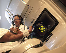
The Radiobiology (also: radiobiology ) examines the biological effects of ionizing radiation , namely alpha, beta and gamma rays and X-rays on living organisms. In addition to acute radiation sickness (e.g. as a result of accidents with nuclear power plants ), the main focus of research is on chronic effects on tumor and normal tissues in connection with radiation therapy .
Effect on cells
Ionizing radiation affecting cells of the body by ionization and excitation of molecules. Due to the much higher concentration, it is not the few direct hits on the macromolecules that are important, but rather the formation of radicals in the tissue water ( radiolysis ). The radicals oxidize important cellular macromolecules and disrupt their function (indirect radiation effect) .
Only the part of the radiated energy absorbed in the tissue is effective. Radiation can be characterized by the amount of energy that a radiation particle emits per micrometer ( linear energy transfer LET, unit k eV / μm). For example, X-rays have an LET of 2.5 keV / μm, fast neutrons of> 20 keV / μm. In radiation biology, the relative biological effectiveness RBW is used instead, a factor that indicates the harmfulness of radiation compared to 250 kV X-rays. In radiation protection , roughly estimated, whole-number quality factors are used instead of the exact RBW.
Radiation damage to biomolecules such as proteins and lipids can be tolerated without any problems; their effects on cell functions are minimal. In contrast, radical reactions with the DNA (genetic material) sometimes lead to cell death or degeneration , since each cell only has two copies and the repair mechanisms have limited capacity. The chromosomes in the cell nucleus are the main target of biological radiation. For every energy absorbed by Gray , around 1000 single-strand and 40 double-strand breaks occur in each cell , almost all of which are, however, repairable.
Unrepaired DNA damage results in cell function disorders, mutation or death of the affected cell. Most radiation effects are only detectable from a certain minimum dose, i.e. radiation in the order of magnitude of the natural background radiation is considered to be harmless in this regard. Since in theory a single mutated cell can grow into cancer or cause an embryonic malformation , there is no known minimum dose for this so-called stochastic radiation damage . It is currently assumed a linear dose-response relationship, for X-rays z. B. 5% cancer deaths per Sievert ; however, this information is the subject of an intense debate.
In addition to mutation and cell death, mammalian cells also experience delays in the cell cycle after irradiation , and stem cells and cancer cells that were previously able to divide indefinitely can differentiate and lose their cloning function after irradiation . In addition to direct cell death , experimental survival curves also record this clonogenic cell death , which plays an important role in radiation therapy . They always have a characteristic, S-shaped shape that can be described mathematically with a linear-quadratic model function .
Effect in the tissue
Different tissues are differently sensitive to radiation. The proportion of dividing cells, the blood flow and the oxygen concentration all contribute to this . The lower the oxygen partial pressure in the tissue, the less sensitive it is to ionizing radiation. It is therefore recommended in radiation therapy to stop smoking and to compensate for any anemia before starting treatment.
The proliferative organization of the tissue is also important: if it has a strictly defined stem cell fraction from which dead cells are replaced (so-called hierarchical or alternating tissue such as blood cells or intestinal mucosa ), then a few days after these stem cells have been destroyed, the entire tissue or tissue is removed Organ perish. Tissues with flexible proliferation do not have a clear separation of stem and functional cells (e.g. liver, lungs, brain) and can recover better from sublethal damage.
Alternating tissues react early (hours up to a maximum of 6 months) after radiation. Flexible tissues can develop late reactions (by definition these are radiation effects that persist after six months). Since most organs are composed of different tissues ( stroma , parenchyma , blood vessels, etc.), the situation in practice is more complicated; every organ can be permanently damaged by the long-term effects of radiation, albeit to a different extent.
The macroscopic structure of the organs also plays a role: organs with a linear structure such as the small intestine or the spinal cord are much more at risk than organs with a parallel structure such as B. glands. On the basis of the long-term effects, which are much more feared than the early effects of radiation, tolerance doses have been defined for most organs and tissues . In the literature, TD 5/5 are usually given, i.e. the dose at which a certain damage occurs in 5% of the test persons within 5 years. For example, the TD 5/5 of the lens for lens opacity is 10 Gy (Emami 1991, PMID 2032882 ). There are globally standardized catalogs of criteria to describe the radiation effects on normal tissues ( CTC for early radiation effects , LENT-SOMA for late effects).
Effect on tumors
The sensitivity of tumors to ionizing radiation is usually higher than that of healthy tissues. They are characterized by a shorter cell cycle time (<2 days) and a higher proportion of dividing cells ( growth fraction > 40%). The dose-effect relationship is S-shaped or sigma-shaped as in normal tissues, but shifted in the direction of the lower dose (to the left). Mathematically, the tumor tissues have a higher α / β ratio (α and β are the coefficients of the linear-square model equation).
The radiation tolerance is higher if the dose rate (= dose per unit of time) is low or if the radiation is distributed over many small treatments. This is due to the tissue reactions that start immediately after exposure to radiation, which the Californian radiation biologist Hubert Rodney Withers summarized as 4 R's in 1975:
- Repair (enzymatic correction of single and double strand breaks and base errors in the DNA)
- Redistribution (continuation of the interrupted cell cycles so that cells from all phases are again available)
- Reoxygenation (increased oxygen supply in the tissue)
- Repopulation (cell regrowth)
One to two treatments per day are common in radiation therapy. There must be a break of at least six hours between treatments. Fast-growing tumors are more amenable to shortened radiation therapy (only the total treatment time is important, not the dose per treatment). On the other hand, the sensitivity of a disintegrating tumor with poor circulation is reduced because of the hypoxia . In addition, normal tissues are always also irradiated, which because of their low α / β ratio are particularly sensitive to long-term effects and should be irradiated more slowly with smaller daily doses. In practice, therefore, a compromise must be made that depends on the particular tumor and the available technical possibilities.
methodology
Radiobiological research works with molecular biological , cytogenetic and cytometric methods on different organisms and cell systems. At the DNA level, radiation-induced mutagenesis and its repair are investigated.
Further topics in radiation biology
- The person in the radiation field:
- Increasing radiation sensitivity to make radiation therapy more effective on tumors
- Increasing the radiation resistance to healthy tissue during radiation therapy to spare
- Basics of radiation risk , radiation damage
- Clinical radiation biology
- Low radiation , negative and positive radiation effects in the low dose range
- Radioactivity in the food chain
- Radiation exposure , radiation protection
- Cell biological basics of radiation therapy
Well-known radiation biologists
- Otto Hug (1913–1978)
- Hedi Fritz-Niggli (1921–2005), founder of radiation biology in Switzerland. She researched the damage caused by low radiation doses, especially in the unborn child and in the sensitive development phase of living things
- Wolfgang Köhnlein (* 1933), University of Münster, retired
- Edmund Lengfelder (* 1943), professor at the University of Munich. He has been researching the health consequences of the Chernobyl disaster since 1986
- Boris Rajewsky (1893–1974)
- Hermann Rink (* 1935), chemist, radiation biologist and emeritus of the Medical Faculty of the University of Bonn
- Christian Streffer (* 1934), University of Essen, retired
- Joachim Wattendorff (1928–2008), biologist and radiation biologist , University of Friborg in Switzerland
- Paul Wels (1890–1963), pharmacologist and radiation biologist. His research interests focused in particular on the effects of X-rays on various cells and of ultraviolet radiation on the skin, as well as the pharmacological effects of irradiated substances
See also
literature
- Eric J. Hall: Radiobiology for the Radiologist . Philadelphia: Lippincott, Williams & Wilkins, 2000 (5th. Ed.), ISBN 0-7817-2649-2
- Thomas Herrmann, Michael Baumann, Wolfgang Dörr: Clinical radiation biology in a nutshell. Urban & Fischer Verlag / Elsevier GmbH; 4th edition 2006, ISBN 3-437-23960-0
- Hedi Fritz-Niggli: Radiation Hazard / Radiation Protection . Verlag Hans Huber, 4th edition 1997
- G. Gordon Steel: Basic Clinical Radiobiology . London: Arnold, 1997 (2nd ed.), ISBN 0-340-70020-3
Web links
- Swiss Society for Radiation Biology and Medical Physics
- The Institute for Radiation Biology at the Helmholtz Research Center for Environment and Health near Munich researches the effects of ionizing radiation on living cells and organisms.
- The SSK Radiation Protection Commission is a body of independent scientists that advises the German Federal Government on all aspects of radiation effects and radiation protection.
Individual evidence
- ↑ Michael Wannenmacher, Jürgen Debus, Frederik Wenz: Radiotherapy . Springer, November 3, 2006, ISBN 978-3-540-22812-7 , pp. 279-82 (accessed February 23, 2013).
- ^ Ralph Graeub: The Petkau Effect , Verlag Zytglogge Gümligen, ISBN 3-7296-0222-5


