Claudine

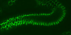
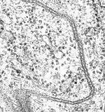
Claudins ( Latin claudere "to close") are a group of proteins that are the most important component of the cell connections , the so-called tight junctions , occurring in the covering tissue ( epithelia ) . Epithelia cover the body surfaces of multicellular animal creatures and also limit the lumen of the organs . Claudins close the spaces between cells in the epithelia and enable the flow of substances through the cell space to be controlled. They thus form a kind of seal between the cells (“paracellular barrier”), which is required in epithelia so that ions and molecules cannot freely pass through the organs. Maintaining a certain milieu of individual compartments (such as pH values of 1 to 2 in the stomach ) would not be possible without these paracellular barriers.
construction
Claudins are small transmembrane proteins that occur in many organisms and are very similar ( conserved ) in their structure, from the nematode Caenorhabditis elegans to humans . They have a size of 20-27 kilodaltons (kDa). However, at the DNA sequence level , the conservation is not very high. They span the cell membrane four times ( tetraspanin ), the N-terminus and the C-terminus are each in the cytoplasm . A claudin has two extracellular (outside of the cell) areas called the smaller and larger extracellular loop. The first extracellular loop consists of an average of 53 amino acids , the second is slightly smaller and has an average of 24 amino acids. The N terminus is usually only very short (4-10 amino acids), the C terminus is more variable in length (between 21 and 63 amino acids). In the first extracellular loop there is an amino acid motif that occurs in all claudins and consists of the amino acids W-GLW-CC. It is assumed that disulfide bridges are formed between the cysteines of one claudin or the cysteines of different claudins . The most highly conserved areas are the transmembrane domains, the most variable area is the C-terminus. With the exception of claudin 12, all human claudins have a PDZ binding motif at the C terminus, with which they bind to PDZ domain proteins. For the C-terminus it could also be shown that it is required for the localization of the claudin molecules in the tight junctions.
So far, 24 different human claudins have been identified and named “Claudin 1” to “Claudin 24”. They are not in a cluster, but are distributed on different chromosomes.
history
Ever since the tight junctions were described and their function for the cells was recognized, the search for factors or structural components of the tight junctions that were required to seal the intercellular spaces has been sought. At the beginning of the 1990s, the Japanese research group headed by Mikio Furuse and Shoichiro Tsukita at the University of Kyoto succeeded in identifying the transmembrane protein occludin as the first integral membrane protein of the tight junction. However, it turned out that occludin is not responsible for the formation and maintenance of the paracellular barrier, since cells lacking occludin can still form tight junction strands. A few years later, the same group discovered the first two proteins actually responsible for the formation of tight junctions and in 1998 gave them the name "Claudin", from the Latin claudere for "close". Some claudin proteins were previously described in other contexts, but their function as sealing components of tight junctions was initially not recognized. When they were classified in the Claudine group, they were renamed accordingly.
A targeted search in the human and mouse genome has so far found a total of 24 proteins which, based on their sequence, structure and expression, could be classified in the claudin group.
Claudins were also discovered in other animal species, for example in all of the mammals examined, in various fish species ( Takifugu rubripes , Danio rerio ), in amphibians ( Xenopus laevis and Xenopus tropicalis ) and, unexpectedly in 2003, even in the nematode Caenorhabditis elegans and the fruit fly Drosophila melanogaster . This discovery was unexpected because both C. elegans and Drosophila have no tight junctions at all, but analogous structures, the septate junctions , in the cells of the epithelia.
Since then, more than 500 studies have been published dealing with claudins, their expression , function and regulation. Since they are the core components of tight junctions, their manipulability would be a major step forward, for example in overcoming the blood-brain barrier , which currently makes drug treatment of brain tumors almost impossible.
Expression

The gene activity of the various claudins in the various tissues was investigated in many different model organisms such as mice, C. elegans , zebrafish ( D. rerio ), D. melanogaster and in some cases also in humans. Each of the 24 known claudins has a specific expression pattern . Various studies on ectopically expressed claudins in cell cultures led to the assumption that every claudin has different properties with regard to the charge selectivity and probably also to the size selectivity of the barrier. The combination of the different claudins that are expressed in an epithelium determines the properties of the barrier. Depending on the demands on the epithelium, claudins are expressed, which lead to a more or less dense barrier. There are Claudine, the ubiquitous , almost in all epithelial tissues are active, such as claudin 1, and others that are very specific expimiert, either spatially, as Claudin 16, exclusively in the ascending part of Henle's loop is found , or temporally like claudin 6, which in mice is only expressed during embryogenesis .
Paracellular barrier
How the claudins interact with each other and how the structure of the tight junctions is built up at the molecular level is not yet known exactly. Cell culture experiments have shown that claudins can enter into both homophilic and specific heterophilic bonds: cells that express claudin 1 with cells that express claudin 3 form tight junction-like structures, but neither does claudin 2 with claudin 3 claudin 1 claudin with 2. a model suggests that Claudine aqueous pores ( aqueous pores ) form, the particular depending on the type of Claudine ions and molecules up to a certain size to pass through (charge selectivity and size selectivity). These properties are determined by charged amino acid residues in the first extracellular loop and are calcium-independent. Mutagenesis experiments in cell culture showed that the reversal of the charge of certain residues in the first extracellular loop leads to a reversal of the charge selectivity of the barrier. This is measured using the TER ( transepithelial electrical resistance ).
Claudin super family
The Claudin family is classified into the PMP22 / EMP / MP20 / Claudin superfamily (pfam00822) by the Pfam protein database of the Sanger Institute in Cambridge, England, which provides alignments of protein sequences. This consists of around 450 proteins from different species, all of which have a similar structure. For some of these proteins, however, completely different functions than those of the actual claudins have been described.
Associated Diseases
Some diseases are associated with changes in claudins: Claudin 3 and 4, for example, are receptors for Clostridium perfringens - enterotoxin (CPE). They were initially not described as claudins, but rather as Rvp.1 ( rat ventral prostrate , later renamed Claudin 3) and CPE-R ( Clostridium perfringens Enterotoxin Receptor , later renamed Claudin 4). The binding of CPE to claudin 3 or 4 leads to lysis of the claudin 3 / claudin 4-expressing cells within ten to twenty minutes and thus to damage to the intestinal epithelium, which manifests itself in severe diarrhea .
Various human diseases can be traced back to mutations in claudin genes. For example, mutations in claudin 16, which is also called paracellin , lead to an increased excretion of calcium in the urine ( hypercalciuria ) and to a decrease in the magnesium content in the blood ( hypomagnesaemia ).
Claudin 14 is expressed in the vertebrates ( vertebrates ) in the liver and kidneys , in the pancreas and in the inner ear . In mice it could be shown that the expression of claudin 14 in the inner ear only begins after birth. Mutations that produce a shift in the open reading frame or a stop codon have been identified in two families living in Pakistan. These mutations cause recessive numbness while kidney and liver functions appear normal. The corresponding mouse mutants also have no defects in kidney or liver function. The phenotype analyzed in more detail in the mouse mutant showed that the hair cells of the inner ear degenerate when claudin 14 is missing in the tight junctions of the inner ear, which leads to loss of hearing function. The tight junction strands between the outer hair cells and the organ of Corti are still present and the cell polarity of the epithelium is not disturbed. Hair cell degeneration is believed to be due to excessive potassium concentrations during early hearing development.
Claudin 11, initially referred to as OSP (oligodendrocyte-specific protein), is mainly expressed in the myelin of the central nervous system (CNS) and in the testes . The mouse mutant shows delayed transmission of stimuli in the nerves and the hind legs are weakened. The males are sterile , but the mutant survives. Freeze fracture electron microscope images show that no tight junctions are found in the CNS myelin or in Sertoli cells . The blood-testicular barrier is defective in these mutants. Claudin 11 interacts with OAP (OSP / Claudin 11-associated protein) and beta-1 integrin in a complex in the cell adhesion and integrin - signaling plays a role.
Claudin 19 is expressed in Schwann's cells in the peripheral nervous system , where it forms tight junction-like structures. Mice that have a deletion in the genomic area of claudin 19 lack these structures and they have impaired stimulus transmission, which affects the musculoskeletal system.
Mice lacking Claudin 1 die a few hours after birth because they become dehydrated. A mutation in the human claudin 1 gene leads to severe skin changes. Mice lacking claudin 5 have a special phenotype : their blood-brain barrier becomes permeable to smaller molecules. Overexpression of claudin 6 leads to malfunction of the epidermis.
In these examples it becomes clear that the claudins play a decisive role in the formation of a functioning paracellular barrier in different epithelia and can be divided into different groups: "housekeeping" claudins, which are largely ubiquitous and basic functions for the formation of the tight- junction ligaments take over, and specialized claudins that are only expressed in certain tissues or do not perform an essential function in all tissues in which they occur.
Individual evidence
- ↑ C. Ruffer, V. Gerke: The C-terminal cytoplasmic tail of claudins 1 and 5 but not its PDZ-binding motif is required for apical localization at epithelial and endothelial tight junctions. in: European journal of cell biology . 2004; 83: 135-144. PMID 15260435 ISSN 0070-2463
- ↑ M. Furuse, K. Fujita, T. Hiiragi, K. Fujimoto, S. Tsukita: Claudin-1 and -2: novel integral membrane proteins localizing at tight junctions with no sequence similarity to occludin. in: Journal of Cell Biology . 1998; 141: 1539-1550. PMID 9647647
- ^ M. Furuse, S. Tsukita: Claudins in occluding junctions of humans and flies. In: Trends in Cell Biology . 2006; 16: 181-188. PMID 16537104
- ^ OR Colegio, C. Van Itallie, C. Rahner, JM Anderson: Claudin extracellular domains determine paracellular charge selectivity and resistance but not tight junction fibril architecture. In: American Journal of Physiology-Cell Physiology . 2003; 284: C1346-C1354. PMID 12700140
- ^ EE Schneeberger: Claudins form ion-selective channels in the paracellular pathway. In: American Journal of Physiology-Cell Physiology . 2003; 284: C1331-C1333. PMID 12734103 ISSN 0363-6143
- ↑ Protein database of the Sanger Institute in Cambridge, England ( Memento of the original from July 7, 2012 in the Internet Archive ) Info: The archive link has been inserted automatically and has not yet been checked. Please check the original and archive link according to the instructions and then remove this notice.
- ↑ T. Ben-Yosef, IA Belyantseva, TL Saunders, ED Hughes, K. Kawamoto, CM Van Itallie, LA Beyer, K. Halsey, DJ Gardner, ER Wilcox, J. Rasmussen, JM Anderson, DF Dolan, A. Forge , Y. Raphael, SA Camper, TB Friedman: Claudin 14 knockout mice, a model for autosomal recessive deafness DFNB29, are deaf due to cochlear hair cell degeneration. In: Human Molecular Genetics . 2003; 12: 2049-2061. PMID 12913076
- ^ A. Gow, CM Southwood, JS Li, M. Pariali, GP Riordan, SE Brodie, J. Danias, JM Bronstein, B. Kachar, RA Lazzarini: CNS myelin and sertoli cell tight junction strands are absent in Osp / claudin- 11 null mice. In: Cell . 1999; 99: 649-659. PMID 10612400
literature
- Christina M. Van Itallie, James M. Anderson: Claudins and Epithelial Paracellular Transport. In: Annual Review of Physiology . 2006; 68: 403-429, doi : 10.1146 / annurev.physiol.68.040104.131404 .
- Julia E. Rasmussen: Claudins and regulation of the paracellular transport system. Dissertation (MS), The University of North Carolina at Chapel Hill, ProQuest Information and Learning, publication number AAT 1432782 . Chapel Hill 2006 (October 27). ISBN 978-0-542-54616-7
- Christina M. Van Itallie, James M. Anderson: Physiology and Function of the Tight Junction . In: Cold Spring Harbor Perspect Biol . August 2009; 1 (2), PMC 2742087 (free full text)

