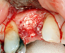
Jaw augmentation

Under jaw augmentation or jaw augmentation ( Latin augmen , `` multiplication '' , `` increment '') are understood surgical procedures in dentistry, oral medicine and maxillofacial medicine that, after a jaw reduction , serve to rebuild the alveolar bone in the edentulous parts of the upper or lower jaw .
Causes of bone loss
The bone loss of the jawbone (jaw loss ) can occur through tooth loss , periodontitis or through the pressure applied by dentures . During this process, osteoclasts release proteolytic enzymes that dissolve the collagenous bone matrix . The collagen fragments released are phagocytosed . In the space between the osteoclast and the bone substance there is a significantly reduced pH value (approx. 4.5), which is maintained by active proton transport and which serves to break down the mineralized matrix components.
Tooth loss
If teeth are lost due to an accident or extraction, the alveolar bone recedes. It is a result of horizontal and vertical resorption processes during the healing process of the alveolus after tooth extraction . Horizontal resorption begins on the thin outer alveolar walls.
Periodontal disease
Periodontitis causes bone loss in the alveoli while the teeth are still anchored in the jawbone. In the event of tooth loss due to periodontitis, the breakdown of the remaining alveoli is accelerated, as the bone volume is already reduced.
Peri-implantitis
Similar to bone loss caused by periodontitis, the implant bed can also become inflamed, which can lead to peri-implantitis with bone loss around the implant. After such an implant has been explanted (removed), the bone usually has to be rebuilt.
Prostheses
In healthy dentition, the teeth are attached to Sharpey's fibers in the alveoli . When the teeth are loaded, tensile forces - and not compressive forces - result on the jawbone. Due to the piezoelectric forces, when the teeth and thus the jawbone are stressed, electrical potentials arise that have a positive effect on bone structure. In edentulous dentition, on the other hand, the pressure load on the dentures acts on the gingiva propria and thus on the jawbone below, which reacts with increased resorption.
In the first year after tooth loss, the alveolar ridge degradation is around 0.5 mm in the upper jaw and 1.2 mm in the lower jaw. In the following years the reduction is 0.1 mm in the upper jaw and 0.4 mm in the lower jaw. The faster breakdown of the lower jaw bone results, among other things, from the fact that the contact surface for a prosthesis is only about half the size of that of the upper jaw. In the upper jaw, the prosthesis also rests on the palate. As a result, the loading forces that act on the lower jaw are twice as great as in the upper jaw. From this it follows that after about 20 years of wearing the prosthesis, the alveolar ridge of the lower jaw has completely broken down and the lower jaw has become flat. It then no longer offers a hold for a full denture .
Absorption classes
The extent of resorption is divided into six classes according to the Cawood and Howell resorption classes.
| class | description |
|---|---|
| class 1 | toothed |
| 2nd grade | immediately after tooth extraction |
| Class 3 | well rounded ridge with adequate height and width |
| Grade 4 | Razor-sharp comb shape with adequate height and width |
| Class 5 | flat ridge with inadequate height and width |
| Grade 6 | highly atrophic comb shape, partly with negative alveolar ridges |
Bone grafting procedure

The bone is built up with different materials. As addition to growth factors, bone morphogenetic proteins ( English Bone morphogenetic proteins, BMP ) used and what the differentiation of mesenchymal stimulate cells into osteoblasts.
Autogenous bone
Fresh autogenous bone is the first choice for bone augmentation. Smaller bone structures can be done with a bone graft from the lower jaw. The sampling points are the oblique line in the retro molar area of the mandible (angle of the jaw) and the mental region (chin area). For this purpose, bone can be removed by means of cylindrical milling or by cutting out a bone block. As a rule, the bone grows back at the removal points. In the case of bone deficits, this procedure is nowadays indispensable for proper implant positioning according to prosthetic and aesthetic aspects.
A rib graft was previously used for larger bone structures. Nowadays monocortical cortico-cancellous bone pieces from the iliac by means of bone flap Method removed, resulting in a second operation area and a general anesthesia makes necessary. The transplant is applied with the cancellous side to the alveolar ridge and fixed in the jawbone with osteosynthesis material such as mini plates, screws or implants. Micromovements in osteoplasty must be completely avoided in order to achieve successful healing.
Guided bone regeneration
In addition to the introduction of bone or bone substitute material is provided a method of guided bone regeneration (GBR) , guided tissue regeneration ' applied. In the GBR procedure, the space that is to be filled with bone is additionally surrounded with a membrane. This has the task of preventing the surrounding cells of the surrounding soft tissue from growing too quickly into the cavity, as this forms faster than bone. Resorbable and non-resorbable membranes can be used. Resorbable membranes are predominantly used, as this avoids a second intervention to remove the membrane and wound healing disorders occur less frequently, especially in the case of a dehiscence of the mucoperiosteal flap , which is used to close the wound. Non-resorbable membranes are made of titanium-reinforced polytetrafluoroethylene (PTFE). Resorbable membranes are made from collagen that has been treated .
Bone splitting
The bone splitting or bone spreading process developed in the 1980s is used for the remaining narrow alveolar ridge. The alveolar ridge is separated into two parts and then stretched to form a gap. The jaw must still have a minimum width of 3 mm so that sufficient peri-implant bone strength is maintained both vestibularly and orally. A residual bone height of 12 mm is mandatory, as a maximum of 70% of the bone height may be used for the splitting process. The blood supply must remain secured through the periosteum .
Alloplastic material
At the end of the 1980s, the use of alloplastic material increased, partly in block form, partly as granules. Here is hydroxyapatite implanted between the bone and mucosa. Other alloplastic materials are, for example, hydroxyapatite, β-tricalcium phosphate , ICBM - Insoluble collagenous bone matrix , copolymers of polylactate / polyglycolic acid and calcium carbonate . When used in the lower jaw, there is a risk of nerve irritation, especially in the area of the mental foramen and the dislocation of the granules towards the lingual.
Allogeneic material
In the 1980s, lyophilized allogeneic cartilage was used to build the alveolar ridge. The process is only of historical importance.
Xenogenic material
Xenogenic bone substitute materials are rarely used for bone formation. These are materials of animal origin (e.g. bovine or porcine). There is a residual risk of the transmission of prions , which are responsible for the transmission of BSE . To reduce the risk of transmission and allergy, deproteinization (withdrawal of protein) takes place. What remains is the inorganic bone part into which new bone sprouts.
Critical appraisal
Different studies conclude that vertical GBR procedures deliver predictable results, while other studies find that a reliable and superior procedure for vertical augmentation in the posterior mandible is not yet available.
Indications

1. Frontal sinus , (green), (Frontal sinus),
2. Ethmoid cells , (purple), (Cellulae ethmoidales)
3. Sphenoid sinus , (red), (Sphenoid sinus)
4. Maxillary sinus , (blue), (Maxillary sinus )
Until the end of the 1990s, the focus was solely on bone augmentation methods in order to improve the prosthesis position and thereby provide dental prostheses with positional stability. Due to the high failure rate with only bone grafting on the one hand and the development of implants with titanium surfaces on the other hand, which have a high stability and longevity through osseointegration , a rethinking took place in the professional world. Since then, bone augmentation procedures in the context of preprosthetic oral surgery have almost exclusively been carried out in combination with dental implants. The objective there is to create a sufficiently large bone bed in which to insert implants. Without implants, complete resorption of the transplanted bone must be assumed within the first three years.
Lower jaw
In the lower jaw, the height of the jawbone is limited in the caudal direction by the mandibular nerve . This must not be touched because otherwise permanent loss of sensitivity can occur, especially in the area of the lower lip and chin. The minimum length of an implant is 8 mm, with implant lengths of 10 to 12 mm being aimed for. If this height is not sufficient, bone augmentation must be carried out. The same applies to an insufficiently wide ridge bone into which the implant is to be inserted.
upper jaw
In the upper jaw, the jaw is built up in the same way as in the lower jaw, with the exception of the area of the upper jaw teeth . In the area of teeth 15-17 and 25-27 , the tooth sockets are (as a rule) only separated from the maxillary sinus by a thin bone lamella, the floor of the maxillary sinus. In the posterior region of the upper jaw, bone loss often occurs as the floor of the maxillary sinus subsides while the external shape of the alveolar ridge is largely unchanged. The thickness of the floor of the maxillary sinus can be reduced to the thickness of paper. In order to be able to insert implants with the corresponding minimum length here, too, bone augmentation must be carried out. This is usually done with a sinus lift.
Sinus lift
To create an implant bed in the posterior region, bone is not built up by placing bones (or bone substitute materials) on the alveolar ridge, but to a certain extent from the inside, namely by placing the bone graft on the floor of the maxillary sinus below the Schneiderian membrane that lines the maxillary sinus. This results in a thickening of the bone within the maxillary sinus.
Nasal floor elevation
In the anterior region, the nasal floor elevation procedure is available, which is similar to a sinus lift.
Trapdoor technology
Special cases arise with the structure of the nasal floor in the front. In the trapdoor technique ( English door trap , trap-door ' ) is inserted an implant with which the turbinate is easily pushed aside. Bone substitute material is poured into the resulting space in order to ensure sufficient stability.
See also
Individual evidence
- ↑ Eckhart Buddecke: Biochemical basics of dentistry . Walter de Gruyter, January 1, 1981, ISBN 978-3-11-085820-4 , pp. 62–.
- ↑ a b c d e f N. Schwenzer, M. Ehrenfeld: Tooth-mouth-jaw medicine . tape 3 : Dental Surgery . Thieme, Stuttgart 2000, ISBN 3-13-116963-X (5 volumes, limited preview in the Google book search).
- ^ JI Cawood, RA Howell: A classification of the edentulous jaws . In: International journal of oral and maxillofacial surgery . tape 17 , no. 4 , August 1988, ISSN 0901-5027 , p. 232-236 , PMID 3139793 (English).
- ↑ K.Müller, R.Streckbein, R.Hassenpflug: Minimally invasive autologous bone transplantation - the domain of maxillofacial surgery or realizable strategic advantages of dental specialization . In: Oral Surgery Journal . February 2004 ( zwp-online.info [PDF; 334 kB ]).
- ↑ P. Coulthard, M. Esposito et al. a .: Interventions for replacing missing teeth: bone augmentation techniques for dental implant treatment . In: Cochrane database of systematic reviews (online) . No. 3 , 2003, ISSN 1469-493X , p. CD003607 , doi : 10.1002 / 14651858.CD003607 , PMID 12917975 (English, review).
- ^ H. Tal, O. Moses: Bioresorbable Collagen Membranes for Guided Bone Regeneration . In: Bone Regeneration . 2012, ISBN 978-953-510-487-2 (English, intechopen.com ). Bioresorbable Collagen Membranes for Guided Bone Regeneration ( Memento of the original from September 27, 2013 in the Internet Archive ) Info: The archive link has been inserted automatically and has not yet been checked. Please check the original and archive link according to the instructions and then remove this notice.
- ^ A. Scipioni, GB Bruschi, G. Calesini: The edentulous ridge expansion technique. A five-year study . In: The International journal of periodontics & restorative dentistry . tape 14 , no. 5 , October 1994, ISSN 0198-7569 , p. 451-459 , PMID 7751111 (English).
- ↑ T. Frauendorf, W. Sümnig: Bone replacement in dental surgery. In: Implantologie Journal 4/2007
- ↑ G. Corinaldesi, F. Pieri u. a .: Evaluation of survival and success rates of dental implants placed at the time of or after alveolar ridge augmentation with an autogenous mandibular bone graft and titanium mesh: a 3- to 8-year retrospective study. In: The International journal of oral & maxillofacial implants. Volume 24, Number 6, 2009 Nov-Dec, pp. 1119-1128, ISSN 0882-2786 . PMID 20162118 .
- ↑ M. Clementini, A. Morlupi et al. a .: Success rate of dental implants inserted in horizontal and vertical guided bone regenerated areas: a systematic review. In: International journal of oral and maxillofacial surgery. Volume 41, Number 7, July 2012, pp. 847-852, ISSN 1399-0020 . doi: 10.1016 / j.ijom.2012.03.016 . PMID 22542079 . (Review).
- ^ I. Rocchietta, F. Fontana, M. Simion: Clinical outcomes of vertical bone augmentation to enable dental implant placement: a systematic review. In: Journal of Clinical Periodontology . Volume 35, Number 8 Suppl, September 2008, pp. 203-215, ISSN 1600-051X . doi: 10.1111 / j.1600-051X.2008.01271.x . PMID 18724851 . (Review).
- ↑ Regina Schindjalova: [Nasal floor elevation as a treatment option for bone loss in the front . Nasal floor elevation as a treatment option for bone loss in the front .] In: DI - Dental Implantologie und Parodontologie . No. 1/2014, 2014, pp. 16-21.
- ^ HH Lindorf, R Müller-Herzog, J Lehner: Lateral nose lift using the trapdoor technique The lateral nose lift using the trapdoor technique . In: ZMK special edition implantology . No. 27, 2011, pp. 6-14.


