
Coats disease
| Classification according to ICD-10 | |
|---|---|
| H35.0 | Retinopathies of the fundus and changes in the retinal vessels |
| ICD-10 online (WHO version 2019) | |


At Coats' disease ( syn. Retinitis exudative retinal telangiectasia and ) is a rare congenital eye disease of the retinal vessels , which usually occurs only on one side and to the deterioration of vision, often until blindness results. The blood vessels are dilated and leaky, so that blood and lipid-containing liquid secretions ( exudates ) can penetrate into and under the retina and its deeper layers. This creates retinal edema , which when viewed from the outside becomes noticeable as a whitish-gray pupil color ( leukocoria ). If left untreated, the exudate leads to progressive inflammatory retinal detachment and irreversible damage.
So far, the reason for the causal vascular changes is not known . Coats disease occurs predominantly in boys and young men. The treatment consists in the obliteration of the vessels by cold treatment ( cryotherapy ) or in a closure of the retinal detachment boundaries by means of laser therapy , which in many cases can prevent complete blindness or at least significantly delay it. In exceptional cases, it may also be necessary to remove the affected eye .
distribution
The Crohn's Coats is a rare congenital eye disease with visible enhancements and changes ( telangiectasia ) of the retinal vessels. There are around 1–9 cases of illness for every 100,000 people. They occur unilaterally in 90% of cases and mostly in boys and young men in the first or second decade of life. The peak of the disease is between six and eight years of age, but the disease can in principle affect anyone between the ages of one month and 79 years. The frequency is about 69% male and 31% female.
Cause and origin
The cause is an endothelial defect in the retinal blood vessels, which results in bulges and aneurysms in these vessels. As a result, both capillary collapse and a disruption of the blood-retinal barrier occur, whereby large amounts of fluid ( exudate ) can escape into the retina. This is where lipid-laden macrophages and cholesterol crystals in the blood are deposited . Over time, these substances accumulate and lead only for thickening of the retina and then ultimately to the - by the exudate-related - detachment with which an increasing loss of vision associated.
The reason for the causal defect in the retinal vessels is still unknown. In some of those affected, increased concentrations of a special signaling molecule , the vascular endothelial growth factor (VEGF) were found. Descriptions of individual cases ( case reports) on the occurrence of the disease together with various other genetic defects are, however, regarded as an indication of genetic involvement. In one case, the X chromosome locus p11.4 was affected. This is also known as the NDP gene (see also Norrie syndrome ) because it is responsible for the formation of Norrin . The latter is a protein that, as a growth factor, is believed to have an impact on the development of retinal vessels. An association with facioscapulohumeral muscular dystrophy has also been described. This change affects chromosome 4 , locus q35.
Clinical manifestations

Typical initial symptoms are secondary squint (strabismus) and the whitish pupil ( leukocoria ). In the case of the latter, the reflex of the fundus of the eye is not red as usual , but whitish-gray in photographs taken with flash light . Patients often see blurred in the affected eye, which also affects their spatial vision . Especially in small children, the loss of vision in one eye can go completely unnoticed. Coats disease is usually painless. However, if the exudate to an increase in intraocular pressure results, it can be a "Green Star" ( glaucoma ) result. Different eye color is also typical. Coats' disease is initially completely asymptomatic in almost ten percent of cases.
Although Coats' disease generally leads to long-term blindness of the affected eye, its course is not the same in all cases. Its progress can spontaneously come to a temporary or permanent standstill. A few cases have been described in which the disease even regressed. However, if a complete retinal detachment occurs, then permanent vision loss can be assumed. In children under five years of age, the clinical picture is generally much more intense than in people over ten years of age.
The vast majority of cases develop profuse subretinal exudate and detachment of the retina . Characteristic consequences are the formation of new vessels on the iris ( rubeosis iridis ), glaucoma and cataracts , as well as inflammation of the middle skin of the eye ( uveitis ) and shrinkage of the eyeball ( phthisis bulbi ). Statistically, the lower outer area of the retina is particularly often affected, which can lead to a medial upper quadrant loss of the visual field. The rate of blindness is particularly high in those patients in whom the subretinal fluid accumulation does not regress after treatment or in whom the retina has large cysts or telangiectasias. The removal of the eye ( enucleation ) is particularly often necessary in patients with glaucoma or rubeosis iridis. Clinically, the clinical picture can look like a retinoblastoma .
Staging
So far, two somewhat different staging have been published. The first comes from Gomez Morales (1964) and divides the course into five stages. Only narrowly limited exudates (stage I) are followed by massive intraretinal exudates (II), then the first small-area exudates (III) and then the complete detachment of the retina (IV). Stage V stands for complications like glaucoma.
A second classification according to Shields et al. a. also describes five stages, however, some of which are underdifferentiated, beginning with stage I, the mere occurrence of retinal telangiectasias, without any evidence of leakage. Stage II describes its exit outside (II A) and inside the fovea centralis (II B). Stage III describes the detachment of the retina, beginning with a partial detachment outside (III A 1) or within the fovea centralis (III A 2) and the entire retina (III B). Stage IV describes the complete detachment with complicating glaucoma and stage V an advanced end stage of the disease.
Clinical examination
The classic leading symptom is leukocoria. Secondary strabismus can develop as a result of the one-sided loss of vision and the resulting disruption of binocular vision ( binocular vision ) . When examining the fundus by means of ophthalmoscopy , the capillary pattern is coarsened and the vessels of the retina are enlarged (dilated) and tortuous. This finding is mostly clearly visible in the temples and peripheral areas. If the disease has broken out, retinal detachments, large-scale, lipid-containing exudates and bleeding from the changed vessels can be found. These vascular changes can be shown particularly clearly by means of fluorescence angiography - if necessary to establish a diagnosis .
Technical examination results

Imaging procedures such as sonography , computed tomography (CT) and magnetic resonance imaging (MRI) can help to establish a diagnosis. Sonographically, Coats' disease appears as an echogenicity in the posterior region of the vitreous without acoustic shadow; Bleeding into the vitreous humor and retina is typical.
Due to the protein-containing exudate , the eyeball appears denser (hyperdense) in the CT compared to healthy subjects. In the advanced stage, the exudate can affect the entire vitreous humor. The anterior border of the subretinal exudate is represented by an increase in contrast. Since the retina is fixed around the optic nerve papilla , advanced detachments are V-shaped.
In the MRI, the exudate detaching the retina shows a high signal intensity in both the T1- and T2-weighted images. In the presence of hemorrhagic fibrosis , it can appear irregular (heterogeneous). The space behind the retina does not expand when using contrast media containing gadolinium . However, there may be a slight expansion between the exudate and the remaining vitreous humor. The exudate shows an extended peak at 1–1.6 ppm on nuclear magnetic resonance spectroscopy .
Differential diagnosis
Differential diagnoses include in particular the retinoblastoma , but also the retinopathy of prematurity , a Persistent hyperplastic primary vitreous and toxocariasis Conditional Marketing Chorioretinitis that idiopathic juxtafoveolare telangiectasia and the damage to the retina ( retinopathy ) due to Leber's Miliaraneurysmen (irregular ectasia to consider retinal vessels). It should also be noted that Leber's miliar aneurysms and Coats’s disease are very similar clinical pictures and are therefore equated by some authors.
pathology
- Pathological preparations in Coats' disease

V-shaped detachment of the retina by the exudate.
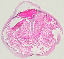
Complete detachment of the retina by the exudate.


A marked detachment of the retina and a yellowish exudate under the retina, which contains cholesterol crystals , are characteristic of the pathological finding.
Under the microscope, the wall of the retinal vessels may appear thickened in some cases and thinned in others. In addition, there is an irregular expansion of the affected vessels. Characteristic is an exudate that consists of cholesterol crystals, macrophages loaded with cholesterol and pigments , as well as red blood cells and hemosiderin . The retina may have a granulomatous reaction caused by the exudate, and in some cases gliosis caused by the injury .
Prevention, Treatment and Prospect for Cure
No preventive measures are known. Especially in the early stages of the disease, cold treatments ( cryotherapy ) and focal laser coagulation can be used to destroy (obliterate) the altered vessels and thus prevent the foreseeable leakage of blood or fluid there. If the disease is more advanced and retinal detachment has occurred, attempts at therapy can be undertaken in the form of partial removal of the vitreous body ( vitrectomy ) or retinal coagulation (connection with the choroid by means of laser coagulation). If blindness has already occurred, removal ( enucleation ) of the affected eye may also be necessary - especially if pain and other complications arise or a retinoblastoma cannot be ruled out with absolute certainty. In the early stages in particular, at least part of the vision can often be preserved through the prompt use of suitable therapeutic measures. A curative treatment is not possible in individual cases, however, a spontaneous regression of the disease is described. Under the assumption that the vascular endothelial growth factor is of considerable importance for the course of the disease, an experimental treatment using bevacizumab or pegaptanib has recently been described in individual cases . However, this approach must be viewed critically.
history
The disease was named after its discoverer, the Scottish ophthalmologist George Coats . He described her on six children in 1908. In 1912, the German ophthalmologist Theodor Carl Gustav von Leber described a disease that was characterized by retinal degeneration due to multiple retinal aneurysms and that occurred primarily in young men. In 1955, Reese showed similarities between the two diseases (Coats' disease and Leber's miliary aneurysms) and assumed that both were merely different manifestations of the same underlying disorder. He used the term Coats' disease for the combination of telangiectasia and retinitis exudativa .
Web links
- Coats disease. In: Online Mendelian Inheritance in Man . (English)
- Katherine B Sims: NDP-Related Retinopathies. GeneReviews entry on NCBI
- Alessandra Del Longo: Coats Disease. Orphanet Encyclopedia. Sept 2004
Individual evidence
- ↑ a b c d e f F. Grehn: Ophthalmology. Verlag Springer, 2008, ISBN 3540752641 , p. 228, limited preview in the Google book search.
- ^ T. Axenfeld, H. Pau: Textbook and Atlas of Ophthalmology. With the collaboration of R. Sachsenweger et al., Gustav Fischer Verlag, Stuttgart 1980, ISBN 3-437-00255-4 , p. 394.
- ↑ a b c Coats disease. In: Orphanet (Rare Disease Database).
- ^ A b J. A. Shields, CL Shields, SG Honavar, H. Demirci: Clinical variations and complications of Coats disease in 150 cases. The 2000 Sanford Gifford Memorial Lecture. In: American journal of ophthalmology Volume 131, Number 5, May 2001, pp. 561-571, ISSN 0002-9394 , PMID 11336930 .
- ↑ LM Smithen, GC Brown, AJ Brucker, LA Yannuzzi, CM Klais, RF Spaide: Coats' disease diagnosed in adulthood. In: Ophthalmology. Volume 112, Number 6, June 2005, pp. 1072-1078, ISSN 1549-4713 , doi : 10.1016 / j.ophtha.2004.12.038 , PMID 15882905 .
- ^ A b c d e f D. P. Edward, MF Mafee, E. Garcia-Valenzuela, RA Weiss: Coats' disease and persistent hyperplastic primary vitreous. Role of MR imaging and CT. In: Radiologic clinics of North America. Volume 36, Number 6, November 1998, x, pp. 1119-31, ISSN 0033-8389 , PMID 9884692 (review).
- ↑ AC Woods, JR Duke: Coats's disease. I. Review of the literature, diagnostic criteria, clinical findings, and plasma lipid studies. In: The British journal of ophthalmology. Volume 47, July 1963, pp. 385-412, ISSN 0007-1161 , PMID 14189710 , PMC 505826 (free full text).
- ↑ a b c H. Heimann, u. a .: Atlas of fundus angiography. Georg Thieme Verlag, 2006, ISBN 3-131-36491-2 , p. 100ff, limited preview in the Google book search.
- ↑ a b c d A. Del Longo: Coats disease. In: Orphanet Encyclopedia. 9, 2004, PDF (English)
- ↑ MM Chang, IW McLean, JC Merritt: Coats' disease. A study of 62 histologically confirmed cases. In: Journal of pediatric ophthalmology and strabismus. Volume 21, Number 5, 1984 Sep-Oct, pp 163-168, ISSN 0191-3913 , PMID 6502405 .
- ↑ a b Y. G. He, H. Wang, B. Zhao, J. Lee, D. Bahl, J. McCluskey: Elevated vascular endothelial growth factor level in Coats' disease and possible therapeutic role of bevacizumab. In: Graefe's archive for clinical and experimental ophthalmology Volume 248, Number 10, October 2010, pp. 1519-1521, ISSN 1435-702X , doi : 10.1007 / s00417-010-1366-1 , PMID 20379736 .
- ^ A b Y. Sun, A. Jain, DM Moshfeghi: Elevated vascular endothelial growth factor levels in Coats disease: rapid response to pegaptanib sodium. In: Graefe's archive for clinical and experimental ophthalmology. Volume 245, Number 9, September 2007, pp. 1387-1388, ISSN 0721-832X , doi : 10.1007 / s00417-007-0559-8 , PMID 17342503 .
- ↑ H. Heimann, u. a .: Atlas of the fundus. Angiography, OCT, autofluorescence and ultrasound. Georg Thieme Verlag, 2009, ISBN 3131463511 , p. 142, limited preview in the Google book search.
- ↑ OMIM Gene map: Xp11.4, NDP to Xp11.3, SLC9A7 , online here
- ↑ 0018 NORRIE DISEASE [NDP, CYS96TRP] dbSNP: rs104894877, online here
- ↑ a b c Coats disease. In: Online Mendelian Inheritance in Man . (English)
- ↑ The OMIM Gene map: 4q35, FSHMD1A to 5p15.3, IRX2 , online here
- ↑ FACIOSCAPULOHUMERAL MUSCULAR DYSTROPHY 1A; FSHMD1A, here online
- ↑ CL Shields, Y. Uysal, R. Benevides, RC Eagle, B. Malloy, JA Shields: Retinoblastoma in an eye with features of Coats' disease. In: Journal of pediatric ophthalmology and strabismus. Volume 43, Number 5, 2006 Sep-Oct, pp. 313-315, ISSN 0191-3913 , PMID 17022167 .
- ↑ B. Förl, I. Schmack, HE Grossniklaus, K. Rohrschneider: Coats disease - important differential diagnosis for retinoblastoma. In: The ophthalmologist. Volume 105, number 8, August 2008, pp. 761-764, ISSN 0941-293X , doi : 10.1007 / s00347-007-1645-3 , PMID 18299842 , PMC 299113 (free full text).
- ↑ JA Shields, CL Shields: Review: Coats disease. The 2001 LuEsther T. Mertz lecture. In: Retina (Philadelphia, Pa.) Volume 22, Number 1, February 2002, pp. 80-91, ISSN 0275-004X , PMID 11884883 (Review).
- ↑ T. Berrocal, A. de Orbe, C. Prieto, I. al-Assir, C. Izquierdo, I. Pastor, J. Abelairas: US and color Doppler imaging of ocular and orbital disease in the pediatric age group. In: Radiographics: a review publication of the Radiological Society of North America, Inc. Volume 16, Number 2, March 1996, pp. 251-272, ISSN 0271-5333 , PMID 8966285 .
- ↑ CM Glasier, MC Brodsky, RE Leithiser, SL Williamson, JJ Seibert: High resolution ultrasound with Doppler. A diagnostic adjunct in orbital and ocular lesions in children. In: Pediatric radiology. Volume 22, Number 3, 1992, pp. 174-178, ISSN 0301-0449 , PMID 1508582 .
- ↑ L. Eisenberg, M. Castillo, L. Kwock, SK Mukherji, DK Wallace: Proton MR spectroscopy in Coats disease. In: AJNR Volume 18, Number 4, April 1997, pp. 727-729, ISSN 0195-6108 , PMID 9127038 .
- ↑ EM Chung, CS Specht, JW Schroeder: From the archives of the AFIP: Pediatric orbit tumors and tumorlike lesions: neuroepithelial lesions of the ocular globe and optic nerve. In: Radiographics Volume 27, Number 4, 2007 Jul-Aug, pp. 1159-1186, ISSN 1527-1323 , doi : 10.1148 / rg.274075014 , PMID 17620473 . (Review).
- ↑ I. Kremer, I. Nissen grain, I. Ben-Sira: Cytologic and biochemical examination of the subretinal fluid in diagnosis of Coats' disease. In: Acta ophthalmologica. Volume 67, Number 3, June 1989, pp. 342-346, ISSN 0001-639X , PMID 2763826 .
- ↑ BF Fernandes, AN Odashiro, S. Maloney, ME Zajdenweber, AG Lopes, MN Burnier: Clinical-histopathological correlation in a case of Coats' disease. In: Diagnostic pathology. Volume 1, 2006, p. 24, ISSN 1746-1596 , doi : 10.1186 / 1746-1596-1-24 , PMID 16942617 , PMC 1564043 (free full text).
- ↑ A. Ramasubramanian, CL Shields: Bevacizumab for Coats' disease with exudative retinal detachment and risk of vitreoretinal traction. In: The British journal of ophthalmology. [Electronic publication before printing] June 2011, ISSN 1468-2079 , doi : 10.1136 / bjophthalmol-2011-300141 , PMID 21653215 .
- ↑ N. Goel, V. Kumar, A. Seth, UK Raina, B. Ghosh: Role of intravitreal bevacizumab in adult onset Coats' disease. In: International ophthalmology. Volume 31, Number 3, June 2011, pp. 183-190, ISSN 1573-2630 , doi : 10.1007 / s10792-011-9436-x , PMID 21437759 .
- ↑ S. Kaul, M. Uparkar, K. Mody, J. Walinjkar, M. Kothari, S. Natarajan: Intravitreal anti-vascular endothelial growth factor agents as an adjunct in the management of Coats' disease in children. In: Indian journal of ophthalmology. Volume 58, number 1, 2010 Jan-Feb, pp. 76-78, ISSN 1998-3689 , doi : 10.4103 / 0301-4738.58480 , PMID 20029154 , PMC 284138 (free full text).
- ^ G. Coats: Forms of retinal disease with massive exudation. In: Royal London Ophthalmic Hospital Reports. Volume 17, Number 3, 1908, pp. 440-525. - Quoted from: www.whonamedit.com: Coats' disease. Last accessed on May 28, 2011
- ↑ T. Leber: About a form of retinal degeneration characterized by the occurrence of multiple miliar aneurysms. In: Arch Klin Ophthalmol. Volume 81, 1912, pp. 1-14.
- ^ AB Reese: Telangiectasis of the retina and Coats' disease. In: American journal of ophthalmology. Volume 42, Number 1, July 1956, pp. 1-8, ISSN 0002-9394 , PMID 13339898 . Quoted from A. Del Longo: Coats disease. In: Orphanet Encyclopedia. 9, 2004, PDF (English)