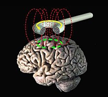
Transcranial magnetic stimulation
The transcranial magnetic stimulation ( transcranial just something like "by the skull") TMS , a technology in which strong with is magnetic areas of the brain both stimulated as can also be inhibited. This makes the TMS a useful tool in neuroscientific research. In addition, transcranial magnetic stimulation is used to a limited extent in neurological diagnostics or suggested for the treatment of neurological diseases such as tinnitus , apoplexy , epilepsy or Parkinson's disease , as well as in psychiatry for the treatment of affective disorders , above all depression , but also from schizophrenia . The first studies carried out do not yet show the extent to which the sometimes quite high clinical expectations of transcranial magnetic stimulation are justified.
History of the TMS
First transcranial ( v. Lat. = Transcranial through the skull through ) magnetic stimulation reach the doctor and physicist Jacques-Arsène d'Arsonval end of the 19th century at the Collège de France in Paris . He used high-voltage coils , such as those used in electrical power stations, to stimulate himself and his test subjects, and was thus able to prove that a changing magnetic field induces a current flow in human tissues . This was followed by experiments with very large coils, mainly carried out in self- experiments , which often completely enclosed the subjects' heads. The test persons saw lively phosphene ( magnetophosphene ) and experienced circulatory disorders and dizziness attacks up to loss of consciousness . Recent research assumes that the observed effects did not come about through stimulation of the brain, but through direct stimulation of the optic nerves and the retina.
At the University of Sheffield , Anthony Barker introduced the modern variant of transcranial magnetic stimulation in 1985. It can be traced back to the technical development of high-performance capacitors and uses significantly smaller coils that only stimulate the cerebral cortex in a small area. Since then, the magnetic stimulation of the cortex near the skull has been virtually uncomfortable for the test subjects or patients and technically (alluding to Sherlock Holmes ) "simplicity itself" .
technical basics
The TMS uses the physical principle of electromagnetic induction. A magnetic coil placed tangentially on the skull generates a short magnetic field of 200 to 600 µs duration with a magnetic flux density of up to 3 Tesla . The resulting change in electrical potential in the cranial cortex causes a depolarization of neurons with the triggering of action potentials . As a first approximation, the strength of this electric field decreases exponentially with the distance from the coil and depends on the properties of the capacitor current and the coil . The current in the coil reaches more than 15,000 amperes . So-called round coils and double coils are used. The latter consist of two round coils that touch or overlap at the edge. As a result, the magnetic field of both sub-coils is superimposed in the middle part of the coil and thus reinforced. Due to their shape, double coils are also known as figure-of-eight or butterfly coils.
In electrical engineering terms, common magnetic stimulators generally distinguish between monophasic and biphasic circuits. An oscillating circuit is closed by a thyristor . After half a period, the direction of the current is reversed. In the monophasic circuit, the capacitor changes polarity after a quarter oscillation and can therefore not be recharged by the current oscillating back. Instead, the current is dissipated via a diode and a resistor . In contrast, in the biphasic circuit, the capacitor is recharged by the current that oscillates back. In the coil, therefore, in the monophasic circuit there results an exponentially decaying current, in the biphasic circuit a current that is similar to a damped sinusoidal oscillation .
A distinction is also made between stimulation with individual magnetic field pulses and stimulation with pulse bursts, so-called repetitive magnetic stimulation (rTMS). Mainly biphasic current pulse forms are used for the rTMS. Today salvos of up to 100 Hz are technically possible. The rTMS is now limited primarily by the heating of the coil. Work is underway on the development of cooled coils.
effect
Magnetic stimulation triggers action potentials in the brain . Despite intensive research since the introduction of the method in 1985, the exact mechanism is still not fully understood.
Above a certain magnetic field strength, a sufficiently strong electric field is generated in the cerebral cortex near the skull to depolarize neurons . This depolarization is most likely to take place on the axon . If the induced electric field runs in the direction of the axon, the required magnetic field strength is smallest. Thus, the direction of depolarization is decisive for preventing large-wave depolarization, which can initiate both the endocrine household and the body's own vasoactive autacoids . The magnetic field strength that is currently required to cause an effect on neuron is called in the neurophysiology excitation charming threshold . Nerve endings, branches and, above all, bends have a particularly low excitation threshold.
application
The TMS is used in neuroscientific research , in neurology and in psychiatry . The short-term disturbance of a small brain region in order to investigate its physiological function is of particular scientific interest. For example, magnetic stimulation can trigger muscle twitching over the motor cortex , and phosphene and scotoma can be generated over the visual cortex . The rTMS of brain regions that are responsible for language can lead to a deterioration in the linguistic expression of the test subjects for a few minutes.
Clinical applications are mostly limited to single pulses over the motor cortex or to repetitive stimulation:
- The triggering of muscle twitches by stimulating the motor cortex is used diagnostically in neurology . It leads to electrical potentials ( motor evoked potentials ; MEP ), which are relatively easy to derive with electrodes. Certain diseases of the brain and the spinal cord such as multiple sclerosis lead to changes in the MEP, which is therefore an important diagnostic tool. The change in stimulus thresholds in various neurological diseases such as migraines or epilepsy is also of diagnostic interest . The use of psychotropic drugs or drugs also leads to changes in the stimulus threshold, which can be measured with the TMS.
- The rTMS can lead to habituation to the stimulation, which can lead to a long-term change in the activity of the cerebral cortex in the stimulated area. For example, rTMS of the motor cortex can impair the mobility of test subjects for a few minutes. One can also change the activity of the prefrontal cortex , which one tries to use in the treatment of depression in psychiatry. The antidepressant effect is said to last for a few days in the patient, but is not sufficiently scientifically proven. In contrast to electroconvulsive therapy (ECT), for plausible reasons, no double-blind study is possible with rTMS: For adjustment, 100 - 110% of the motor threshold is used (depending on the study) . With the rTMS one tries - without the risks of ECT - to treat therapy-refractory depression with a frequency of 10 Hz (corresponds to the alpha rhythm of the brain waves in the relaxation state) in different "trains" (sequences) with different numbers of sessions. In the case of schizophrenia, a stimulation frequency of around 1 Hz is used. (See abstracts via pubmed.gov)
In scientific research , the range of applications is greater.
A principal problem with stimulation by TMS is the spatial resolution. It is unclear to what extent connected regions are stimulated by the stimulation of a target region. Thus it is difficult to make statements about the exclusive role of a stimulated brain area. Another problem arises from the fact that TMS stimulations cannot be standardized with regard to their intensity at the moment: The standardization of the stimulation using the above-mentioned relation to the motor threshold is questionable, since this limit value does not show any correlation in other brain regions within the same head. So you don't know how strongly a certain area was stimulated, not even if the motor threshold is given as a reference. When using the stimulation protocols detailed below, there are often conflicting results, which can vary from study to study as well as from subject to subject. The complex structures of the brain are probably influenced in many ways by different protocols, so that precise statements about the mode of action of individual protocols have not yet been possible:
- Using single pulses, brain areas can be influenced in a well-defined and controlled manner. This allows to interfere directly with certain processing steps (e.g. in the visual system) and thus to determine these processing steps precisely in terms of time (relative to the stimulus presentation). The disadvantage of the single pulse is its low energy, so that often only very weak stimuli can be disrupted in their processing or the disruption is very minor.
- With a double pulse (paired pulse) a large part of the temporal precision is retained, the influence on the neural processing is much greater.
- The so-called theta-burst stimulation has proven useful in the past because of its suitability for long-term potentiation in order to improve the strength of neuronal connections. Theta-burst stimulation consists of several short bursts (from 50–100 Hz for 100–1000 ms), which are separated from one another by a longer time interval (seconds). Brain regions are presumably part of a network when, after theta burst stimulation, their activity is more synchronized than before.
- Repetitive stimulation (rTMS) is used in research similar to that in clinical application.
- Another possibility, which in turn can consist of any of the listed applications, is the simultaneous stimulation of different brain areas with two or more coils in order to be able to study the influence of the areas on one another or their role in a network.
Risks and Side Effects
Test subjects and patients facing TMS should speak to their treating physician about risks and side effects . The risks and side effects described here can only provide an overview. The attending physician will have to decide in each individual case whether a person is suitable for TMS or not.
Since the introduction of magnetic stimulation in 1985, hardly any side effects have been observed. The most common side effect is a temporary headache, which occurs mainly when muscles are stimulated. In particular, the very rare triggering of an epileptic seizure should be avoided in rTMS. Therefore, in 1998, in a consensus of various scientists, strict application regulations for the TMS were drawn up in order to minimize the risks. B. by excluding people at risk from scientific experiments. However, newer protocols with a stronger effect, such as theta-burst stimulation, are not yet taken into account in this consensus, making the risks of such stimulations more difficult to calculate.
literature
- AT Barker, R. Jalinous, IL Freeston: Non-invasive magnetic stimulation of human motor cortex. In: The Lancet . 1, 1985, pp. 1106-1107.
- S. Groppa, M. Peller, HR Siebner: Functional diagnosis of the corticomotor pathways with transcranial magnetic stimulation: an introduction. In: Klin Neurophysiol. 41 (1), 2010, pp. 12-22.
- T. Zyss: Will electroconvulsive therapy induce seizures: magnetic brain stimulation as hypothesis of a new psychiatric therapy. In: Psychiatr Pol. 26 (6), 1992, pp. 531-541.
- G. Höflich et al .: Application of transcranial magnetic stimulation in treatment of drug-resistant major depression: a report of two cases. In: Hum Psychopharmacol. 8, 1993, pp. 361-365.
- P. Fox et al .: Imaging human intracerebral connectivity by PET during TMS. In: Neuroreport. 8, 1997, pp. 2787-2791.
- SA Brandt, CJ Ploner, BU Meyer: Repetitive transcranial magnetic stimulation. In: Neurologist. 68, 1997, pp. 778-784.
- T. Paus et al .: Dose-dependent reduction of cerebral blood flow during rapid-rate transcranial magnetic stimulation of the human sensorimotor cortex. In: J Neurophysiol . 79 (2), 1998, pp. 1102-1107.
- A. Post, MB Muller, M. Engelmann, ME Keck: Repetitive transcranial magnetic stimulation in rats: evidence for a neuroprotective effect in vitro and in vivo. In: European Journal of Neuroscience. 11 (9), 1999, pp. 3247-3254.
- GW Eschweiler, C. Plewnia, M. Bartels: Which Patients with Major Depression Benefit from Prefrontal Repetitive Magnetic Stimulation. In: Fortschr Neurol Psychiatr. 69 (9), 2001, pp. 402-409.
- S. Evers, K. Hengst, PW Pecuch: The impact of repetitive transcranial magnetic stimulation on pituitary hormone levels and cortisol in healthy subjects. In: J Affect Disord. 66 (1), 2001, pp. 83-88.
- AP Strafella, T. Paus, J. Barrett, A. Dagher: Repetitive transcranial magnetic stimulation of the human prefrontal cortex induces dopamine release in the caudate nucleus. In: J Neurosci. 21 (15), 2001, p. RC157.
- S. Smesny et al: Repetitive Transcranial Magnetic Stimulation (rTMS) in acute and long-term therapy in therapy-resistant depression. In: Neurologist. 72 (9), 2001, pp. 734-738.
- MP Szuba et al: Acute mood and thyroid stimulating hormone effects of transcranial magnetic stimulation in major depression. In: Biol Psychiatry. 50 (1), 2001, pp. 22-27.
- W. Peschina, A. Conca, P. Konig, H. Fritzsche, W. Beraus: Low frequency rTMS as an add-on antidepressive strategy: heterogeneous impact on 99m Tc-HMPAO and 18 F-FDG uptake as measured simultaneously with the double isotopic SPECT technique . Pilot study. In: Nucl Med Commun. 22 (8), 2001, pp. 867-873.
- S. Cohrs, F. Tergau, J. Korn, W. Becker, G. Hajak: Suprathreshold repetitive transcranial magnetic stimulation elevates thyroid-stimulating hormone in healthy male subjects. In: J Nerv Ment Dis. 189 (6), 2001, pp. 393-397.
- F. Manes et al .: A controlled study of repetitive transcranial magnetic stimulation as a treatment of depression in the elderly. In: Int Psychogeriatr. 13 (2), 2001, pp. 225-231.
- AM Catafau et al: SPECT mapping of cerebral activity changes induced by repetitive transcranial magnetic stimulation in depressed patients. A pilot study. In: Psychiatry Res. 106 (3), 2001 May 30, pp. 151-160.
- O. Seemann, G. Köpf: The use of repetitive transcranial magnetic stimulation in psychiatry. In: NeuroDate. 3, 2002, pp. 25-27.
- GW Eschweiler, C. Plewnia, M. Bartels: Similarities and differences between therapeutic transcranial magnetic stimulation and electroconvulsive therapy. In: Neurology. 22, 2003, pp. 189-195.
- G. Hajak, F. Padberg, U. Herwig, GW Eschweiler, S. Cohrs, B. Langguth, C. Schönfeldt-Lecuona, AJ Fallgatter, J. Höppner, C. Plewina, P. Eichhammer: Repetitive Transcranial Magnetic Stimulation (PDF; 605 kB). In: Neurology. 1, 2005, pp. 46-58.
- A. Erhardt et al: Repetitive transcranial magnetic stimulation increases the release of dopamine in the nucleus accumbens shell of morphine-sensitized rats during abstinence. In: Neuropsychopharmacology . 2004; Jun 9, pp. 2074-2080.
- O. Seemann: repetitive transcranial magnetic stimulation. In: NeuroDate. 2006; 6, pp. 13-14.
- H. Siebner, U. Ziemann (ed.): The TMS book: Transkranielle Magnetstimulation . Springer-Verlag, 2007, ISBN 978-3-540-71904-5 .
- LM Stewart et al: Motor and phosphene thresholds: a transcranial magnetic stimulation correlation study. In: Neuropsychologia. Volume 39, Issue 4, 2001, pp. 415-419.
- JP Lefaucheur et al: Evidence-based guidelines on the therapeutic use of repetitive transcranial magnetic stimulation (rTMS). In: Clin Neurophysiol. 125 (11), 2014 Nov, pp. 2150-2206. doi: 10.1016 / j.clinph.2014.05.021 .
Individual evidence
- ↑ MC Ridding, JC Rothwell: Is there a future for therapeutic use of transcranial magnetic stimulation? In: Nature Reviews Neuroscience . 8, 2007, pp. 559-567.
- ↑ LA Geddes: d'Arsonval, Physicial and Inventor. In: IEEE Engineering in Medicine and Biology. July / August 1999, pp. 118-122.
- ↑ AT Barker, R. Jalinous, IL Freeston: Non-invasive magnetic stimulation of human motor cortex. In: Lancet. 1, 1985, pp. 1106-1107.
- ↑ M. Van den Noort, S. Lim, P. Bosch: Recognizing the risks of brain stimulation In: Science . 346, 2014, p. 1307.
Web links
- Review on the subject of rTMS (PDF file; 108 kB) No confirmed effectiveness in depression
