Venous leg ulcer

| Classification according to ICD-10 | |
|---|---|
| I83.2 | Varices of the lower extremities with ulceration and inflammation |
| ICD-10 online (WHO version 2019) | |
The term ulcus cruris venosum ( Latin : ulcus = ulcer , crus = calf , vena = blood vein ) describes a ulcus cruris ("open leg", lower leg ulcer ), which as a result of an advanced venous disease such as the ulcus cruris varicosum as a sign of a so-called chronic venous Insufficiency has occurred. It is a chronic wound that is usually found in the lower third of the lower leg, at the level of the inner ankle. Ultimately, responsible for its occurrence is the particularly high pressure of the blood in the veins as part of the venous disease in this area . The term "ulcus" translated into everyday language means something like "ulcer". This is somewhat misleading, because a venous leg ulcer is a substance defect in the tissue that extends at least into the dermis . This is basically always colonized by bacteria and often heals only very poorly and slowly.
The venous leg ulcer is by far the most common cause of leg ulcers. It is treated by reducing the congestion of blood in the venous system of the legs through compression therapy , reducing inflammatory changes and stimulating the granulation of the tissue.
distribution
In 37 to 80 percent of all chronic leg ulcers it is a venous leg ulcer . Elderly patients are most commonly affected. On average, around 0.6 percent of all people between the ages of 18 and 79 were affected at least once, and from the 8th to 9th decade the incidence is around 1 to 3 percent. The so-called "Bonn Vein Study" by the German Society for Phlebology , which surveyed the spread of venous leg ulcers in 2003, estimated the number of those affected at 80,000. Almost half a million people in Germany also have a venous leg ulcer that has already healed. The risk of developing such a leg ulcer increases with age.
Cause and origin
The cause of a venous leg ulcer is a chronic venous insufficiency (CVI). In particular, defective venous valves lead to an increase in pressure in the veins. Since people stand upright, the lower extremities are particularly affected due to the hydrostatic pressure . This is where the pressure of the blood in the veins ( venous hypertension ) is highest and the veins are particularly full there (venous hypervolemia).
This high pressure in the large veins of the legs also increases the pressure in the venous (post-) capillary vessels . As a result, more proteins flow from it into the tissue and lead to edema there . At the same time, the capillary vessels are damaged and show a long-term microangiopathic remodeling; As a result, the tissue is no longer supplied (there are indications that, for example, oxygen diffusion is decreasing), initially reacts with scarring processes and then slowly dies. "Atrophy blanche" (capillaritis alba) and "dermatoliposclerosis" can appear as preliminary stages of a venous leg ulcer .
Blood coagulation disorders that are independent of the pathological changes in the venous system can make the process significantly more difficult and accelerate it. As fibrinogen increases in the blood, its viscosity and the extent to which the red blood cells tend to clump increases. There are also indications that neurological disorders can occur: For example, a lengthening of the nerve conduction speed of the nervus peroneus together with a reduction in the perception of cold and heat as well as vibrations could be demonstrated. The neural control of the capillaries is also measurably changed and the outflow of lymph via the lymph vessels is reduced.
A venous leg ulcer , which occurs mainly as a result of varicose veins , is also known as a varicose leg ulcer . If a post-thrombotic syndrome is the main cause, it is also called post-thrombotic leg ulcer . If CVI and peripheral arterial occlusive disease (PAD) together lead to the development of an ulcer, one speaks of a mixed leg ulcer . The mixed leg ulcer accounts for about 15% of all leg ulcers.
Clinical manifestations
Typically a venous leg ulcer appears as an inflamed and often painful substance defect of the tissue on the distal lower leg in patients with chronic varicose veins or after a thrombosis . It is most often found in the area of the inner ankle, more rarely also on the outer ankle, or around the lower leg (gaiter ulcus). It only occurs in exceptional cases before the age of 40. A venous leg ulcer is usually relatively flat in the initial stage, and the floor is typically covered with “greasy-purulent”. Other changes caused by chronic venous insufficiency such as atrophy blanche , hyperpigmentation and dermatosclerosis are usually found in its vicinity .
Investigation methods
Ultimately, the classic leg ulcer is a "visual diagnosis", for the assurance of which the guideline-compliant diagnosis of chronic venous insufficiency is necessary, especially with regard to the detection of possible differential diagnoses of a leg ulcer . Typical changes in the environment with the absence of other risk factors for the development of a leg ulcer and its location are groundbreaking for diagnostics. Apparative procedures ( e.g. Doppler sonography ) are also useful to confirm the diagnosis of CVI.
To document the healing process, the ulcer can be measured or documented photographically. If the healing process is noticeable, in some cases (not routinely) a tissue removal to rule out a malignancy or a smear for bacteriological testing is indicated.
Necroses and chronic infiltrations can be found in the fine tissue processing of a venous leg ulcer . There is no evidence of malignancy .
treatment
The treatment is primarily aimed at reducing the congestion (pressure and volume overload) in the venous system and thus especially in the venous hair vessels as well as the inflammatory changes and then promoting the granulation of the tissue.
In addition to conservative compression therapy , which reduces the pressure in the veins, anticoagulant, wound cleansing and surgical procedures are also suitable. When pain is a stage-is pain appears.
Procedures dealing with pressure and volume overload
The possible operational actions include unblocking actions, such as the removal of varicose veins (superficial veins) and associated transfascial perforating (which Unterschenkelfaszie piercing veins), the reconstruction and transplantation of venous valves in the deep venous system and the surgical treatment of Unterschenkelfaszie (paratibial fasciotomy and fascination ectomy ) .
Another method that reduces pressure and volume overload in the venous system is sclerotherapy . Basically, of course, all methods that reduce or completely interrupt the pathological blood flow (from the body towards the toes) are suitable for this. Here, the deep be saphenous veins of the legs closed, which a foaming, for example by injecting agent ( "foam sclerotherapy") by laser technology or by surgical procedure - the so-called "vein stripping by Babcock can be performed -". With the latter two procedures, there are fewer subsequent recurrences . The sustainability of the success of foam sclerotherapy, however, depends on the diameter of the treated vein and is all the more significant the smaller it is. The advantage of this method is that there is less pain associated with it after the procedure.
The effectiveness of early endovenous ablation, for example by radio frequency ablation or ultrasound-guided foam sclerotherapy, was shown in the British EVRA study. In patients with venous leg ulcers, sclerosing procedures can at least for a short time normalize the circulatory situation and promote healing of the wound.
Functional support of the muscle pump of the lower leg and the ankle pump by increasing the patient's mobilization is also an important support for the therapy of venous leg ulcers. Exercises in vein or vascular sports, as well as observing the motto " Sitting and standing is bad, prefer to run and lie " (3S-3L rule) are established basic measures in the therapy of people with chronic venous insufficiency. Other complementary measures include physical therapy using manual lymphatic drainage and intermittent pneumatic compression .
Local wound treatment

The venous leg ulcer is one of the so-called chronic wounds and in most cases requires a correspondingly longer healing time of about one year in 30-66% of cases. The healing process takes two years for 20% of those affected and up to five years for 8%. If the factors that lead to ulcer formation are adequately treated, local therapy of the ulcer can support the healing process. Basically, suitable local therapy must allow undisturbed wound healing. However, it cannot accelerate wound healing per se (the body's own process). A moist wound treatment is preferable to a dry one. Various dressing materials are offered by the industry. These include gauzes, foams, calcium alginate wadding or compresses, hydrogels, hydrocolloids and hydroactive dressings. So far it has not been proven that a certain type of wound dressing is fundamentally superior to another. The attending physician selects the local therapy that appears most suitable for the individual case according to the stage.
Numerous procedures that complement local treatment have been studied to date. There were only a few indications of (more or less pronounced) effectiveness. These include topical application of zinc , factor XIII , collagen , amelogenin, and hydrogen peroxide .
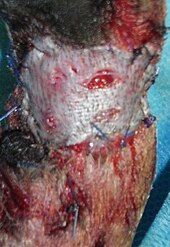
The ulcers can also be treated with debridement and excision and then covered with skin transplantation (for example split skin or mesh graft plastic ) , especially if there is an insufficient healing tendency . Debridement of the wound can also be carried out with medication, for example using salicylic petroleum jelly or enzymatic wound cleaning. The use of antibiotics , local antiseptics (e.g. iodine-PVP ) or wound dressings that release silver ions are particularly geared towards the inflammatory changes . So far, however, there is no reliable proof of effectiveness for any local antiseptic. For the topical application of agents intended to relieve pain, no evidence has been found to promote healing. However, a mixture of lidocaine and prilocaine appears to temporarily relieve pain. With local therapy in particular, there is a challenge in the fact that patients tend to be particularly often and extensively sensitized to locally used substances. These should therefore be as inert as possible. Because of the development of resistance, antibiotics should only be used for clinically manifest infections and not for bacterial colonization typical of ulcers.
Drug therapy (systemic)
Drug therapy does not replace decongestive or local treatment. The possible undesirable effects and possible contraindications also limit its use.
A positive active detection is previously for acetylsalicylic acid , pentoxiphylline , iloprost , prostaglandin E1 and the Flavoinoide diosmin / hesperidin (combination), coumarin / troxerutin (combination), Sulodexide and Buckeye - extract before. In addition, a substitution of zinc, iron, folate, albumin, vitamin C and selenium in malnutrition states can support healing.
prevention
In order to prevent the formation of a venous leg ulcer , a pre-existing chronic venous insufficiency must be consistently treated.
Since thrombophilia is an essential additional factor, it can also be useful to assess the risk of close relatives of patients with a venous leg ulcer as part of prevention . For this purpose, statistically significant laboratory changes in venous leg ulcers compared to the normal population are suitable . These may depend on the individual case , testing for Factor V Leiden , antithrombin deficiency, protein C deficiency, protein S deficiency , lupus anticoagulant and anti- cardiolipin antibodies be displayed.
Prospect of healing
The chances of recovery are generally good, although the treatment can also be very tedious. An ulcer is considered to be resistant to therapy if, under optimal therapy, it does not show the first tendency to heal after three months or if it has not healed within twelve months. Once it has healed, consistent prevention is required to prevent it from breaking open again.
Historical aspects
Traditionally, long-standing festering wounds on the lower extremities are called "open legs". An early medical treatise on the treatment of ulcus cruris (venosum), Middle High German old (er) schaden , is the book of old damage , which was created around 1450 in southwest Germany and is probably the oldest special recipe for varicose veins in the legs. Until the 19th century there was an opinion in folk medicine that ulcers should not be allowed to heal if there was a simultaneous heart disease. In 1877 Max Schede introduced the venous surgical treatment of the venous leg ulcer using a procedure named after him.
Individual evidence
- ↑ a b c d e f g h i j k l m n o S3 guideline of the German Society for Phlebology for the diagnosis and therapy of venous leg ulcers on the AWMF website , (online)
- ↑ a b c d e f g h i P. Altmeyer et al.: Basic knowledge of dermatology: A presentation accompanying the lecture. Verlag W3l, 2005, ISBN 3-937137-95-5 , p. 377, (online)
- ↑ Bonn Vein Study of the DGP (online) Eberhard Rabe et al., Phlebology 32 (1), page 1-14, Schattauer of 2003.
- ↑ a b A. Berger et al .; Plastic surgery: basics, principles, techniques. Springer, 2002, ISBN 3-540-42591-8 , p. 64, (online)
- ↑ J. Braun et al: Clinical guidelines for internal medicine. Urban & Fischer-Verlag, 2009, ISBN 978-3-437-22293-1 , pp. 180ff., (Online)
- ↑ a b S. O'Meara et al: Compression for venous leg ulcers. In: Cochrane Database Syst Rev. 2009 Jan 21, PMID 19160178
- ↑ a b J. E. Jones et al.: Skin grafting for venous leg ulcers. In: Cochrane Database Syst Rev. 2007 Apr 18; (2), PMID 17443510
- ↑ Manjit S. Gohel, Francine Heatley, Xinxue Liu, Andrew Bradbury, Richard Bulbulia: A Randomized Trial of Early Endovenous Ablation in Venous Ulceration . In: New England Journal of Medicine . April 24, 2018, doi : 10.1056 / NEJMoa1801214 ( nejm.org [accessed December 13, 2018]).
- ↑ Joachim Dissemond: Ulcus cruris - Genesis, Diagnostics and Therapy , Uni-Med Verlag, Bremen a. a. 2007, ISBN 978-3-89599-298-8 , page 105
- ^ A b S. O'Meara et al.: Antibiotics and antiseptics for venous leg ulcers. In: Cochrane Database Syst Rev. 2010 Jan 20; (1), PMID 20091548 .
- ↑ M. Briggs et al .: Topical agents or dressings for pain in venous leg ulcers. In: Cochrane Database Syst Rev. 2010 Apr 14; 4, PMID 20393931
- ^ Official journal for the administrative district of Düsseldorf, Verlag Bagel, 1866, p. 395, (online)
- ↑ Joachim Peters (Ed.): The 'book of old damage', Part I: Text. Medical dissertation, Bonn 1973 (commissioned by Königshausen & Neumann, Würzburg).
- ↑ Ingrid Rohland: The 'Book of Old Damage'. Part II: Commentary and Dictionary. (Medical dissertation Würzburg) Pattensen near Hanover (now: Verlag Königshausen & Neumann, Würzburg) 1982 (= Würzburg medical historical research. Volume 23).
- ^ Gundolf Keil : 'Book of old damage'. In: Burghart Wachinger et al. (Hrsg.): The German literature of the Middle Ages. Author Lexicon . 2nd, completely revised edition, Berlin / New York 1978–2008, ISBN 3-11-022248-5 , Volume 1 ( 'A solis ortus cardine' - Colmar Dominican chronicler. ), 1978, Colmar . 1080 f.
- ↑ Wolfgang Wegner: 'Book of old damage'. In: Werner E. Gerabek u. a. Encyclopedia of Medical History. 2005, p. 218.
- ↑ W. Hach and others: Perspectives on the history of medicine in the 19th century. Schattauer Verlag, 2007, ISBN 978-3-7945-2592-8 , p. 20, (online)
- ↑ W. Hach et al.: Vein surgery: Guide for vascular surgeons, angiologists, dermatologists and phlebologists. Schattauer Verlag, 2007, ISBN 978-3-7945-2570-6 , p. 68, (online)
literature
- Kerstin Protz: Modern wound care practical knowledge, standards and documentation , 8th edition, Elsevier Verlag, Munich 2016, ISBN 978-3-437-27885-3
