epithelium
The epithelium [ epitel ] ( Greek ἐπί epi on ',' on 'and thele "mother's breast," "nipple") is a biological collective term for cover fabric and gland ngewebe. These are single or multi- layer cell layers that cover all inner and outer body surfaces of the multicellular organisms (exception: joint capsules and bursa of the musculoskeletal system).
In addition to muscle , nerve and connective tissue, the epithelium is one of the four basic types of tissue .
construction
Epithelia are clearly separated from the connective tissue by the basement membrane and contain no blood vessels .
Another characteristic common to all epithelial cells is their polarity :
- The outer, apical side faces the outside (e.g. in the case of the skin) or the lumen (e.g. in the case of the intestine or the glands).
- The basal side is connected to the underlying tissue by a basal lamina .
The polarity of epithelial cells is also characterized by structural and functional differences between the apical and basal membrane of the epithelial cells. In this context, one speaks of an apical and basolateral domain .
Furthermore, epithelial cells have an adhesive complex ( final ridge complex ) consisting of zonula occludens ( tight junction ), zonula adhaerens ( adherens junction ) and desmosome ( macula adhaerens ). On the one hand, the adhesive complex represents a physico-chemical barrier and, on the other hand, connects adjacent epithelial cells with one another.
The cells are close together and are rich in cell contacts . As a result, the tissue has only small intercellular spaces with correspondingly little intercellular substance . With the help of emperipolesis , other cells penetrate the epithelia.
Classification of the epithelia
Epithelia are specifically differentiated in many ways and depending on the organ. First of all, one can distinguish between surface epithelia and glandular epithelia:
- Surface epithelia primarily have a protective function (e.g. the skin ). They can absorb substances ( resorption , e.g. intestinal mucosa ) and form a barrier that separates the respective organ from the environment (especially through the cell contacts mentioned above, the tight junctions ).
- Glandular epithelia determine the function of all glands ( secretion , excretion ). They produce secretions of all kinds (including in the salivary glands and sweat glands or in the intestinal mucosa ).
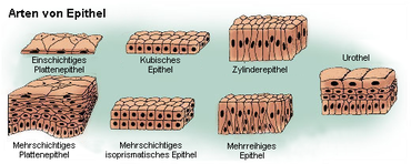
To distinguish between the numerous epithelial types, it has proven useful to emphasize two features: on the one hand, the number of cell layers and, on the other hand, the shape of the cells in the superficial cell layer (see below).
Monolayer epithelia
Simple epithelia
- monolayer squamous epithelium: Such epithelia are mainly used for the smooth lining of inner surfaces. Because they are very thin, single-layer squamous epithelia enable an exchange of substances (e.g. gas exchange in the alveoli ). Examples:
- Endothelium (epithelial lining of the blood and lymph vessels)
- Mesothelium ( pleural , pericardial , peritoneal epithelium ( serous membranes ))
- single-layer isoprismatic epithelium (also cubic epithelium): The epithelial cells have an almost cube-shaped shape. These larger cells are metabolically active and take on active transport tasks in the sense of secretion / absorption . Examples:
- Renal tubules
- Submandibular gland ( salivary glands )
- Bile ducts
- Ovarian epithelium
- single-layer, highly prismatic epithelium ("columnar epithelium" or "columnar epithelium"): Elongated, columnar cells with a lively metabolism take on barrier and transport functions. Examples:
Multi-row epithelia
The multi-row epithelium is also still single-layered, all cells are anchored to the basal lamina as in the single-layer epithelium , but not all of them reach the lumen . Highly prismatic cells fulfill the actual function, while small basal cells are available as a reserve for submerged cells. The cell nuclei are at different heights and thus form apparent layers (rows).
- Ciliated epithelium in the trachea and the other airways up to and including the segment bronchi.
- Vas deferens
- Epididymis
- Eustachian tube
Layered epithelia
In the multilayered epithelium, many (more than ten) layers of cells lie on top of one another. Basically, a three-way division can be made: Cell divisions take place in the basal layer, which is anchored to the basal lamina. The cells ascend and differentiate in a specific way in a middle or intermediate layer. Eventually they reach the surface or superficial layer.
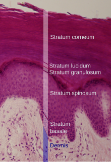
- multilayered squamous epithelium: This epithelium is of great importance and can be found wherever mechanical stress is high. The cytoskeleton and cell contacts are tailored to this load. In regions that are constantly humidified, the multilayered squamous epithelium remains keratinized , where it is exposed to the air, it becomes cornified .
- multilayered uncornified squamous epithelium:
- Oral cavity , esophagus , anal canal
- vagina
- Cornea and conjunctiva of the eye
- in the male urethra just before the outer mouth
- Multilayered, keratinized squamous epithelium: Another protective function is the death and keratinization of the outer cell layers. The cells are solidly anchored to each other with desmosomes and with hemidesmosomes in the basal lamina:
- in humans, the epidermis is the only keratinizing squamous epithelium
- in ruminants it also comes in the reticulum , omasum and rumen ago
- multilayered uncornified squamous epithelium:
- multilayer isoprismatic epithelium: ovarian follicles that have reached the stage of secondary follicle have such an epithelium.
- two-layer isoprismatic epithelium: This form of epithelium is found in the ducts of the sweat glands . The ciliary body is also covered by such an epithelium, although it is part of the retina .
- multi-layer high prismatic epithelium: This less common epithelial form is to be distinguished from the much more important multi-row high prismatic epithelium. It only occurs in three places on the human body:
- in the male urethra in its course from the prostate to just before the outer mouth
- in the main ducts of the large salivary glands (two-layer)
- in the conjunctival fornix , a reserve fold of the conjunctiva
Transitional epithelium ("urothelium")
The transitional epithelium ("urothelium") is a special epithelium of the urinary tract ( renal pelvis , ureter , urinary bladder ) that has multiple rows or layers, depending on the bladder filling (or stretching of the urothelium) . The deck / umbrella cells are particularly important here. They form the so-called crusta , which has the task of protecting uric acid. In contrast to the squamous epithelium, the upper cell layer is more cubic.
Functions of the epithelia
Protective function
The epithelium basically fulfills two different protective functions: On the one hand, purely mechanical protection, primarily through the multilayered epithelia. The epidermis of the skin must have sufficient tear resistance and must not detach from the connective tissue underneath. On the other hand, the epithelium must seal the inner body orifices: stomach and intestinal contents must be used in a controlled manner (highly prismatic epithelium), the urine must remain in the bladder and ureter (transitional epithelium ), the blood-brain barrier must be preserved (capillary endothelium). Of course, mechanical loads have to be withstood here as well, but the tight junctions that occur more frequently in such cells are decisive for the seal .
Absorption
Resorption is the transport of precisely defined substances from apical to basal. The classic example is the absorption of nutrients in the intestinal mucosa. The apical surfaces are often differentiated, for example, an epithelia cell can enlarge its surface through the formation of numerous microplicae (folds) or microvilli . The exact mechanisms ( transport , phagocytosis , pinocytosis , lysosomes ) are the subject of other articles.
secretion

All secretion processes of the body take place from the glandular epithelia. Accordingly, there is a great variety here, from the individual goblet cells of the intestinal mucous membrane to the sweat glands of the skin to entire organs such as the salivary glands or the pancreas . Glands are organs made up of specialized epithelial cells; they are used for secretion. One differentiates:
- exocrine glands that bring their secretions to the surface through an excretory duct. They excrete on inner or outer surfaces (e.g. lacrimal gland , salivary gland , sweat gland ), and
- endocrine glands that deliver their secretions directly to the surrounding extracellular fluid and have no duct. Often the secretions (hormones) then diffuse into blood vessels and are distributed throughout the entire organism (e.g. thyroid , pituitary ).
One can also distinguish between the secretion route, that is
- holocrine (cell disintegrates to produce secretions, typical of the sebum glands of the skin),
- apocrine (constriction of the vesicles, e.g. lactating mammary gland ),
- merocrine (by exocytosis ) and
- ekkrin (by transporter ),
the latter being divided into, according to the composition of the secretion
- serous (thin fluid, protein-rich, sometimes containing digestive enzymes, narrow gland lumen, e.g. parotid gland , pancreas ),
- mucous (viscous, slimy; serves to form transport mucus, wide lumen, e.g. Brunner's glands in the duodenum ) and
- Seromucous (mixed - secretion is both serous and mucous; this case is the most common, e.g., mandibular salivary gland ).
In addition, a distinction is made between intraepithelial and extraepithelial glands:
- Intraepithelial glands are single cells embedded in the cover epithelium (e.g. the mucous goblet cell of the intestine).
- Extraepithelial glands are multicellular organs that therefore no longer have any space in the epithelium itself and have been shifted into the deeper tissue layers. They consist of glandular end pieces that form the secretion. A distinction is made between tubular (tubular), alveolar (bubble-shaped) and acinous (bubble-shaped; however, thicker "wall" and smaller lumen) and mixed forms of extraepithelial glands. Switching points take the secretion from the end pieces and guide it into the strip pieces / secretion tubes (made of cylindrical epithelium); many secretion tubes collect to the secondary ducts n, which open into the main duct, which finally the secretion on an epithelial surface, z. B. the intestinal mucosa releases.
Sensory function
Most of the human sensory cells are embedded in epithelial cell structures. This construction is recommended because epithelia, as superficial cell layers, naturally assume a mediating position between inside and outside. Examples:
- Retina (retina) of the eye
- inner and outer hair cells of the inner ear, whereby in the case of the outer hair cells the sensor system and the change in length of the cell body are directly coupled
- The olfactory mucous membrane (olfactory epithelium) in the nasal mucosa
- Taste cells of the back of the tongue
- Merkel cells ( mechanoreceptors ), as well as pain and temperature receptors in the epidermis
Transport function
Some epithelia also have cilia on their surface, which have a transport function. They can smuggle foreign bodies out of the organism with their powerful blow.
See also
- Neuroepithelium
- Epithelialization
- Ussing chamber (for measuring epithelial properties)
Individual evidence
- ↑ Becher, Lindner, Schulze (ed.): Latin-Greek vocabulary in medicine . 3. Edition. Verlag Gesundheit GmbH, Berlin 1991, ISBN 3-333-00627-8 .


