Musculature


The musculature is an organ system in tissue animals and describes the muscles. The term refers e.g. B. with the terms abdominal muscles or back muscles on the muscle groups of the respective body section and their interaction.
A muscle ( Latin musculus 'Mäuschen', Middle High German also mūs - a tense muscle looks like a mouse under the skin) is a contractile organ that can move internal and external structures of the organism through the sequence of contraction and relaxation . This movement is the basis of the active movement of the individual and the change in shape of the body as well as many internal body functions.
The basic classification of the muscles in mammals including humans is based on the histological structure and the mechanism of contraction. Accordingly, a distinction is made between smooth muscles and striated muscles. The latter can be further divided into the heart muscles and the skeletal muscles . Further differentiation options result from the shape, the fiber types and functional aspects (see below). The underlying tissue of a muscle is the muscle tissue , which consists of characteristic muscle cells. In skeletal muscle, the muscle cells are called muscle fibers .
Comparison of muscle types
| Smooth musculature | Heart muscles | Skeletal muscles | |
|---|---|---|---|
| construction | |||
| • Motorized end plate | no | no | Yes |
| • fibers | fusiform, short (<0.4 mm) | branched | cylindrical, long (<15 cm) |
| • Mitochondria | few | lots | few to many (depending on muscle type) |
| • Cell nuclei / fiber | 1 | 1 | lots |
| • Sarcomeres | no | yes, max. Length 2.6 µm | yes, max. Length 3.7 µm |
| • Syncytium | no (single cells) | no (but functional syncytium) | Yes |
| • sarkopl. Reticulum | little developed | moderately developed | strongly developed |
| ATPase | little | medium | much |
| function | |||
| • Pacemaker | spontaneously active (slowly) | Yes fast) | no (needs nerve stimulus) |
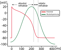
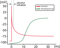
| • Stimulus response | graduated | "All or nothing" | "All or nothing" |
| • Tetanisable | Yes | No | Yes |
| • Workspace | Force / length curve is variable | in the rise of the force / length curve | at the maximum of the force / length curve |
| Stimulus response |

|

|

|
histology
The designation of the cytological structures of the muscle cells is subject to a specific nomenclature for the muscles and is therefore partly different from that of other cells :
| Muscle cell | other cells of the organism |
|---|---|
| Sarcoplasm | cytoplasm |
| sarcoplasmic reticulum | smooth endoplasmic reticulum |
| Sarcosome | Mitochondrion |
| Sarcolemma (a) | Cell membrane |
- Skeletal muscles are the arbitrarily controllable parts of the muscles and ensure mobility. They are also called striped - or striated muscles, as their myofibrils, in contrast to the smooth muscles, are arranged very regularly and thus create a recognizable ring pattern of red myosin filaments and white actin filaments . All skeletal muscles are assigned to the somatic muscles .
- The heart muscle is working rhythmically, can not cramp , has its own conduction system can depolarize spontaneously, contains the cardiac isoform of troponin I and T. It has the striation of skeletal muscles, however involuntarily primarily through the sinus node -controlled and thus represents a own muscle type.
- The smooth muscles are not subject to conscious control, but are innervated and controlled by the autonomic nervous system. This includes, for example, the muscles of the intestine . All smooth muscles are assigned to the visceral muscles .
The striated muscles come from the myotomes of the somites of the abdominal wall, the smooth ones from the mesoderm of the splanchnopleura , so that these are also referred to as the viscera. In the area of the head intestine , the visceral muscles are innervated by the cranial nerves and are striated across, while the remaining intestinal muscles are made up of smooth muscle fibers .
Other categorization options
A muscle can be classified in different ways, although this classification is not direct and unambiguous. Often the properties overlap. Depending on the perspective, they are differentiated by their cell structure, shape or function. Furthermore, types of muscle fibers can be distinguished that are mixed up in a muscle.
Anatomically
- Examples: ciliary muscle to deform the lens of the eye , sphincter muscles around the anus , mouth , eye, bladder outlet and stomach outlet ( pylorus )
- Examples: esophagus , stomach , intestines , heart
- spindle-shaped muscles
- Example: soleus muscle
- feather-shaped muscles
- multi-bellied muscles
- Example: rectus abdominis muscle
- multi-headed muscles
It is also divided into: cytological (see above) and functional (see below)
Classification of muscle fiber types
According to enzyme activity
- Type I fibers: SO ( Engl. S low o xidative fibers = slow oxidative fibers')
- Type II fibers:
- Type II-A-fibers: FOG (engl. F ast o xydative g lycolytic fibers =, fast oxidative / glycolytic fibers')
- Type II X-fibers: FG (engl. F ast g lycolytic fibers = fast glycolytic fibers'). There are different types (B or C) depending on the species.
According to contraction property
Extrafusal fibers (also twitch fibers ) (work muscles)
- ST fibers ( s low t witch fibers = 'slowly twitching fibers') are very persistent, but do not develop high forces (corresponds to SO).
- Intermediate type (corresponds to FOG)
- FT fibers (engl. F ast t witch fibers =, fast-twitch fibers') can develop high forces, however, quickly become fatigued (corresponding to FG).
- Tonus fibers can only exert a slow, worm-shaped contraction. They occur rarely, for example in the external eye muscles, in the tensor tympani muscle and in muscle spindles.
Intrafusal fibers ( muscle spindles ) serve as stretch receptors and to adjust the sensitivity of the muscle spindles.
By color
- Red muscles (corresponds to SO)
- White muscles (corresponds to FG)
- They are light in color in many animals (but not in humans) because of their low myoglobin content .
The ratio of the composition of a skeletal muscle from different muscle fiber types is largely genetically determined and can be changed to a limited extent through targeted endurance or strength training. This does not change the relationship between type I and type II fibers, but it does change that between type II-A and type II-X. II-A fibers are formed from many II-X fibers (e.g. in the trapezius muscle during strength training II-A content from 27% to up to 44% of all fibers). The distribution of the various muscle fibers in a muscle is not homogeneous, but rather different at the origin, insertion or inside and on the surface of the muscle.
Muscle contraction
description
The contraction is a mechanical process triggered by a nerve impulse . In the process, protein molecules ( actin and myosin ) slide into one another. This is made possible by rapidly successive conformational changes in the chemical structure, whereby the extensions of the myosin filaments - comparable to rapid rowing movements - pull the myosin filaments into the actin filaments. If the nerve stops supplying the muscle with impulses, the muscle slackens, which is referred to as muscle relaxation .
Types of contraction
Depending on the change in force (tension) or length of the muscle, several types of contraction can be distinguished:
- isotonic ("tensioned in the same way"): The muscle shortens without any change in force.
- isometric (“equal measure”): The force increases with the muscle length remaining the same (holding-static). In the physical sense, no work is done because the distance covered is zero.
- auxotonic ("differently tensioned "): both strength and length change. This is the most common type of contraction in everyday movements.
More complex forms of contraction can be put together from these elementary types of contraction. They are most commonly used in everyday life. These are z. B.
- the support twitch: first isometric, then isotonic contraction. Example : lifting a weight off the floor and then bending the forearm.
- the stroke: first isotonic, then isometric contraction. Example : chewing motion, slap in the face.
With regard to the resulting change in length of the muscle and the speed at which this takes place, contractions can e.g. B. can be characterized as follows:
- isokinetic ("equally fast"): The resistance is overcome with a constant speed.
- concentric: the muscle overcomes the resistance and thus becomes shorter (positive-dynamic, overcoming). The intramuscular tension changes and the muscles shorten.
- eccentric: whether intentionally or not, the resistance is greater than the tension in the muscle, which stretches the muscle (negative, dynamic, yielding). There are changes in tension and lengthening / stretching of the muscles.
Structure and function of the striated skeletal muscles
Each muscle is encased by an elastic covering of connective tissue ( fascia ), which encloses several meat fibers (also secondary bundles ) , which in turn are enclosed and held together with connective tissue ( perimysium externum and epimysium ), which is penetrated by nerves and blood vessels and is attached to the fascia attached. Each meat fiber is subdivided into several fiber bundles (also primary bundles), which are mounted so that they can be moved relative to one another so that the muscle is flexible and clinging to the body. These fiber bundles are a union of up to twelve muscle fibers that are united with capillary vessels by means of fine connective tissue.
The muscle becomes active by tensing ( contraction ), then relaxing again, exercising a movement and a force. Muscle contraction is triggered by electrical impulses ( action potentials ) that are sent out by the brain or spinal cord and passed on via the nerves .
The muscle fiber is a syncytium, i.e. a cell that arises from several determined precursor cells ( myoblasts ) and therefore contains several nuclei . The muscle fiber can reach a considerable length of more than 30 cm and about 0.1 millimeter in thickness. It is incapable of dividing, which is why when the fiber is lost, no replacement can grow back and when the muscle grows, only the fiber thickens. This means that the upper limit of the muscle fibers is set from birth. In addition to the usual components of an animal cell , myofibrils , which are the finest fibers, make up around 80 percent of the fiber mass. The membrane covering of muscle fibers is called a sarcolemma.
Functional classification of the skeletal muscles
In terms of their cooperation, muscles are divided into opposing and cooperating. Agonists (players) and antagonists (opponents) have opposite effects. Synergists, on the other hand, have the same or a similar effect and therefore work together in many movement sequences.
- Example: Antagonists: biceps and triceps ;
- Example: Synergists: for push- ups you need the triceps and the chest muscles ( pectoralis major , - minor ).
- Muscles that pull extremities towards the body are called adductors (pullers), their antagonists, the abductors (pullers), ensure that the extremities are spread apart from the body.
Example: outer and inner muscles of the thigh, with which one can spread the legs and bring them together.
- Flexors, on the other hand, buckle the fingers and toes, their antagonists are the extensors (extensors).
- Rotators (perform rotary movements, e.g. of the forearm or head)
Human skeletal muscles
Every healthy person has 656 muscles, with men making up about 40% and women about 32% of the total body mass, but the overall muscularity depends on the lifestyle.
The largest muscle in humans in terms of area is the large back muscle (Musculus latissimus dorsi) , the largest muscle in terms of volume is the Musculus gluteus maximus (largest gluteal muscle), the strongest the mastication muscle ( Musculus masseter ), the longest the Tailor's muscle ( Musculus sartorius ), the most active are the eye muscles and the smallest are the stapes muscles ( Musculus stapedius ). Because of the amount of mechanical work that muscles have to do, they are one of the main consumers of body energy, along with the nervous system .
Postnatal Development
In the newborn, the muscles in the trunk are more developed than those in the extremities. The muscle percentage is about 21 percent of the body weight. During growth, the muscle mass in men increases by about 32.8 times, but the total body mass only about 19.4 times. In men, muscle development ends between the ages of 23 and 27, and in women between the ages of 19 and 23. The muscle mass in men is around 37-57%, while in women it is around 27-43%.
| Age | man | woman |
|---|---|---|
| 10-19 a | 43-57 | 35-43 |
| 20-49 a | 40-54 | 31-39 |
| 50-100 a | 37-48 | 27-34 |
As the muscles grow older, they return to a state similar to that before they were fully developed. So this mainly concerns a breakdown of the muscles in the legs.
Physiological muscle failure
Due to its microscopic anatomy, a muscle cannot contract completely (the sarcomere can only shorten by about 30%), nor can it stretch indefinitely (the sarcomere would otherwise tear). This results in two different forms of physiological insufficiency of a muscle:
- Active muscle failure occurs when the agonist can no longer contract because it is already maximally contracted.
- Passive muscle failure occurs when the agonist cannot continue to contract because its antagonist is already maximally stretched.
With two-joint muscles, it is possible to counteract the muscle insufficiency (with regard to the muscle effect on one joint) by stretching the muscle in the other joint (or shortening the antagonist). For example, the biceps brachii muscle acts more strongly in terms of its flexion force in the elbow joint when the arm is retroverted ( i.e. the elbow joint behind the body), since the active insufficiency of the muscle is now counteracted by stretching the shoulder joint (the long biceps head covers both joints).
Musculoskeletal disorders and injuries
|
|
See also:
See also
literature
- Schmidt, Unsicher (Ed.): Textbook Preclinical - Part B , Deutscher Ärzte-Verlag Cologne, 2003, ISBN 3-7691-0442-0
- Frédéric Delavier: The new muscle guide. Targeted strength training, anatomy (OT: Guide des mouvements de musculation ). BLV, Munich 2006, ISBN 3-8354-0014-2
- Sigrid Thaller, Leopold Mathelitsch: What can an athlete do? Strength, performance and energy in the muscle . Physics in Our Time 37 (2), pp. 86-89 (2006), ISSN 0031-9252
- Detlev Drenckhahn (Ed.): Anatomy Volume 1 . Urban & Fisher, Munich 2008
Web links
- Uni Mainz: The human muscles in tables
- on contracture, see the article at Pflegewiki contracture and contracture prophylaxis
Individual evidence
- ↑ Jürgen Martin: The 'Ulmer Wundarznei'. Introduction - Text - Glossary on a monument to German specialist prose from the 15th century. Königshausen & Neumann, Würzburg 1991 (= Würzburg medical-historical research. Volume 52), ISBN 3-88479-801-4 (also medical dissertation Würzburg 1990), p. 153 ( mus : muscle, especially on the upper arm).
- ↑ Franz Daffner: The growth of people. Anthropological study . 2nd Edition. Engelmann, Leipzig 1902. p. 342.


