Megaesophagus
The megaesophagus (from the Greek μέγας mégas , German 'large' , and οισοφάγος oisophágos , German ' esophagus ' ) is a disease of the esophagus that occurs in various domestic animals and can either be hereditary or as a result of an underlying disease. A megaesophagus can be congenital, but can in principle also appear again at any age. The dog is most commonly affected ; however, the disease has also been described in cats , cattle and sheep . In human pathology, the term is used in connection with esophageal achalasia , especially in its secondary form through infection with Trypanosoma cruzi in Chagas disease.
Pathophysiology
Congenital megaesophagus
A congenital megaesophagus is due to a mechanical or functional disorder in the patency of the esophagus, either as a result of an existing stricture or a neuromuscular problem. This means that the food ingested does not reach the stomach through normal peristalsis, as usual , but remains in the esophagus. Due to a lack of muscle tone and / or the pressure of the contents of the esophagus on the esophageal wall, the organ becomes widened ( dilated ) over time, which in turn increasingly restricts peristalsis and thus functionality ( vicious circle ).
Persistent right aortic arch
The persistent right aortic arch is a special case of congenital stricture, which symptomatically resembles the arteria lusoria in humans. In embryonic development, the aorta does not develop from the left, as usual, but from the right fourth branchial arch artery . This causes the esophagus to be pinched between the aorta, pulmonary trunk and ligamentum arteriosum , which leads to a mechanical narrowing of the esophagus, which in turn leads to mechanical dilatation as soon as the affected puppies are weaned and the solid food cannot pass the constriction .
Acquired megaesophagus
An acquired megaesophagus can occur as a result of a mass (e.g. from tumors or abscesses ) that presses on the esophagus, from an acquired stricture, for example as a result of scarring , or as a result of an acquired neurological or muscular disorder ( polymyositis , polymyopathy ) be. In dogs, it most often develops idiopathically , i.e. without an identifiable cause, but acquired myasthenia gravis , systemic lupus erythematosus , hiatal hernia , lead poisoning , esophagitis , hypothyroidism , thymoma or adrenal insufficiency can also cause a megaesophagus.
clinic
Signal element
Puppies with primary congenital megaesophagus are usually presented at the age of a few weeks to a few months, partly due to persistent regurgitation (often interpreted by the owner as vomiting) and weight loss, partly also due to acute aspiration pneumonia . Breeds that are more frequently affected by congenital megaesophagus are Shar-Pei , Fox Terriers , German Shepherds , Great Dane , Irish Setters , Labrador Retrievers , Miniature Schnauzers , Newfoundlands and Siamese cats .
Puppies with a persistent right aortic arch develop symptoms when they switch from milk to solid food that gets stuck at the constriction and mechanically expands the esophagus. Breeds that are more frequently affected by this problem are Boston Terriers , German Shepherds and Irish Setters.
Acquired megaesophagus can occur either as an idiopathic disorder or secondary to other diseases at any age . There are no age or race predispositions .
Symptoms
The main symptom of a megaesophagus is regurgitation of food from the esophagus. It must be differentiated from vomiting , when food is expelled from the stomach . This usually happens during the medical history, during which the owner is asked about signs of vomiting (signs of nausea, belching, abdominal cramps). Regurgitation does not have to take place immediately after ingestion, but can also occur with a time delay.
Chronic megaesophagus leads to weight loss and emaciation . Aspiration pneumonia can also often occur as a complication when regurgitated food enters the windpipe and is aspirated.
diagnosis
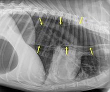
The suspicion of megaesophagus arises from the anamnesis and the clinical picture. If pressure is applied to the abdomen, the esophagus in the throat can visibly expand. A radiograph of the chest shows a widened, filled with air and / or feed esophagus. In congenital or acquired megaesophagus, the widening is usually even; in megaesophagus as a result of a persistent right aortic arch or other stricture, the esophagus is only dilated in front of the constriction.
Therapy and prognosis
A causal treatment is possible in the persistent right aortic arch as well as in the secondary acquired megaesophagus: In the first case, the mechanical narrowing of the esophagus is remedied by surgical severing of the ligamentum arteriosum, in the second the underlying disease (e.g. myasthenia gravis ) is treated. If an underlying disease is successfully treated, the prognosis is good. If the right aortic arch is persistent, surgical intervention is carried out in good time, and the megaesophagus can regress. If the intervention occurs too late, the widening can be retained despite the removal of the bottleneck.
Congenital megaesophagus occasionally resolves spontaneously before 6 months of age. Medicinal or surgical treatment is not possible. By trial and error, the owner can find the consistency of food that the affected dog can best tolerate. Feeding should be from an elevated position and the dog should be held in the same position for 10-15 minutes after feeding so that gravity can assist the food passage into the stomach. Frequent small meals seem to work best. A so-called Bailey Chair , a chair in which dogs can eat while sitting upright, can also help .
Most animals with a congenital megaesophagus that does not resolve spontaneously eventually die from acute aspiration pneumonia or from pulmonary fibrosis as a result of frequent aspiration pneumonia.
Genetics and Breeding Hygiene
The congenital megaesophagus is hereditary, although the inheritance patterns can differ depending on the race. In the fox terrier the congenital megaesophagus is described as autosomal recessive , in the miniature schnauzer as autosomal recessive with 60% penetrance . It can also occur as part of a congenital diffuse disease of the nervous system, for example in the Dalmatian . Affected animals and their parents are to be excluded from breeding.
The persistent right aortic arch is an inhibitory malformation of the branchial arch arteries that involves both genetic factors and environmental factors. The inheritance is polygenic . Affected animals should be excluded from breeding; a breeding value estimation would be desirable.
The acquired megaesophagus is usually not directly hereditary. However, breeds with an increased risk of hypothyroidism (underactive thyroid gland ) and Addison's syndrome also have an increased risk of acquired megaesophagus due to these diseases.
literature
- A. Herzog: Pareys Lexicon of Syndromes - Hereditary and breeding diseases of domestic and farm animals. Parey Buchverlag, Berlin 2001, ISBN 3-8263-3237-7 , pp. 288-289.
Web links
- Megaesophagus in a puppy (autopsy findings)
Individual evidence
- ↑ Gerd Herold and colleagues: Internal Medicine 2020. Self-published, Cologne 2020, ISBN 978-3-9814660-9-6 , pp. 234, 434 and 435.
- ↑ a b A. Herzog: Pareys Lexicon of Syndromes - Hereditary and breeding diseases of domestic and farm animals. Parey Buchverlag, Berlin 2001, ISBN 3-8263-3237-7 , pp. 24-25.
- ↑ Ulrich Möhnle u. a .: Megaesophagus as a consequence of Addison's disease - case report. In: Small Animal Medicine. No. 6/13, pp. 271-275.
- ↑ a b c d The Merck Veterinary Manual. 9th edition. Whitehouse Station NJ, ISBN 978-0-911910-50-6 , p. 134.
- ↑ a b c d e The Merck Veterinary Manual. 9th edition. Whitehouse Station NJ, ISBN 978-0-911910-50-6 , p. 316.
- ↑ Bailey Chair for feeding dogs with megaesophagus
- ↑ A. Herzog: Pareys Lexicon of Syndromes - Hereditary and breeding diseases of domestic and farm animals. Parey Buchverlag, Berlin 2001, ISBN 3-8263-3237-7 , pp. 288-289.
