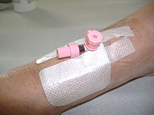
Peripheral venous catheter
The peripheral venous catheter , the peripheral venous catheter , the peripheral venous access , the PVK , the PVVK , the peripheral indwelling venous cannula , the peripheral indwelling venous catheter (depending on the manufacturer also Abbokath , Braunüle , Flexüle , Venflon , Vygonüle ; colloquially also Viggo , venule , needle or (venous) access , in Austria also simply conduit ) is a special form of vascular access. The catheter is usually inserted into a vein in the crook of the elbow or on the back of the hand. It is used for parenteral fluid therapy or the intravenous administration of medication without stressing the patient with multiple punctures. Also, blood transfusions are generally administered via peripheral venous catheter. Such an (intravascular) catheter can generally be used for several days. On the other hand, a surgically implantable long-term catheter (e.g. port catheter ) is used for permanent access to the blood circulation .
Placing a peripheral venous catheter through a peripheral venous puncture (or, if central venous puncture is not possible, a bloody vein opening, the sectio vein ) is usually reserved for doctors . The doctor can also delegate this measure to specialist personnel such as nurses , midwives , paramedics or paramedics or emergency paramedics , whereby the latter may also work independently under certain conditions.
construction
The peripheral venous catheter consists of a steel cannula (steel mandrin ) and a surrounding transparent plastic catheter made of silicone or polyurethane.
A venous catheter is structured as follows (numbers in brackets see illustration):
- IV cannula (1)
- Venous catheter (1a)
- Wing to fix with plaster (1b)
- Injection valve, also called injection valve, in color coding of the cannula size, here 20G (1c)
- Luer lock connector for an infusion system (1d)
- Steel mandrin, here half withdrawn (2)
- Protective cap, removed here (3)
use
Intravenous application
Selection of the vein
Suitable veins for a peripheral access are in principle all superficial veins, but usually the veins of the forearm, the back of the hand and the crook of the elbow. In the case of missing upper limbs or unsuitable veins and in babies, the peripheral venous access is placed on the back of the foot, in the latter also on the temple or forehead. Punctures of veins in the joint area should, however, be avoided as far as possible due to the mechanical stress caused by joint movements and the significantly reduced comfort for the patient. Often neglected, but sometimes successfully punctured areas are above the elbow in the upper arm area, where the tourniquet is usually placed . However, like skin veins running near the elbow , these require a certain amount of practice and good persuasion to the patient and are therefore unsuitable for beginners.
Selection of the puncture site
- Each puncture site should be selected as distal as possible, i.e. as far away as possible from the center of the body or from an organ. This has z. B. advantages if the vein becomes inflamed as a result of the peripheral venous catheter or as a result of the infusions ; the vein remains usable “above” the inflamed area for further punctures for venous catheters (possibly with restrictions).
- In principle, puncture sites should preferably be placed on the non-dominant half of the body , as they tend to affect the patient less there than on their dominant side.
Puncture
First a suitable vein is punctured and the venous catheter is carefully pushed a short distance into the vascular lumen . If the puncture is successful, a transparent chamber at the end of the puncture needle fills with blood . After pulling out the loose “rear part” one by one while holding the “front part”, the catheter can now be advanced completely into the vein via the puncture needle. This minimizes the risk of the vascular lumen being punctured by the pointed steel cannula.
Fixation
Now the established access has to be secured with a special plaster. You must not accidentally hit the rear end of the cannula because of the risk of embolism mentioned above. In the case of a position near the hand or joint, a bandage or small additional plasters, which are usually located on the plastic packaging of the cannula protective plaster, without applying too much pressure, for the purpose of fixation and preventing premature dislocation of the needle and increasing patient comfort and safety when changing clothes, arm movements and while walking.
Applying syringes, blood-free work
Before pulling it out completely, a swab should be placed directly under the rear catheter opening on the skin or a cellulose pad on the potential drip point, in order to catch the blood, which sometimes flows back quickly, and to prevent stains on the patient's clothing, floor or bed. During the subsequent rapid attachment of syringes, blood collection adapters, mandrels or connectors while compressing the punctured vein further proximally , it must be ensured that no swab material gets between the adapter and the catheter opening when screwing on.
Patency check
Before administering important or aggressive infusion solutions, the patency and the correct position and stability of the access should also be checked. This is done by means of specially drawn up, so-called NaCl syringes, usually filled with 5 to 10 milliliters of physiological saline solution, which are injected with a certain pressure while simultaneously observing the skin area around the catheter and the effort required to empty the syringe . Bulging, stiffness or persistent injection pain in the patient indicate a paravenous (from Latin para , "next to") location, i.e. injection into perivascular tissue. This makes a complete new installation of the peripheral venous catheter absolutely necessary. Colloquially, this is called "para-running". In general, of course, general hygiene regulations must be observed.
Another way to check the correct position of the access is to do a return test. The infusion already connected to the indwelling cannula is kept below heart level and a check is made to see whether blood is flowing back into the infusion tube. If this is the case, a correct position is very likely. If no blood runs back, this indicates a paravenous location or a location in front of a venous valve .
disposal
Just like cannulas, the steel mandrels used are disposed of in cannula disposal boxes.
Renewal, reopening
Renewing an indwelling peripheral venous catheter is a common routine task in hospitals. Aggressive infusion drugs such as B. prostaglandin E 1 derivatives or blood products can lead to the formation of clots and blockages of the catheter tip in the blood vessel. In addition, venous valves or walls can attach themselves to the opening over time and reduce or completely prevent the flow of newly attached infusion solutions. Simple methods of reopening are to gently pull or lift the catheter attached to the plaster while observing the drip chamber of the attached infusion. If the number of drops increases noticeably, placing swabs underneath, sticking the peripheral venous catheter that has been pulled out with an additional plaster or putting a bandage around the puncture site and then clamping it in front of or behind the upwardly protruding additional syringe inlet cover of the catheter can permanently maintain this effect - depending on the place of adhesion. The fault in this case is a catheter that has been pushed forward a little too far, pulling it a little out of the vessel and thus loosening an adhesion to a wall. Replacing a connector that is often used these days and is relatively expensive can "save" a catheter that no longer appears to be continuous.
If this does not help, you can, if necessary, with the infusion tubes attached, kink a section of the tube near the catheter and press the kinked part or push the vented tube under pressure with your thumb and forefinger towards the catheter opening in order to apply pressure to the front via the fluid in the lumen To create an opening that can displace any clots or venous valves. This is of course problematic with long clots that could lead to an embolism. For this reason, this measure may only be carried out by experienced doctors and, if possible, a new system should be preferred.
A classic mistake when a new peripheral catheter is to be inserted is that it is already in place, preferably on the other arm, but has not been seen by the staff. Here, too, it is advisable to first inspect the patient carefully and release both arms or, rarely, both feet. This then only needs to be tested for consistency.
Blood collection
A blood sample is, in particular already longer lying venous catheters, not always possible and not always recommended, although at the same time easily fluid can be injected intravenously. Blood tests can possibly be falsified by blood components or the dilution effects of previous infusions. However, this problem does not exist if blood is taken directly after a venous catheter has been inserted.
Piercing
Peripheral venous catheters are also used for piercing purposes other than their original purpose . The venous catheter is pierced through the skin and the underlying fatty tissue at previously marked puncture and exit points and the steel cannula is then removed. The jewelry to be used is placed in the end of the plastic sleeve and pulled through the skin when it is pushed out.
variants
Venous catheters are color-coded; Like the cannulas, they are available in several sizes. Their diameter is given in millimeters , often also in the non-SI unit of gauge (see table below). Depending on the diameter, the corresponding plastic catheter has a different length within the body (25–50 mm). With the diameter and length of the catheter, the flow rate changes; it ranges from 22 ml / min for 0.7 mm catheters to 330 ml / min for 2.2 mm access points (for aqueous infusions). While the sizes 0.7 mm to 1.1 mm are used for children due to the thin vessels, the sizes 1.1 mm and 1.3 mm are usually used for adults. In situations in which the infusion of large amounts of infusion or blood is necessary in a short time ( shock , multiple trauma ), sizes 1.5 mm to 2.2 mm are used due to the high flow rate.
If access is not required for a short period of time, according to the current recommendation of the Commission for Hospital Hygiene and Infection Prevention, it should no longer be closed with plastic mandrels. Instead, the catheter is flushed with sterile saline solution and sealed with a stopper to prevent blood clotting in the catheter.
| Color coding of venous catheters | |||||||
|---|---|---|---|---|---|---|---|
| Size in gauge | 24 | 22nd | 20th | 18th | 17th | 16 | 14th |
| colour | yellow | blue | pink | Green-white / green |
White | Gray | Orange-brown |
|
Outside diameter in mm |
0.7 | 0.9 | 1.1 | 1.3 | 1.5 | 1.7 | 2.2 |
|
Inside diameter in mm |
0.4 | 0.6 | 0.8 | 1.0 | 1.1 | 1.3 | 1.7 |
|
Flow rate in ml / min |
22nd | 36 | 61 | 103/96 | 128 | 196 | 343 |
|
Flow in l / h |
1.32 | 2.16 | 3.66 | 6.18 / 5.76 | 7.68 | 11.76 | 20.58 |
|
Stitch length in mm |
19th | 25th | 33 | 33/45 | 45 | 50 | 50 |
The 1.3 mm peripheral venous catheter is available in two different designs (green and green / white); the only difference is the stitch length, i.e. the length of the plastic catheter in the vein.
A venous catheter for pediatrics in sizes 0.7 mm and 0.45 mm with and without injection valve in the materials PUR (polyurethane) and FEP ( fluoroethylene propylene ) is offered by some manufacturers.
Safety catheter
The use of safety catheters has been prescribed for the prevention of needlestick injuries since TRBA 250 (Technical Rules for Biological Agents in Health Care and Welfare Care) came into force in May 2014. With these catheters, immediately after removing the steel mandrel from the plastic tube, which is located in the vein, a protection is placed over the tip of the cannula. This can be done in the form of a small metal clip that is pushed over the steel mandrel when it is pulled out, depending on the manufacturer, in some other variants . Thus, the risk of is needlestick injuries and resulting infections with, for example, HIV , hepatitis C or hepatitis B reduced. The material of the plastic catheter is partly provided with X-ray contrast strips.
history
The inventor of the first permanent indwelling catheter made of plastic was David J. Massa from the Mayo Clinic in 1950 . Bernhard Braun , doctor, chemist and entrepreneur, developed a German version in 1962 .
See also
Web links
- Entry on peripheral venous catheters in the Flexikon , a Wiki of the DocCheck company
- Peripheral venous access . CareWiki
Individual evidence
- ^ Marianne Abele-Horn: Antimicrobial Therapy. Decision support for the treatment and prophylaxis of infectious diseases. With the collaboration of Werner Heinz, Hartwig Klinker, Johann Schurz and August Stich, 2nd, revised and expanded edition. Peter Wiehl, Marburg 2009, ISBN 978-3-927219-14-4 , pp. 49-53 ( catheter-associated infections ).
- ^ Rainer Fritz Lick , Heinrich Schläfer: Accident rescue. Medicine and technology . Schattauer, Stuttgart / New York 1973, ISBN 978-3-7945-0326-1 ; 2nd, revised and expanded edition, ibid 1985, ISBN 3-7945-0626-X , p. 182 f.
- ↑ U. Kamphausen, N. Menche, K. Protz: Healing methods and tasks of nurses in therapy. In: Care Today. Urban & Fischer Verlag at Elsevier GmbH, Munich 2011; P. 605.
- ^ University of Cologne, Medical Faculty: Indwelling venous cannula. 2nd Edition. Cologne 2011, p. 11 ( PDF; 553 KB ).
- ↑ Prevention of infections caused by vascular catheters. Accessed February 1, 2020 .
- ↑ baua.de (PDF).
- ↑ https://www.bbraun.de/de/products/b0/vasofix-braunuele.html
- ↑ Massa DJ, Lundy JS, Faulconer A, Jr, Ridley RW: A plastic needle. Proc Staff Meet Mayo Clin 1950, 25 (14): 413-415 PMID 15430460 .


