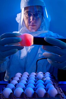
Embryonated chicken egg
The embryonated chicken egg (also egg culture, incubated chicken egg or chicken embryo culture) is a special laboratory technique in virology for the multiplication and isolation of viruses . In this process, the appendix organs of a pre-incubated chicken embryo or, rarely, the embryo itself are inoculated with virus-containing material and after a few days the newly created viruses are recovered and further propagated or examined. Other pathogens that multiply intracellularly are also rarely reproduced in hen's eggs, such as rickettsia and chlamydia . The embryonated chicken egg method was developed by Ernest Goodpasture in 1932 and optimized for virus replication in 1946. Until the development of cell culture technology in the 1950s, this was the only method for the targeted replication of viruses in the laboratory, only the infection of test animals was possible until then. Today, the cultivation in hen's eggs has largely been replaced by cell culture and is limited to a few viruses for which no or insufficient reproduction in cell cultures is possible. The use in virus diagnostics is limited to individual cases or pathogens of animal diseases for which an examination of the embryonated hen's egg is still required by law. The embryonated hen's egg is still most important in the cultivation of orthomyxoviruses , especially in the industrial production of vaccines and the isolation of influenza viruses .
Technique of the embryonated chicken egg
Only chicken eggs from infectiously controlled chicken populations are used to multiply viruses, as the embryo must not be pre-infected with natural pathogens that cause chicken diseases. These eggs are also called specified pathogen-free (SPF eggs). In order to obtain a favorable size of the embryo, the fertilized chicken eggs are further incubated in special incubators for 5 to 14 days at 38 ° C and a relative humidity of 60%, depending on the infection technique selected later . These incubators also have a turning device that moves the eggs regularly. With a special fluorescent screen, the development of the embryo can be observed after the eggs have been removed (" sheer "), dead or non-fertilized eggs are sorted out. CEF cells are also produced from embryonated chicken eggs .
Inoculation of the appendage organs
Usually it is not the embryo itself that is inoculated, but its appendage organs. Depending on the virus species or the question (reproduction or virus detection), these are the yolk sac , the amnion , the allantois or the chorionic allantoic membrane (CAM). Since these show an optimal size for inoculation and susceptibility to viruses at different times during embryonic development, the length of the incubation time for the eggs used is also different. The best time for infections of the yolk sac is 5 to 7 days after fertilization, for amnion and allantois 10 days, for CAM 11 to 12 days. If the inoculation is done too early, the appendix organ is not large enough, if it is done too late, increasingly inhibitory influences of the immune defense (including the formation of immunoglobulin Y ) or a decreasing susceptibility of the organs can prevent a successful infection.
Inoculation is carried out under sterile conditions to avoid bacterial contamination. After disinfection, the egg shell is opened with a small punch. When inoculating the CAM, this takes place on the side of the egg on which the embryo rests on the inside; in all other appendage organs, the blunt egg pole is opened. About 0.1 to 0.2 ml of virus-containing liquid ( inoculum ) is injected into the corresponding structure using a cannula . Then the bowl is closed with an adhesive or wax. After one day of further incubation, the eggs are checked with a light device and dead embryos are sorted out as inoculation damage. After a further one to six days, the multiplied viruses can be obtained from the allantoic or amniotic fluid ( egg white ) or the CAM and yolk sac membrane. Before that, the egg is stored overnight at 4 ° C so that the embryo dies and the blood vessels contract. Usually, further purification steps follow, such as filtration or ultracentrifugation , in order to obtain virus preparations that are as pure as possible.
Assessment of embryo development
The inoculated eggs are x-rayed regularly, usually daily, and the development status of the embryo is assessed. If bacterial contamination and inoculation damage are excluded, the death of the embryo may indicate virus replication, this can be observed , for example, after inoculating the yolk sac with herpes simplex viruses . Other macroscopically visible signs of virus replication are increased vascular markings in the embryo, signs of inflammation , petechial bleeding or malformations . When vaccinated with smallpox viruses , herpes simplex viruses and the Rous sarcoma virus , dark, blotchy changes known as "pocks" appear on the CAM . These are so characteristic that they can be used to make a diagnosis when a virus is detected. In most cases, various further examinations follow to identify the pathogen, for example an electron microscopic examination, a hemagglutination test or the direct detection of virus components ( antigens and nucleic acid ) for precise typing.
Importance in virus diagnostics
The importance of the embryonated hen's egg for virus isolation and virus diagnosis is limited, in the case of human viruses it is now limited to the isolation and typing of influenza viruses. When isolating unknown, new pathogens, in addition to molecular methods, cell culture and electron microscopy, embryonated hen's eggs are also used. In veterinary medicine , a number of pathogens are also grown in egg culture, including representatives of the smallpox viruses , orthomyxoviruses , herpes viruses , togaviruses and various avian viruses. The following table gives an overview of the most important pathogens and their behavior in embryonated hen's eggs. Some viruses can only be propagated successfully and with an acceptable yield in hen's eggs if the wild type has been adapted to the hen's egg through several passages ( blind passages ).
| virus | Virus family | Inoculation | Changes | Removal (harvest) |
|---|---|---|---|---|
| Herpes simplex viruses | Herpesviridae | Yolk sac | Embryo death | |
| Herpes simplex viruses | Herpesviridae | CAM, allantois | large "pocks", proliferative herd | CAM |
| Chicken herpes virus 1 ( infectious laryngotracheitis virus ) | Herpesviridae | CAM, allantois | large spots on the CAM | CAM |
| Pig herpes virus 1 ( Aujeszky's disease virus ) | Herpesviridae | CAM | no visible changes | CAM (after adaptation) |
| Orthopoxvirus bovis ( cowpox virus ) | Poxviridae | CAM | Hemorrhagic foci | CAM |
| Vaccinia virus | Poxviridae | CAM | Necrosis | CAM, allantois |
| Ectromelia virus (mouse pox virus) | Poxviridae | CAM | small proliferative spots | CAM |
| Fowlpox virus | Poxviridae | CAM | large proliferative spots | CAM (slow growth) |
| Myxomatosevirus (Leporipoxvirus myxomatosis) | Poxviridae | CAM | smallest spots | CAM |
| Encephalomyocarditis virus (avian encephalomyelitis, chick encephalitis) | Picornaviridae | Amnion, allantois | Embryopathy | Amniotic and allantoic fluid |
| Infectious bursitis virus | Birnaviridae | |||
| Vesicular stomatitis virus | Rhabdoviridae | CAM, allantois | Embryo death, CAM | CAM, allantoic fluid |
| Rabies virus | Rhabdoviridae | CAM (adaptation) | no visible changes | CAM, allantoic fluid (after adaptation, several passages) |
| Avian Infectious Bronchitis Virus | Coronaviridae | Allantois | Embryo death, dwarfism | Allantoic fluid |
| Equine encephalomyelitis viruses | Togaviridae | Allantois | Embryo death | CAM, allantoic fluid |
| TBE virus | Flaviviridae | Allantois, CAM | CAM, allantoic fluid | |
| Sendai virus | Paramyxoviridae | Allantois | Allantoic fluid | |
| Newcastle Disease Virus | Paramyxoviridae | Amnion, CAM | Embryo death, CAM | Allantoic fluid |
| Canine distemper virus | Paramyxoviridae | CAM (adaptation) | smallest spots on the CAM | CAM, allantoic fluid |
| Influenza viruses (A, B, and C) | Orthomyxoviridae | Amnion, allantois | partial embryo death | Allantoic fluid, amniotic fluid |
Vaccine production in the embryonated chicken egg
The great advantage of virus replication in embryonated hen's eggs is the high virus yield. This was used in the production of various vaccines as early as the 1960s, for example to obtain enough raw material for dead vaccines ( split vaccines ) against the measles virus ( measles vaccines ) and various influenza viruses ( influenza vaccines ). Especially with the latter, the reproduction in cell cultures has not yet reached sufficiently high virus concentrations, so that the embryonated hen's egg still determines the industrial production of influenza vaccines to this day. The problem with this method, however, is the time-consuming cleaning of the egg material, which always leaves traces of egg proteins and therefore cannot be administered if there is a chicken protein allergy .
swell
- SJ Flint, LW Enquist, VR Racaniello, AM Skalka: Principles of Virology. Molecular Biology, Pathogenesis, and Control of Animal Viruses. 2nd Edition. ASM-Press Washington DC 2004, ISBN 1-55581-259-7 , p. 30.
- Frank Fenner , BR McAuslan and others: The Biology of Animal Viruses. 2nd Edition. Academic Press, New York NY et al. 1974, ISBN 0-12-253040-3 , pp. 42f.


