Angina pectoris
| Classification according to ICD-10 | |
|---|---|
| I20 | Angina pectoris |
| I20.0 | Unstable angina pectoris |
| I20.1 | Angina pectoris with proven coronary spasm |
| I20.8 | Other forms of angina pectoris |
| I20.9 | Angina pectoris, unspecified |
| ICD-10 online (WHO version 2019) | |
The angina pectoris (abbreviation AP ; literally "chest congestion"; synonyms stenocardia , mutatis mutandis "heart tightness" of batteries heart tan and heart anxiety ) is a paroxysmal pain in the chest , by a temporary circulatory disorder of the heart, typically in the context of a coronary heart disease triggered (CHD) becomes. Mostly this is based on a constriction of one or more coronary vessels . Angina pectoris is therefore not a disease, but a symptom or the name for the clinical symptoms of acute coronary insufficiency. Drugs used to treat angina are called antianginosa .
Epidemiology
In 2003 the cause of death statistics in Germany showed 10.9 % of the deaths for CHD and 7.5% for acute myocardial infarction . Patients with stable angina pectoris have a mortality rate of 2-3% annually.
According to the Federal Statistical Office, the causes of death in 2014 were as follows:
- I25 - Chronic ischemic heart disease (CHD): 69,890 cases in 8.0%,
- I21 - Acute myocardial infarction (heart attack): 48,181 cases in 5.5%
- I50 - heart failure : 44,551 cases in 5.1%
Pathophysiology
Angina pectoris is caused either by physical or emotional or psychological stress. Most patients have coronary sclerosis . Under the load, there is a reduced blood flow ( ischemia ) of the tissue and thus the typical symptoms. A special form is Prinzmetal's angina , in which a temporary ischemia of the myocardium is triggered by a spasm of the coronary arteries. The duration of an attack ranges from seconds to minutes. The " Cardiac Syndrome X " also triggers angina pectoris.
A distinction is made between the occurrence of AP at rest ( resting AP ) or under stress ( exertion AP ). The resting AP poses an immediate risk of heart attack.
A differentiation between AP and myocardial infarction is possible in some cases using the drug glycerol trinitrate . In the event of an AP attack, the drug will significantly reduce the pain and tightness in the chest. However, this type of distinction is considered unreliable.
Symptoms
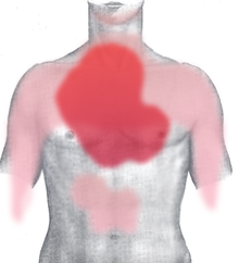
Symptoms typically start suddenly and last for seconds, minutes, and rarely hours. They are often described as burning sensation, " heartburn ", tearing or cramp-like pressure in the area of the heart ( cardialgia ) and are often felt as a retrosternal tightness behind the breastbone ( retrosternal ) (see main article Head's zone ) . The pain often radiates to both sides of the chest, more rarely to both shoulders and upper arms, to the upper abdomen and back, over the neck to the lower jaw and into the entire left arm and into the hand. They can also appear just between the shoulder blades, in the stomach area, and in the right half of the chest. In addition, those affected often complain of breathlessness or tightness of the chest. In addition, they mostly suffer from anxiety and sweats.
In the case of toothache-like complaints in the lower jaw, preferably on the left side, the patient may first visit a dentist. If, by chance, there is a dental finding that is also a possible cause (e.g. a purulent tooth), in rare cases this can lead to coronary heart disease being overlooked as the primary cause (see Buddenbrook syndrome ).
classification
Angina pectoris is divided into stable angina pectoris and unstable angina pectoris depending on symptoms, occurrence and prognosis. Microvascular angina pectoris is a special form.
Stable angina pectoris
A stable AP is present in patients who complain of constant symptoms that are reproducible under similar circumstances over a longer period of time. Symptoms typically subside after a few minutes of rest and can be improved with sublingual nitroglycerin administration . ECG changes usually do not occur. The cause are usually stenoses of the coronary arteries . In some patients, the symptoms may improve after a few minutes if the exertion that triggered it was continued. One then speaks of a migration phenomenon . This is explained with bypass circuits on the myocardium. The variant angina is a special form of stable angina pectoris. It is spastic vasoconstriction caused to the coronary arteries and often occurs independent of the load on. In the case of Prinzmetal's angina, ECG changes such as B. ST segment elevations occur.
Stable angina pectoris is divided into four degrees of severity using the CCS classification (classification of the Canadian Cardiovascular Society):
| stage | definition |
|---|---|
| CCS I | no restriction of normal physical activity Angina pectoris only with strong, rapid or prolonged exertion |
| CCS II | slight restriction in normal physical activity Angina pectoris when walking or climbing stairs at high speed or after meals, |
| CCS III | significant reduction in normal physical activity Angina pectoris when walking less than 100 m or after climbing stairs from a floor at normal speed |
| CCS IV | Angina pectoris with any physical exertion or even at rest |
Unstable angina pectoris
Unstable angina pectoris is the simplest form of acute coronary syndrome . It goes hand in hand with a great risk of heart attack. Characteristic are changes in the symptoms, such as the first occurrence of angina pectoris complaints, the occurrence of complaints at rest or the increase in the duration of the seizure, frequency of seizures or pain intensity. Angina pectoris complaints before ( pre-infarct angina or pre-infarct syndrome as a sub-form of unstable angina) and within two weeks after a myocardial infarction are also referred to as unstable. Typically, the effect of nitroglycerin is also reduced.
The unstable AP is usually caused by coronary vascular disease and arteriosclerosis . In this context, arteriosclerotic plaques in the coronary arteries are torn locally. This leads to a mechanical partial relocation and reflex narrowing of the arteries due to vasospasm . A forming thrombus can trigger an acute myocardial infarction . The angina decubitus (including angina nocturna ) is a form of unstable angina with particular night occurring while lying chest pain. The reason for this is the overloading of the previously damaged heart muscles with increased venous blood return while lying down.
| Braunwald classification of unstable angina pectoris | |||
|---|---|---|---|
| stage | Clinical circumstances | ||
|
A Secondary angina pectoris as a result of non-cardiac disease |
B Primary angina |
C Unstable angina within 2 weeks of myocardial infarction |
|
|
Degree of severity I New angina pectoris when exercising without discomfort at rest |
IA | IB | IC |
|
Severity II angina at rest within the past two months but not within the past 48 hours |
IIA | IIB | IIC |
|
Severity III angina at rest within the last 48 hours |
IIIA | IIIB | IIIC |
|
Subdivision according to drug therapy: Stage 1 : No medication • Stage 2 : Oral medication with β-blockers , nitrates , calcium antagonists • Stage 3 : Maximum drug therapy (e.g. nitroglycerin iv) |
|||
|
Subdivision according to ECG findings: Patients with ST segment changes in a complaint state • Patients without ST segment changes in a complaint state |
|||
Microvascular angina pectoris
In microvascular angina pectoris (also cardiac syndrome X ), an increased flow resistance of the arterioles in the heart leads to a local circulatory disorder. Women are more often affected than men. There is an increased risk in patients with diabetes mellitus or high blood pressure . Patients with microvascular angina pectoris have a good prognosis and no increased risk of myocardial infarction.
treatment
In 2005 there were 316,000 admissions to hospitals in Germany. Angina pectoris was the leading cause of patient admission to clinics. In the case of an acute attack, physical rest is the first priority. If the symptoms persist for more than 15 minutes at rest (" acute coronary syndrome "), we recommend positioning the patient with the upper body raised by 30 °, making a complete electrocardiogram as quickly as possible and uninterrupted monitoring of the heart rhythm.
To alleviate the symptoms and to obtain information about a possible infarction, glycerol trinitrate is usually given first. The release of nitrogen monoxide and the resulting vasodilatation often lead to an improvement in angina pectoris, but almost never in a heart attack. In addition to nitroglycerin how in stable angina treatment with blood pressure-lowering agents and frequency beta blockers , central calcium antagonists or ranolazine and ivabradine done. If the examination reveals evidence of a heart attack, treatment with oxygen, heparin, and acetylsalicylic acid is initiated.
For long-term therapy of the underlying disease, further diagnostics and therapy using a cardiac catheter examination and possible dilatation (expansion) of the bottleneck ( PTCA ) are usually required.
Differential diagnosis
| Differential diagnoses in patients with chest pain in the general practitioner's office in% | |||
| Diagnoses | Klinkman n = 396 |
Lamberts n = 1875 |
Svavarsdottir n = 190 |
| breathing | 5 | 3 | 6th |
| Cardial | 16 | 22nd | 18th |
| Pulmonary embolism | 2 | ||
| Gastrointestinal tract | 19th | 2 | 4th |
| Orthopedic | 36 | 45 | 49 |
| Psychosomatic | 8th | 11 | 5 |
| Others | 16 | 17th | 16 |
Usually the main symptom is chest pain . Coronary artery disease was diagnosed in around 15% of chest pain patients, 3.5% of them had acute coronary syndrome. Psychogenic disorders were blamed for the pain in 9.5%. Gastrointestinal diseases are also relevant for differential diagnosis. For example, gastrointestinal reflux was found at a rate of 3.5%. In the differential diagnosis of angina pectoris, the following diseases must be taken into account:
- Gastrointestinal disorders like
- Cardiovascular diseases like
- Orthopedic diseases like
- Cervical spine - thoracic spine syndrome
- Intercostal neuralgia
- Mental causes like
- Cardiophobia (fear of heart disease)
- Panic attacks
- Somatoform disorder
- Pulmonary diseases like
See also
- Stress cardiomyopathy ( broken heart syndrome )
- Cardiac asthma
Individual evidence
- ↑ Angina Pectoris, the. In: duden.de
- ↑ DIMDI - ICD-10-WHO Version 2016.
- ↑ Norbert Donner-Banzhoff: National care guidelines for chronic CHD. Long version. Volume 1. Deutscher Ärzteverlag, Cologne 2007, ISBN 978-3-7691-0544-5 , p. 12 ( preview in Google book search).
- ↑ a b c d Erland Erdmann : Clinical cardiology: diseases of the heart, the circulatory system and the vessels near the heart. Springer Verlag, Berlin / Heidelberg 2011, ISBN 978-3-642-16481-1 , p. 14 ( preview in Google book search).
- ↑ Table of the 10 most common causes of death. Federal Office of Statistics
- ↑ a b c d Wolfgang Gerok : The internal medicine: reference work for the specialist. 11., completely reworked. and exp. Edition. Schattauer Verlag, Stuttgart 2007, ISBN 978-3-7945-2222-4 , p. 142, urn : nbn: de: 101: 1-2015060920056 ( preview in Google book search).
- ↑ a b c d What Is Coronary Microvascular Disease? National Heart, Lung, and Blood Institute; Retrieved December 10, 2012.
- ↑ Grading of angina pectoris. (PDF) Canadian Cardiovascular Society, 1976, accessed August 25, 2015 .
- ↑ a b Harald Lapp, Ingo Krakau: The heart catheter book: Diagnostic and interventional catheter techniques. 3rd, completely revised and exp. Edition. Georg Thieme Verlag, Stuttgart / New York, NY 2009, ISBN 978-3-13-112413-5 , p. 202 ( preview in the Google book search).
- ↑ Reinhard Larsen: Anesthesia and intensive medicine in cardiac, thoracic and vascular surgery. (1st edition 1986) 5th edition. Springer, Berlin / Heidelberg / New York a. a. 1999, ISBN 3-540-65024-5 , p. 180 f.
- ↑ a b Vinzenz Hombach: Interventional cardiology, angiology and cardiovascular surgery: technology, clinic, therapy. Schattauer Verlag, Stuttgart 2001, p. 324 ( preview in the Google book search).
- ↑ Gerd Herold : Internal Medicine . Cologne 2007, p. 210 .
- ↑ C. Thomas: Special Pathology . Schattauer, Stuttgart / New York 1996, ISBN 3-7945-1713-X , p. 172 ( preview in Google Book search).
- ^ E. Braunwald: Unstable angina. A classification. In: Circulation . Volume 80, Number 2, August 1989, ISSN 0009-7322 , pp. 410-414, PMID 2752565 .
- ↑ Doctors newspaper , August 2, 2007, p. 9.
- ↑ CW Hamm: Guidelines: Acute Coronary Syndrome (ACS) - Part 1: ACS without persistent ST elevation. In: Journal of Cardiology . 93, 2004, pp. 72–90 ( leitlinien.dgk.org [there link to PDF download; 398 kB]).
- ↑ a b Management of Chest Pain (ESC Clinical Practice Guidelines) . ( Memento of December 16, 2013 in the Internet Archive ) European Society of Cardiology guidelines on chest pain from 2002; Retrieved December 5, 2012.
- ↑ MS Klinkman, D. Stevens, DW Gorenflo: Episodes of care for chest pain: a preliminary report from MIRNET. Michigan Research Network. In: The Journal of family practice. Volume 38, Number 4, April 1994, ISSN 0094-3509 , pp. 345-352, PMID 8163958 .
- ^ AE Svavarsdóttir, MR Jónasson, GH Gudmundsson, K. Fjeldsted: Chest pain in family practice. Diagnosis and long-term outcome in a community setting. In: Canadian family physician Médecin de famille canadien. Volume 42, June 1996, ISSN 0008-350X , pp. 1122-1128, PMID 8704488 , PMC 2146490 (free full text).
- ↑ Guideline No. 15 (PDF; 415 kB) of the German Society for General Medicine and Family Medicine (DEGAM). March 2011 (short version, valid until: December 31, 2015).
- ↑ Ilse Voget and Eilert Voget: From Heberden to Prinzmetal: Angina pectoris Synonyma: terms - explanations - definitions. Medikon Verlag, Munich 1987, ISBN 3-923866-16-X .
- ↑ Guideline Chest Pain. (PDF; 1.4 MB) long version. (No longer available online.) In: leitlinien.degam.de. DEGAM, March 2011, pp. 96, 102 , archived from the original on December 16, 2013 ; accessed on July 31, 2019 (valid until December 31, 2015).

