
Filariasis
| Classification according to ICD-10 | |
|---|---|
| B73 | Onchocerciasis |
| B74 | Filariasis (excl. Onchocerciasis, tropical (pulmonary) eosinophilia triggered by filariae) |
| B74.0 | Filariasis due to Wuchereria bancrofti ( Elephantiasis due to Wuchereria bancrofti , lymphatic filariasis) |
| B74.1 | Filariasis due to Brugia malayi |
| B74.2 | Filariasis due to Brugia timori |
| B74.3 | Loiasis including African eyeworm disease, calabar swelling, loa-loa filariasis |
| B74.4 | Mansonelliasis ; Infection by Mansonella ozzardi , Mansonella perstans , Mansonella streptocerca |
| B74.8 | Other filariasis including dirofilariasis |
| B74.9 | Filariasis, unspecified |
| ICD-10 online (WHO version 2019) | |
As filariasis (syn. Filariasis ) various diseases are named that go back to the infection with parasitic roundworms , the filaria (representatives of the Filarioidea). Accordingly, they belong to the worm diseases . Filariasis manifests itself, depending on the species, primarily in the lymphatic system or in the superficial and deeper connective tissue.
Pathogens and vectors
Pathogen
Filiariosen causative agent of various parasitic nematodes from the group consisting of filarial (Filarioidea). Depending on how they live, different types cause different types of filariasis. Wuchereria bancrofti and Brugia malayi lead to lymphatic filariasis through colonization of the lymphatic vessels. Loa loa lives as a wandering filament in the subcutaneous tissue , the subcutis, and triggers loiasis , which can also manifest itself in the eye ("eye worm"). The onchocerciasis pathogen , Onchocerca volvulus , also lives in the subcutaneous tissue and can lead to so-called river blindness. The filariae lead an endosymbiotic way of life with bacteria of the genus Wolbachia , they are essential for the existence of worms.

Wuchereria bancrofti

Brugia malayi

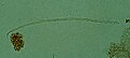
Microfilariae of Loa loa (right) and Mansonella perstans (left) in the same blood smear
Onchocerca volvulus


Vectors

The filaria larvae, known as microfilariae, are transmitted by various blood-sucking insects. It is transmitted through the bite of the insect, which acts as an intermediate host and vector . The insects ingest the larvae of the filariae, the microfilariae, with the blood of their host and at the same time release the developed filariae into the blood and lymphatic system.
Mosquitoes play a central role in the transmission of the pathogen causing lymphatic filariasis. Thus Mangifera by mosquitoes of the genus Aedes and Culex transmit Brugia malayi of species of the genus Anopheles and Mansonia . Loiasis vectors are horseflies of the genus Chrysops , and Onchocerca volvulus is only transmitted by individual species of black flies of the genus Simulium . Blood-sucking mites or ticks can also play a role in filariae pathogenic to animals, for example in Litomosoides carinii , which infects rats.
Occurrence and epidemiology
The spread of the diseases depends on the range of the respective vectors. Most filariasis is transmitted by insects in tropical countries. It is estimated that around 100 to 200 million people worldwide are infected with roundworms. The population in developing countries is particularly affected.
Classification and clinical picture
Filariasis are differentiated according to the different lifestyles of the filariae and the associated symptoms of the disease. The lymphatic forms, triggered by species living in the lymphatic system, are regarded as filariasis in the strict sense of the word.
Lymphatic filariasis

The lymphatic filariasis is also considered filariasis in the true sense and above all by the filarial Mangifera , Brugia malayi and Brugia timori triggered.
The first infection occurs in the tropical epidemic areas in early childhood. The filariae develop and multiply in the lymph nodes of the infected person and the microfilariae migrate to the peripheral lymphatic vessels, where they can be periodically detected. During this time, the infected body increasingly develops an immune defense, which manifests itself in an increased number of eosinophilic granulocytes in the blood count ( eosinophilia ) with fever and intermittent lymphangitis . At the same time, there is a strong increase in the number of microfilariae ( microfilaremia ) with a time-of-day ( circadian ) periodicity.
With increasing manifestation of the disease, various long-term effects develop, which are mainly due to the increase in living and dead microfilariae in the lymphatic system. Inflammation and swelling of the testicle ( orchitis ), the epididymis ( epididymitis ) and the spermatic cords ( funiculitis ), water fractures in the testicles ( hydrocele ) and lymphedema of the scrotum (lymphatic scrotum ) occur particularly in the male testicle area . After several years of progression and increasing clogging of the lymph vessels ( obstruction ) and subsequent obliteration by dead microfilariae, hardening of the lymph vessels (lymph varicoses ), chyluria and chylothorax due to lymph drainage disorders and the development of elephantiasis , especially of the legs, due to lymphatic collections in the tissue.

Elephantiasis of the leg caused by microfilariae

Elephantiasis of the leg caused by microfilariae

Africans with genital elephantiasis, 1906


Loiasis
The loiasis is only by Loa loa triggered and their distribution is limited to tropical rain forests of Africa. In contrast to lymphatic filariasis, the filariae migrate in the subcutaneous tissue (wandering filariasis), in the connective tissue under the skin and below the conjunctiva of the eye (subconjunctival). If loa loa occurs in the eye, it is referred to as an "eye worm".
The symptoms of loiasis are mainly due to immune reactions and allergic reactions. Sharply demarcated and severely itchy swellings form on the skin, which are present for a few days and recur (recur) at irregular intervals and are known as calabar bumps or cameroon bumps. The larvae (microfilariae) are released into the lymphatic system and can thus be detected accordingly, but have no symptoms analogous to lymphatic filariasis.
Onchocerciasis and river blindness

Onchocerciasis, also known as nodular filariasis, is caused by infection with Onchocercas volvulus . As with loiasis, the adult worms also live in the subcutaneous connective tissue in this infection. However, they do not migrate, but stay locally in nodes and release their larvae into the adjacent connective tissue. The larvae destroy the elastic fibers here and lead to chronic itching , skin inflammation ( dermatitis ), lichenification , atrophy of the affected tissue, depigmentation of the skin and detachment of skin folds in the connective tissue ( presbyteria ).
In the epidemic areas, up to 10% of the cases of infection lead to blindness due to microfilariae infestation of the eyes. This can happen both through the involvement of the cornea and the eye chamber as well as through the involvement of the rear areas of the eye near the retina and the optic nerve . The infestation in the front sections leads to local inflammation of the cornea ( keratitis ), the conjunctiva ( conjunctivitis ), the iris and ciliary body ( iridocyclitis ) and the average eyeball ( uvea , uveitis ) and above to photophobia (photophobia), glaucoma (green Star ) and cataract (cataract). In the back of the eye, the infection leads to allergic reactions, inflammation of the retina and choroid ( chorioretinitis ) and the optic nerve ( neuritis nervi optici ) or optic atrophy . These forms of blindness triggered by the infestation with the microfilariae of Onchocercus volvulus are summarized as river blindness.
Mansonelliasis
The Mansonella filaments also live in the connective tissue. Here, find Mansonella ozzardi and Mansonella Perstans in the connective tissue of the peritoneum (peritoneum) and Mansonella streptocerca in the subcutaneous tissue. Unlike other filaria, they are largely non-pathogenic, so they do not trigger any diseases and symptoms that go beyond worm infection.
Other filariasis

In addition to the filariasis described, other human filariasis are summarized in the International Statistical Classification of Diseases and Related Health Problems . This mainly includes zoonoses , i.e. infections with species in which humans are not the actual host ( false host ). Examples of such zoonoses are infections with Dirofilaria immitis and Dirofilaria repens , Acanthocheilonema reconditum and Brugia pahangi . In addition to humans, these species mainly affect dogs (→ heartworm disease , cutaneous dirofilariasis ), cats (→ dirofilariasis ), ungulates and primates.
Diagnosis
Depending on the type and location of the manifestation of the filariasis, the diagnosis is also carried out differently. In lymphatic filariasis, the microfilariae occur in the blood count and the lymphatic fluid and can be detected there, whereby the microfilariae of Loa loa and Wuchereria bancrofti can be detected at specific times of the day. They usually occur at night and are therefore tailored to the blood-sucking behavior of the vectors. Lymphatic Mansonella species, on the other hand, show no periodic occurrence. The microfilariae of Onchocercas volvulus and Mansonella streptocerca occur in the skin.
Various serodiagnostic procedures also provide information on infection with filaria, especially the immunofluorescence test (IFT), the complement fixation reaction (KBR) and the enzyme immunoassay (EIA) for the detection of antibodies and antigens in blood serum .
treatment
Drug treatment and temporary prophylaxis for loiasis and lymphatic filariasis is provided by the administration of diethylcarbamazine , an anthelmintic . Ivermectin , an avermectin , is recommended against microfilariae and albendazole , also an anthelmintic, against adult filariae. Other drugs can be used in the treatment of onchocerciasis.
Due to the good results of studies in which ivermectin is used, the WHO changed its recommendations in 2017 and now recommends a triple combination of ivermectin, diethylcarbamazine and albendazole for the treatment and prophylaxis of lymphatic filariasis, which has a high success rate even with simple mass distribution. In addition to lymphatic filariasis, this combination is also effective against accompanying intestinal helminth infections and head lice .
However, the administration of diethylcarbamazine is not indicated in areas where onchocerciasis is also endemic, as the intake of diethylcarbamazine can lead to serious side effects in the event of an infection. Likewise, ivermectin must not be given in the event of an infestation with Loa loa , as this can trigger acute severe side effects, especially in the case of high parasitaemia . In both cases, the intake of medication can lead to an acute massive death of the parasites and the release of toxins, which can trigger a severe inflammatory reaction that can lead to multiple organ failure and death . In many African countries in particular, all three parasites are endemic.
The long-term effects, especially the hydrocele and in some cases elephantiasis, can be treated surgically. However, filariasis also leads to considerable psychological and social consequences, so that the WHO classifies lymphatic filariasis as the second most severe "disabling" disease.
Eradication of the lymphatic filariasis
At the World Health Assembly of the WHO in 1997 the elimination of lymphatic filariasis was proclaimed a world health goal, from which the Global Program to Eliminate Lymphatic Filariasis (GPELF) emerged in 2000 ..
In addition to vector control measures, prophylactic and therapeutic mass treatments are particularly recommended. The aim in endemic areas is to achieve one-time treatment of at least 65% of the population annually for five years in order to prevent further spread - but this does not benefit people with permanent disabilities. The standard is an annual dose of diethylcarbamazine and albendazole for all residents, except pregnant women, children under two years of age and the seriously ill. Only in countries with a similarly high prevalence of onchocerciasis, i.e. in large parts of Africa and in Yemen, is a combination of ivermectin and albendazole issued instead (also to the entire population, with the exception of pregnant women, children under, because of the possible undesirable effects of diethylcarbamazine with simultaneous river blindness) 90 cm and the seriously ill).
By 2018, 6.7 billion single doses had been issued under the GPELF program and 850 million people in 66 countries had been treated at least once. For 2016, the WHO reported coverage of almost 58% of people in need of therapy or prophylaxis (with almost 500 million treated in forty countries at the time). As a result, the bulk dispensing could be stopped in twenty countries, nine countries had achieved the required coverage. 500 million people no longer need any therapy or prophylaxis. It is estimated that by 2015 the mass treatment prevented or cured 97 million infections, 18 million cases of hydrocele and at least 5.5 million cases of lymphedema. Nevertheless, in 2018 there were still 852 million people in 52 countries in need of treatment.
See also
Trade journals
- Filiaria Journal , in Open Access , published from 2002 to 2008, since then part of Parasites & Vectors
Literature and evidence
- ↑ a b c d e f g h i Keyword "filariasis" in Pschyrembel dictionary of sexuality. Walter de Gruyter, Berlin 2006, ISBN 3-11-016965-7 , p. 473.
- ↑ Achim Hörauf, Sabine Mand, Dietrich W. Büttner: Doxycycline for chemotherapy of filariasis: Elimination of Wolbachia, essential bacterial endosymbionts in worms. In: Dtsch Arztebl. 2003; 100 (37), pp. A-2383 / B-1988 / C-1875.
- ^ A b Keyword “Filariasis” in Heinz Mehlhorn (Ed.): Encyclopedic Reference of Parasitology. Biology, Structure, Function Springer Verlag, Berlin, Heidelberg, New York 2001, ISBN 3-540-66239-1 .
- ↑ a b Keyword "Loiasis" in Pschyrembel Dictionary Sexuality. Walter de Gruyter, Berlin 2006, ISBN 3-11-016965-7 , pp. 893-894.
- ↑ a b Keyword "Ochozercose" in Pschyrembel Dictionary Sexuality. Walter de Gruyter, Berlin 2006, ISBN 3-11-016965-7 , p. 1104.
- ↑ keyword "Mansonella ozzardi" in Pschyrembel Dictionary sexuality. Walter de Gruyter, Berlin 2006, ISBN 3-11-016965-7 , p. 937.
- ^ Keyword "Mansonella perstans" in the Pschyrembel Dictionary Sexuality. Walter de Gruyter, Berlin 2006, ISBN 3-11-016965-7 , p. 937.
- ↑ Keyword "Mansonella streptocerca" in Pschyrembel Dictionary Sexuality. Walter de Gruyter, Berlin 2006, ISBN 3-11-016965-7 , p. 937.
- ^ E. Palumbo: Filariasis: Diagnosis, Treatment and Prevention. ( Memento of the original from February 1, 2012 in the Internet Archive ) Info: The archive link was inserted automatically and has not yet been checked. Please check the original and archive link according to the instructions and then remove this notice. (PDF; 54 kB) In: Acta bio-medica. Volume 79, Number 2, August 2008, pp. 106-109, ISSN 0392-4203 . PMID 18788504 . (Review).
- ^ World Health Organization : Global Program to eliminate lymphatic filiariasis: progress report. Wkly Epidemiol Rec 2017; 92: 594-607
- ↑ WHO page on the elimination of lymphatic filariasis (EN)
- ↑ David H. Molyneux: Advancing toward the Elimination iof lymphatic filiariasis New England Journal of Medicine 2018, Volume 379, Issue 19 of November 8, 2018, pages 1871-1872, doi: 10.1056 / NEJMe1811455 .
Web links
- filariasis.net - an open access portal on lymphatic filariasis
- Interactive world map and overview (English) of the World Health Organization on lymphatic filariasis