Green fluorescent protein
| Green fluorescent protein | ||
|---|---|---|

|
||
| Belt model according to PDB 1EMA | ||
|
Existing structural data: see UniProt entry |
||
| Mass / length primary structure | 238 amino acids , 26.9 kDa | |
| Secondary to quaternary structure | Monomer or dimer | |
| Identifier | ||
| Gene name (s) | GFP | |
| External IDs | ||
| Occurrence | ||
| Parent taxon | Aequorea victoria | |
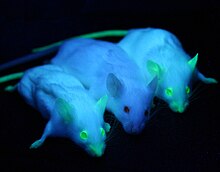
The green fluorescent protein (abbreviation GFP ; Engl. Green Fluorescent Protein ) is a first time in 1962 by Osamu Shimomura described protein from the jellyfish Aequorea victoria , the green when excited by blue or ultraviolet light fluoresces . Its inestimable importance in biology , especially cell biology , lies in the ability to fuse GFP with any other protein in a gene- specific manner. Due to the fluorescence of the GFP, the spatial and temporal distribution of the other protein in living cells, tissues or organisms can be observed directly.
In 2008 the Nobel Prize in Chemistry was awarded to Osamu Shimomura , Martin Chalfie and Roger Tsien for the “discovery and further development of the green fluorescent protein” .
properties
The primary structure consists of 238 amino acids with a molecular mass of 26.9 kDa . The actual fluorophore of GFP evidently forms autocatalytically from the tripeptide sequence Ser 65 –Tyr 66 –Gly 67 within the polypeptide chain. This fluorescence is not based on a remodeling by an external enzyme or subsequently integrated substances, so it works completely without any cell-specific processing systems.
In its original organism, GFP receives its excitation energy through radiation-free energy transfer from the photoprotein aequorin . In applications, GFP is always optically stimulated. The unmodified, naturally occurring GFP has two excitation maxima . The first is at a wavelength of 395 nm, the second at 475 nm. The emission wavelength is 509 nm.
application
Douglas Prasher isolated and sequenced the DNA of GFP in 1992 . Since Prasher succeeded in using GFP as a marker for other proteins in 1994, this technique has become a standard method in cell biology in just a few years. To produce GFP fusion proteins, the DNA of the protein to be examined is linked to the GFP DNA and brought into a form (see vector ) that can be taken up by the cell so that it produces the fusion protein independently ( transfection / or transformation at not cell cultures). In many cases, the protein to be examined is still transported to the correct location in the cell , and the GFP can provide information about the temporal and spatial localization of the target protein in the cell using fluorescence microscopy .
GFP can be classified as non-toxic in almost all eukaryotic cells and is therefore perfectly suited for the investigation of biological processes in vivo . If the expression is very high, the only problem can be the formation of peroxide during the formation of the fluorophore, which could put the cell under stress and damage it.
Modern fluorescence microscopy methods, such as Vertico-SMI , STED microscopy , 3D SIM microscopy and photoactivated localization microscopy, can resolve structures marked with GFP derivatives or photoactivatable fluorescent proteins beyond the optical resolution limit.
Applications that differ from laboratory methods, such as B. a luminous rabbit or a genetically manipulated zebrafish ( Danio rerio ) available in the US in the pet trade under the name GloFish can be found.
variants
There are now various modified versions of the original GFP that show different fluorescence spectra. Depending on the color, they are called, for example, CFP ( cyan ) or YFP ( yellow ). With skillful use, individual cell organelles can be colored differently and then separately observed by means of segregation (spectral deconvolution). The development of enhanced variants such as enhanced GFP (EGFP) or enhanced YFP (EYFP) is also to be seen as crucial. RoGFP , rxYFP and HyPer are redox- sensitive variants of GFP. Voltage-dependent GFP variants are e.g. B. PROPS or the VSFP .
Fluorescent proteins from corals ( Anthozoa ) are also becoming increasingly important . The zoanFP (from Zoanthus sp.) Or the red fluorescent protein drFP583 (from Discosoma ), trade name DsRed, should be mentioned here. Many of these proteins have already been mutated and changed in their properties in order to gain other properties. Many fluorescent proteins are tetramers. This is used by incorporating monomers that develop at different speeds. So the color of the protein changes over time. Such fluorescence timers are useful, for example, to determine the age of organelles . The DsRed mutant E5, for example, has this property.
The green fluorescent protein in art

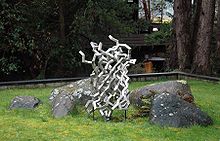
The German-American artist Julian Voss-Andreae , who specializes in “protein sculptures”, created a sculpture in 2004 that is based on the structure of GFP.
swell
- ↑ Osamu Shimomura, FH Johnson and Y. Saiga: Extraction, purification and properties of aequorin, a bioluminescent protein from the luminous hydromedusan, Aequorea . In: Journal of Cellular and Comparative Physiology . Volume 59, 1962, pp. 223-239. PMID 13911999 doi : 10.1002 / jcp.1030590302
- ↑ Osamu Shimomura: The discovery of aequorin and green fluorescent protein . In: Journal of Microscopy . Volume 217, 2005, pp. 1-15. PMID 15655058 doi : 10.1111 / j.0022-2720.2005.01441.x
- ↑ UniProt P42212
- ^ I. Moen, C. Jevne, J. Wang, KH Kalland, M. Chekenya, LA Akslen, L. Sleire, PO Enger, RK Reed, AM Oyan, LE Stuhr: Gene expression in tumor cells and stroma in dsRed 4T1 tumors in eGFP-expressing mice with and without enhanced oxygenation. In: BMC Cancer . Volume 12, 2012, p. 21, doi: 10.1186 / 1471-2407-12-21 . PMID 22251838 . PMC 3274430 (free full text).
- ↑ DC Prasher, VK Eckenrode, WW Ward, FG Prendergast and MJ Cormier: Primary structure of the Aequorea victoria green-fluorescent protein . In: Genes . Volume 111, 1992, pp. 229-233. PMID 1347277
- ↑ M. Chalfie, Y. Tu, G. Euskirchen, WW Ward and DC Prasher: Green fluorescent protein as a marker for gene expression . In: Science . Volume 263, 1994, pp. 802-805. PMID 8303295 doi: 10.1126 / science.8303295
- ↑ Transgenic aquarium fish. Telepolis, November 23, 2003.
- ↑ VV Verkusha and KA Lukyanov: The molecular properties and applications of Anthozoa fluorescent proteins and chromoprotein. In: Nature Biotechnology . Volume 22, 2004, pp. 289-296. PMID 14990950 doi : 10.1038 / nbt943
- ↑ A. Terskikh et al. : "Fluorescent Timer". Protein That Changes Color With Time. In: Science. Volume 290, 2000, pp. 1585-1588. PMID 11090358 doi : 10.1126 / science.290.5496.1585
- ^ Julian Voss-Andreae Sculpture . Retrieved June 14, 2007.
- ^ J Voss-Andreae: Protein Sculptures: Life's Building Blocks Inspire Art . In: Leonardo . 38, 2005, pp. 41-45. doi : 10.1162 / leon.2005.38.1.41 .
- ↑ Alexander Pawlak: Inspiring Proteins . In: Physics Journal . 4, 2005, p. 12.
literature
- RY Tsien : The green fluorescent protein. In: Annual Review in Biochemistry . Volume 67, 1998, pp. 509-544. PMID 9759496 doi : 10.1146 / annurev.biochem.67.1.509
- Martin Chalfie and Steven Kain: GFP: Properties, Applications and Protocols. 2nd edition, Wiley & Sons, Hoboken 2005, ISBN 0-471-73682-1
- M. Zimmer: Glowing Genes. A Revolution in Biotechnology . Prometheus Books, Buffalo, NY 2005, ISBN 1-59102-253-3
- Daniel Veith, Martina Veith: Biology of fluorescent proteins: A rainbow from the ocean Biology in our time , 6/2005, pp. 394–404. doi : 10.1002 / biuz.200410295
- Michael Groß: Current research: green light for biologists. In: Spectrum of Science Dec. 2008 p. 14
- Manuel Gunkel, Fabian Erdel, Karsten Rippe, Paul Lemmer, Rainer Kaufmann, Christoph Hörmann, Roman Amberger and Christoph Cremer : Dual color localization microscopy of cellular nanostructures In: Biotechnology Journal, 2009, 4, 927-938. ISSN 1860-6768
See also
- GFP cDNA
- Modern examination methods in cell biology : FRET
- Luminous rabbit (application in art)
Web links
- Green Fluorescent Protein - History (Engl.)
- Protein Database: Molecule of the Month - Green Fluorescent Protein (GFP )
- Interpro: Protein of the Month: Green Fluorescent Protein. (engl.)
- Environmental research center: microbiologists make bacteria glow. (PDF file; 1.40 MB)
- The Nobel Prize in Chemistry in 2008 (Engl.)
- Proteopedia: Green Fluorescent Protein (Engl.)
- Nature Reviews Poster: Fluorescent Proteins Illuminate Cell Biology (English; PDF file; 1.62 MB)
- GFP - GFP from A to Z, with fantastic pictures and the latest scientific information


