Amygdala


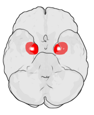
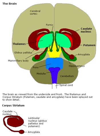
The amygdala is a pair of core areas of the brain in the medial part of the respective temporal lobe . It is part of the limbic system . The name of the amygdala (technical plural: amygdalae) is derived from the Latin amygdala , from ancient Greek ἀμυγδάλη , almond (kernel) ' . It is also known as the almond kernel or the amygdaloid corpus .
The amygdala is involved in fear conditioning and generally plays an important role in the emotional evaluation and recognition of situations as well as the analysis of possible dangers : It processes external impulses and initiates the vegetative reactions to them. Research results from 2004 show that the amygdala in the perception of any form of excitement , so emotionally - or relish emphatic feelings , essential and perhaps the sex drive is involved. The amygdala is important for the sensation of fear or fear: Patients with Urbach-Wiethe syndrome , in whom the amygdalae are damaged on both sides, do not show fear reactions even in potentially life-threatening or traumatic situations. So far, the only known fear-inducing stimulus for these patients is feelings of suffocation.
Anatomical and functional organization of the amygdala
A distinction is made between three different areas of the tonsil nucleus complex: On the one hand, the centromedial core group, including the central and medial nuclei - both descendants of the striatum . Then the basolateral complex, whereby the nuclei nucleus lateralis, nucleus basalis - which also splits into a small-cell inner and a large-cell lateral part - and the nucleus basolateralis should be mentioned. And third, the cortical nucleus group with the cortical nucleus.
Interconnection of the amygdala
The amygdala consists of 13 individual nuclei (some of which are still divided into subunits) and receives a great deal of information from higher brain centers via fiber connections. This structural division of the amygdala into individual nuclei is associated with different connection and functional profiles in mammals and humans.
The medial core is in connection with the olfactory cortex areas that are important for odor perception ; a phylogenetic relic when, for example, the predators made themselves noticeable through their weather (but especially pheromone perception). The basolateral and phylogenetically younger core group gets its information primarily from the posterior core group of the thalamus (nuclei posteriores). Important reflexes are mapped there, and important information arrives here from almost all sensory cortex areas via sensations such as smell, taste, sight, hearing and touch.
With a startle reaction , an organism reacts to a surprisingly perceived, potentially threatening stimulus. Responsible for this reaction is a neural connection between the amygdala , such as the central nucleus, and the basal ganglia , which connect the amygdala to the motor system.
Afferents
In contrast to the afferents of the hypothalamus (with one exception) all afferents to the amygdala are heavily preprocessed, i.e. the information has already been processed or connected thalamically in secondary visual, sensory and auditory areas of the cerebral cortex . The afferents mainly reach the basolateral core complex of the amygdala. The exception here is the sense of smell . It releases collaterals via the olfactory bulb directly without thalamic switching to the medial amygdala .
Efferents
The central core of the amygdala receives most of the efferents of the basolateral complex and in turn sends efferents to:
- the middle hypothalamus to activate the sympathetic nervous system ,
- the reticular core ( Formatio reticularis ) to strengthen reflexes ,
- the motor nucleus of the trigeminal nerve and the motor nucleus of the facial nerve to trigger anxious facial expressions ,
- the parabrachial nucleus to stimulate breathing ,
- the paraventricular nucleus of the hypothalamus to stimulate ACTH -Ausschüttung in the pituitary gland ( stress response , " stress response "),
- the nucleus dorsalis of the vagus nerve to influence the gastrointestinal tract and
- the locus caeruleus , the nucleus tegmentalis lateralis dorsalis and the area tegmentalis ventralis (VTA) for the production of the neurotransmitters acetylcholine , adrenaline and dopamine . This increases vigilance and alertness.
Medical importance
In humans, a variety of specific phenomena and symptoms such as memory disorders , the inability to emotionally assess situations, autism , depression , narcolepsy , post-traumatic stress disorder and phobias and the like can occur. a. indicate malfunction of the amygdala. These disorders can be caused by damage, developmental problems or an imbalance of the neurotransmitters or, on the contrary, they can also be the result of a situation-appropriate functioning of the amygdala. The amygdala links events with emotions and stores them. Over time, the trigger threshold for assessing stimuli as dangerous decreases, and generalization occurs. Here the amygdala is overexcited. If an event was associated with danger , pain or suffering , situations considered to be similar can trigger strong somatic reactions ( e.g. panic , nausea , apathy , fainting ), regardless of whether they are objectively comparable and even regardless of whether a (conscious) Memory of the original event exists. Therefore, the term body memory often appears in this context . Provoking situations for this often dramatic reliving be trigger ( Engl. For "trigger") or called Restimulator.


The conditioning of animals to combine certain “neutral” stimuli with fear changes the information stored in the amygdala , as experiments by Joseph LeDoux and other scientists have shown. It serves as a simple Pavlovian learning machine that links aversions with neutral events and thus helps to react to the environment. Without an amygdala, animals lose the ability to conditioning to fear stimuli.
In animal experiments it was found that electrical stimulation of various points in the amygdala can cause a wide variety of reactions. Signals in the central core lead to anger or flight reactions . In other places, vegetative reactions , for example an increase in the pulse , but also the eating behavior and sexuality can be triggered.
Primates with their amygdala removed can see objects without recognizing their emotional significance. They also lose all aggression . After Heinrich Klüver and Paul Bucy discovered this in 1937, consideration was given to undertaking such an intervention to treat crime ( psychopathy research ).
Research from 2009 shows that autistic children have an enlarged amygdala as early as the second year of life. The enlargement was still preserved in the fourth year of life.
The Urbach-Wiethe syndrome , which occurs in around 100 people - mostly in South Africa - is a genetic, selective calcification of the basolateral amygdala. It causes a lack of trust and mistrust.
There is some thought that adults in second language acquisition may not have access to the amygdala's implicit memory and therefore find it more difficult to connect emotionally to words.
Research at the University of Vienna in 2015 questioned previous studies on excitation patterns in the amygdala. Data previously generated by functional magnetic resonance imaging , which were believed to be amygdala activities, probably only represented the blood flow in the Rosenthal vein .
literature
- Eric R. Kandel, James H. Schwartz, Thomas M. Jessell : Principles of Neural Science . 4th edition. McGraw-Hill Medical, 2000, ISBN 0-8385-7701-6 (English).
- Purves et al .: Neuroscience Including Sylvius . 3. Edition. Sinauer, 2004, ISBN 0-87893-725-0 (English).
- HT Blair, GE Schafe, EP Bauer, SM Rodrigues, JE LeDoux: Synaptic plasticity in the lateral amygdala. A cellular hypothesis of fear conditioning . In: Learn Mem . tape 8 , 2001, p. 229-242 (English).
Web links
- Function of the amygdala - in a nutshell at Wissenschaft-online.de
- When reason and feeling work in a team on Wissenschaft.de
Individual evidence
- ↑ Patricia H. Janak, Kay M. Tye: From circuits to behavior in the amygdala . In: Nature . tape 517 , no. 7534 , p. 284–292 , doi : 10.1038 / nature14188 , PMID 25592533 , PMC 4565157 (free full text) - ( nature.com ).
- ^ Ralph Adolphs: Emotional vision. In: Nature Neuroscience. 7, 2004, pp. 1167-1168, doi: 10.1038 / nn1104-1167 .
- ↑ Feinstein, JS, Adolphs, R., Damasio, A., & Tranel, D. (2011). The human amygdala and the induction and experience of fear. Current biology, 21 (1), 34-38.
- ↑ Justin S Feinstein, Colin Buzza, Rene Hurlemann, Robin L Follmer, Nader S Dahdaleh: Fear and panic in humans with bilateral amygdala damage . In: Nature Neuroscience . tape 16 , no. 3 , p. 270–272 , doi : 10.1038 / nn.3323 , PMID 23377128 , PMC 3739474 (free full text).
- ↑ D. Bzdok, A. Laird, K. Zilles, PT. Fox, S. Eickhoff: An investigation of the structural, connectional and functional sub-specialization in the human amygdala. Human Brain Mapping, 2012.
- ↑ MW. Mosconi, H. Cody-Hazlett, MD Poe, G. Gerig, R. Gimpel-Smith, J. Piven: Longitudinal study of amygdala volume and joint attention in 2- to 4-year-old children with autism. Arch Gen Psychiatry . 2009; 66 (5): 509-516 PMID 19414710
- ↑ Lisa A. Rosenberger, Jack van Honk (2019). The human basolateral amygdala is indispensable for social experiential learning. Current Biology . DOI: 10.1016 / j.cub.2019.08.078
- ↑ fMRI measurements of amygdala activation are confounded by stimulus correlated signal fluctuation in nearby veins draining distant brain regions. Nature , May 21, 2015, accessed August 27, 2015 .
- ↑ Neuroscience: One mistake challenges thousands of brain studies. Profile , June 24, 2015, accessed August 27, 2015 .
