Nile blue
| Structural formula | |||||||||||||||||||
|---|---|---|---|---|---|---|---|---|---|---|---|---|---|---|---|---|---|---|---|

|
|||||||||||||||||||
| General | |||||||||||||||||||
| Surname | Nile blue | ||||||||||||||||||
| other names |
|
||||||||||||||||||
| Molecular formula | C 20 H 20 N 3 O + | ||||||||||||||||||
| Brief description |
|
||||||||||||||||||
| External identifiers / databases | |||||||||||||||||||
|
|||||||||||||||||||
| properties | |||||||||||||||||||
| Molar mass | 318.39 g mol −1 | ||||||||||||||||||
| Physical state |
firmly |
||||||||||||||||||
| Melting point |
> 300 ° C |
||||||||||||||||||
| solubility |
soluble in water (50 g l −1 at 25 ° C) |
||||||||||||||||||
| safety instructions | |||||||||||||||||||
|
|||||||||||||||||||
| As far as possible and customary, SI units are used. Unless otherwise noted, the data given apply to standard conditions . | |||||||||||||||||||
Nile blue , often referred to as Nile Blue A (usually only the hydrogen sulphate ), is a fluorescent phenoxazine - dye .
As an indicator dye , Nile blue shows a blue color in an acidic environment and is red in an alkaline environment.
Boiling a solution of Nile blue with sulfuric acid produces the dye Nile red .
properties

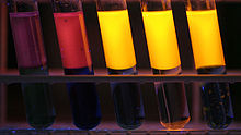
V. l. n.r .: 1000 ppm , 100 ppm, 10 ppm, 1 ppm, 100 ppb.

V. l. n.r .: 1. methanol , 2. ethanol , 3. tert -butyl methyl ether , 4. cyclohexane , 5. n -hexane , 6. acetone , 7. tetrahydrofuran , 8. ethyl acetate , 9. dimethylformamide , 10. acetonitrile , 11. toluene , 12. Chloroform
Nile blue is a fluorescent dye . The fluorescence shows a high quantum yield , especially in apolar solvents :
The absorption and emission maxima of Nile blue are strongly dependent on the pH value and the solvent used ( solvatochromism ).
| solvent | Absorption λ max (nm) |
Emission λ max (nm) |
|
|---|---|---|---|
| toluene | 493 | 574 | |
| acetone | 499 | 596 | |
| Dimethylformamide | 504 | 598 | |
| chloroform | 624 | 647 | |
| 1-butanol | 627 | 664 | |
| 2-propanol | 627 | 665 | |
| Ethanol | 628 | 667 | |
| Methanol | 626 | 668 | |
| water | 635 | 674 | |
| 0.1 N hydrochloric acid | (pH = 1.0) | 457 | 556 |
| 0.1 N sodium hydroxide solution | (pH = 13.0) | 522 | 668 |
| Ammonia water | (pH = 11.0) | 524 | 668 |
The fluorescence duration of Nile blue was determined to be 1.42 ns in ethanol. This is shorter than the corresponding value of Nile Red at 3.65 ns. The fluorescence duration is relatively invariant to dilutions in the range of 10 −3 - 10 −8 mol · dm −3 .
The Nile blue coloring
Nile blue is used for the histological staining of biological specimens. A distinction is made between neutral lipids ( triglycerides , cholesterol esters , steroids ), which are colored pink, and acidic ( fatty acids , chromolipids , phospholipids ), which are colored blue.
Kleeberg's Nile blue staining requires the following chemicals:
- Nile blue A.
- 1% acetic acid
- Glycerine or glycerine gelatin
The workflow
The preparation is in formalin fixed. This will be frozen sections or teased preparations made. They are then immersed in the Nile blue solution for 20 minutes and then rinsed off with water. For better differentiation, it is immersed in 1% acetic acid for 10–20 minutes until the colors are pure. This can u. This may already be the case after 1–2 minutes. Then watering is carried out thoroughly in several changes of water (one to two hours). The stained specimen can then be drawn onto a slide and the excess water sucked off. The preparation can be enclosed in glycerine or lukewarm glycerine gelatine.
The result
Unsaturated glycerides are pink, kernels and elastics are dark blue, fatty acids and numerous fatty substances and fat mixtures are blue to purple.
Example: Detection of poly-β-hydroxybutyrate granules (PHB)
The PHB granules in the cells of Pseudomonas solanacearum can be made visible by staining with Nile blue A. The PHB granules of the stained smears show under an epifluorescence microscope at 450 nm excitation wavelength with oil immersion, at 1000-fold magnification a strong orange-colored fluorescence.
Nile blue in oncology
Derivatives of Nile blue are potential photosensitizers in photodynamic therapy (PDT) of malignant tumors . These dyes are highly concentrated through dye aggregation in the tumor cells, especially in the lipid membranes and / or sequestered in the subcellular organelles .
With the Nile blue derivative N- ethyl-Nile blue (EtNBA), it was possible to differentiate between normal and premalignant tissue in animal experiments by means of fluorescence imaging or fluorescence spectroscopy. EtNBA shows no phototoxic effects.
Manufacturing
Nile blue and related napthoxazinium dyes can be made in a number of ways.
- Acid-catalyzed condensation of 5- (dialkylamino) -2-nitrosophenols with 1-naphthylamine
- 3- (Dialkylamino) phenols with N -alkylated 4-nitroso-1-naphthylamines
- N , N -dialkyl-1,4-phenylenediamines with 4- (dialkylamino) -1,2-naphthoquinones
Alternatively, the product of an acid-catalyzed condensation of 4-nitroso- N , N- dialkylaniline with 2-naphthol in the presence of amines can be oxidized and a second amine substituent can be introduced in the 5-position. The following equation shows the first possible synthesis
Individual evidence
- ↑ a b Entry on Nilblau A. In: Römpp Online . Georg Thieme Verlag, accessed on June 1, 2014.
- ↑ a b Data sheet Nilblau (PDF) from Merck , accessed on June 3, 2008.
- ↑ Data sheet Nile Blue A from Sigma-Aldrich , accessed on May 9, 2017 ( PDF ).
- ↑ a b c Jiney Jose and Kevin Burgess: Benzophenoxazine-based fluorescent dyes for labeling biomolecules , in Tetrahedron , 2006 , 62 , pp. 11021-11037; doi : 10.1016 / j.tet.2006.08.056 .
- ^ Roche Lexicon, accessed June 25, 2007 .
- ^ Benno Romeis , Microscopic Technology , 15th edition, R. Oldenbourg Verlag, Munich 1948.
- ↑ 97/647 / EG: Decision of the EU Commission of September 9, 1997 on a preliminary test program for the diagnosis, detection and identification of Pseudomonas solanacearum (Smith) Smith in potatoes , accessed on June 27, 2007 .
- ↑ Lin CW, Shulok JR, Kirley SD, Cincotta L., Foley JW: Lysosomal localization and mechanism of uptake of Nile blue photosensitizers in tumor cells , in: Cancer Research , 1991 , 51 , pp. 2710-2719; PMID 2021950 .
- ↑ HJ van Staveren: Fluorescence imaging and spectroscopy of ethyl nile blue A in animal models of (pre) malignancies , in: Photochemistry and photobiology , 2001 , 73 , pp. 32-38; PMID 11202363 .
- ^ Andreas Kanitz, Horst Hartmann: Preparation and Characterization of Bridged Naphthoxazinium Salts. In: European Journal of Organic Chemistry. 1999, pp. 923-930, doi : 10.1002 / (SICI) 1099-0690 (199904) 1999: 4 <923 :: AID-EJOC923> 3.0.CO; 2-N .
literature
- FJ Green: The Sigma-Aldrich Handbook of Stains, Dyes and Indicators , Aldrich Chemical Company, Milwaukee, 1990.
- J. Rao, A. Dragulescu-Andrasi, H. Yao: Fluorescence imaging in vivo: recent advances , in: Current Opinion in Biotechnology , 2007 , 18 , pp. 17-25; PMID 17234399 .


