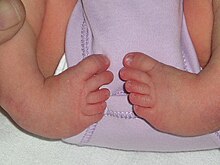
Clubfoot

| Classification according to ICD-10 | |
|---|---|
| Q66.0 | Congenital deformities of the feet |
| M21.5 | Acquired claw hand, club hand, acquired claw foot and club foot |
| ICD-10 online (WHO version 2019) | |
Under clubfoot ( pes equinovarus formerly, pes varus and crooked foot called) comprises various foot deformities. In addition to the most common, the innate form, there is also the acquired form, the so-called neurogenic clubfoot , which is usually caused by a disruption of the nerve supply.
The congenital clubfoot ( Pes equinovarus et plantiflexus adductus congenitus ) belongs to the group of extremity malformations and is a combination of various deformities of the foot , usually accompanied by an inward twist ( supination ) of the foot (the sole of the foot points inward) and anomalies of the lower leg muscles . As a rule, several malpositions come together:
- the so-called supination or varus position of the rear foot ( pes varus )
- Sickle foot position of the forefoot ( pes adductus )
- Equinus ( equinus )
- Splayfoot ( Pes supinatus )
- Archesus ( Pes excavatus , Pes cavus )
This is associated with a shortening of the Achilles tendon . The therapy is usually started as soon as possible after birth. Treatment is often difficult and lengthy, even if it is started early.
causes
Clubfoot can be congenital or acquired. On average, around one in 1000 children is born with this peculiarity, with boys being affected twice as often as girls. The cause of the congenital clubfoot has not been definitively established.
The following are possible:
- unfavorable position of the embryo in the uterus
- Amniotic band syndrome
- strong and comparatively long-lasting decrease in the amount of amniotic fluid ( oligohydramnios )
- Concomitant with Neuralrohrfehlbildungen as a result of paralysis of the muscles of the lower leg
- Result of taking folic acid - antagonists such. B. aminopterin or methotrexate in the 4th to 12th week of pregnancy ( aminopterin syndrome , aminopterin embryopathy )
The cause of the acquired clubfoot is the weakening of the musculus peroneus longus and the musculus peroneus brevis , which are innervated by the superficial peroneal nerve, which can also be the result of injuries or infections. The tibialis posterior muscle , which brings the foot into supination and plantar flexion, is called the "clubfoot muscle " .
Clinical picture
The lumpy shape shows the following picture in the congenital clubfoot:
- Equinus foot: plantar flexion in the upper ankle
- The hindfoot is more supinated than the forefoot
- Forefoot adduction
Developmental disorder in the lower part of the spinal cord , therefore weakening of the calf muscles ( gastrocnemius muscle ) and excess weight of the posterior tibial muscle ( tibialis posterior muscle ); further shortening of the lateral bands between fibula ( fibula ) and ankle bone ( talus ) and the heel bone ( calcaneus ): These belts prevent the frontward migration of the fibula against the talus at the dorsal flexion , so that an increasing equinus formed.
A talocalcaneal angle of less than 30 ° is typical in the infant's lateral x-ray.
treatment
Treatment depends on the etiology and severity of the deformity . Early therapy is important.
Operational
At the age of three months, all conservatively non-redressible structures should be corrected by surgery. This age is now generally considered to be the ideal time. The operation consists of an elongation of the Achilles tendon , a so-called extended "posterior release"; the straightening between the talus and calcaneus is also corrected.
In the case of subluxation of the navicular bone , a "medial release" must also take place, with the ligaments between the talus , navicular and medial cuneiform bone , repositioning of the navicular bone and possibly lengthening of the tendon of the posterior tibial muscle . The aim of the operation is to reposition all components as completely as possible.
Conservative
Clubfoot plaster: for modeling, classic plaster is superior to modern plastics. With clubfoot you always put on thigh casts and not lower leg casts , as on the one hand a better redression of the foot is possible, on the other hand the lower leg cast (especially with the existing equinus foot) slips down slightly and then causes pressure points. The knee joint must be bent by at least 60 ° so that the foot as a whole can be redressed outwards in relation to the thigh. The equinus foot position is initially retained. The final correction of the equinus is usually done by lengthening the tendon of the tibialis posterior muscle .
Clubfoot treatment according to Ponseti
The treatment according to Ignacio Ponseti provides a special manual reduction with step-by-step correction according to anatomical criteria. As a rule, a complete correction can be achieved without surgery after three to eight plaster casts. After the plaster reduction has been completed, a special splint (Dennis Brown splint or Alfa Flex splint) is placed for three months, which initially has to be worn all day. Gradually, the wearing time is then shortened during the day, so that after another three months the splint only has to be worn at night or during an afternoon nap, up to the age of four. This phase is necessary for the therapy to be successful. The parents are usually taught how to use the splint consistently. Children treated according to this method usually accept wearing the splint mentioned without any problems.
Clubfoot treatment according to Bonnet Dimeglio, also known as the "French method"
The French method is dynamic movement therapy. During the first few days, the newborn child is entrusted by the orthopedic surgeon to a specialized children's physiotherapist, whose commitment is crucial. The foot is initially corrected four to five times a week and then carefully treated three to four times a week after the correction. The position is changed in the smallest steps over two to three months, the stuck tissue is loosened and the muscles are stimulated through movement. After manual therapy, the feet are fixed in taping bandages and lower leg plaster casts. The therapy frequency is reduced to twice a week if the result is successful. It is an important aspect that the newborn child retains freedom of movement in the knees and hips. If it is not possible to adjust the heel correctly, a tenotomy of the Achilles tendon is performed at the age of three to four months.
The French method works in great detail and precisely on the individual misalignments of the clubfoot. It is demanding for the therapist and therefore difficult to reproduce. Therefore this form of therapy can only be offered selectively by a few, but well-trained specialists.
Clubfoot treatment according to Zukunft-Huber
This type of therapy was developed in the 1990s and is based on the manual therapeutic criteria of normal foot development in the child in the first year of life. It offers alternative starting points for therapy for sickle, serpentine, lumpy, buckled, flat and spastic pointed-buckle flat feet. Bandages are applied to the infant and the parents are instructed accordingly. The advantage is that you don't need splints, surgery or plasters. The child also does not wear heavy splints or plasters that restrict movement. The disadvantage is the time required, as a 30-minute therapy must be performed independently three times a day. Grade II and III clubfeet intended for surgery often did not have to be operated on, but this depends on the severity of the malformation and is not always possible.
insoles
Correction in the 3-point correction system:
- Medial heel
- Cuboid bone base metatarsal V.
- Head metatarsal 1 and big toe
Important: The more physiologically the correction impressions are adjusted, the better the correction option. After adjusting the aid, it is necessary to check the overall position again.
Anti-varus shoe
The anti-varus shoe is medially narrower than a normal shoe and thus corrects the abduction position of the shoe between the shoe joint and the toe cap correction position against sickle foot ( pes adductus ).
Characteristics of the anti-varus shoe:
- Heel stiffener
- pronating foot position
- slight wing heels
Fixing and functional orthotics
Guidelines
If, in addition to the clubfoot deformity, there is an inner gyration in the lower leg, the thigh must be included in the correction.
Model acceptance
Model acceptance in the absolutely best possible corrective position of the foot should be strictly adhered to when adjusting orthoses , night splints and insoles to assess the possibility of correction.
Execution of the orthosis
If possible made of a light, flexible material such as polyethylene . These materials allow the foot slight micro-movements and at the same time make the correction easier to tolerate. Closed-cell, cross-linked foam made of polyethylene is recommended as the inner material; However, this can lead to severe skin irritation in children who are under six months old . Therefore, deer skin or terry cloth lining is often used for such children. The locks to fix the leg in the orthosis must always be well padded and, above all, wide enough, otherwise the lymphatic and blood circulation could be impaired.
There are different types of orthoses available. There are some with:
- plantar joint
- Extensions - flexion joint in the upper ankle
- Supination - pronation joint
- Abduction - adduction joint in the forefoot
- Correction moves
- Lower leg shell (splint)
- Upper and lower leg shell (splint),
the most common prescription is the thigh splint. The upper and lower leg splint is particularly suitable for retention in small children . The knee joint must always be flexed in the splint so that the foot can be held in the direction of abduction. The splint is usually only put on at night, as this is where the children grow the most. The abduction position of the forefoot is usually actively maintained by the muscles, as the shortened medial muscles ( M. tibialis anterior and posterior , but also M. adductur hallucis ) are overactive compared to the lateral muscle group (especially the peronaei muscle ).
Corrective principles
Foot leg:
- With the knee bent approx. 70 ° to 90 ° (severe cases 90 °, mild cases 70 °)
- Redression of the foot in maximum pronation and abduction.
The aim is to achieve the anatomical position in the forefoot, midfoot and rearfoot and to enable mobility in the upper ankle joint , so that with optimal care, apart from the scars and calf atrophy of the affected extremity, no deformity can be detected.
Historical
There are already clubfoot representations on Egyptian works of art , and the treatment of clubfoot redressing was already described very precisely by Hippocrates of Kos (370 BC) and in the Corpus Hippocraticum . There is also a description of how to put on bandages and reducing shoes. Tenotomy (cutting through the Achilles tendon) has also been known since ancient times, but it did not become established as a treatment method until the first half of the 19th century. A first detailed description of measures to correct clubfoot was given in 1574 by Francisco Arceo. The French orthopedic surgeon Vincent Duval (also the literary source for Dr. Bovary, Madame Bovary's husband ) received an award for his clubfoot operations in 1835.
literature
- S1 guideline for congenital clubfoot of the German Society for Orthopedics and Orthopedic Surgery (DGOOC). In: AWMF online (as of 2012)
- Jürgen Krämer, Joachim Grifka: Orthopedics, trauma surgery. Springer, 2007, ISBN 978-3-540-48498-1 , pp. 311-314.
- K. Gray, V. Pacey, P. Gibbons, D. Little, C. Frost, J. Burns: Interventions for congenital talipes equinovarus (clubfoot). In: Cochrane database of systematic reviews (online). Volume 4, 2012, p. CD008602. doi: 10.1002 / 14651858.CD008602.pub2 . PMID 22513960 . (Review).
- G. Ulrich Exner u. a .: clubfoot. Pathoanatomy, manual-functional and operative treatment. Steinkopff, Darmstadt 2005, ISBN 3-7985-1485-2 .
- A. Ficklscherer: BASICS Orthopedics and Trauma Surgery. Elsevier, 2012, ISBN 978-3-437-42208-9 .
- Doris Schwarzmann-Schafhauser: Clubfoot. In: Werner E. Gerabek u. a. (Ed.): Encyclopedia of medical history. De Gruyter, Berlin / New York 2005, ISBN 3-11-015714-4 , p. 764 f.
- Weimann-Stahlschmidt K, Krauspe R, Westhoff B: Current status of the treatment of children's clubfoot (PDF) OUP 2014; 1: 027-033, doi : 10.3238 / oup.2014.0027-0033 (currently unavailable)
Web links
Individual evidence
- ↑ G. Ulrich Exner, Friedrich Anderhuber, Verena Haldi-Brändle, Hilaire AC Jacob, Gunther Windisch: Club foot. Pathoanatomy. Manual-functional and operative treatment . In: Deutsches Ärzteblatt , 2005, 102 (51–52), p. A-3580
- ↑ Foot deformities: the clubfoot. Verlag Springer, accessed on July 8, 2009 .
- ↑ F. Hefti: Pediatric Orthopedics in Practice. Springer 1998, ISBN 3-540-61480-X .
- ^ B. Stephens Richards, Shawne Faulks, Karl E. Rathjen, Lori A. Karol, Charles E. Johnston, Sarah A. Jones: A Comparison of Two Nonoperative Methods of Idiopathic Clubfoot Correction: The Ponseti Method and the French Functional (Physiotherapy) Method. In: The Journal of Bone and Joint Surgery. Vol. 90, November 2008, ISSN 1535-1386 , pp. 2313-2321, doi: 10.2106 / JBJS.G.01621 .
- ↑ Barbara Zukunft-Huber: The little foot, really big . Three-dimensional manual foot therapy for children's foot malpositions. 3. Edition. Urban & Fischer, 2017, ISBN 978-3-437-55082-9 .
- ↑ Physiotherapy: foot therapy according to Zukunft-Huber. (PDF; 68 kB) University Children's Clinic Mainz, Department of Physiotherapy, September 2006, accessed on March 11, 2018 .
- ↑ Hippocrates on the clubfoot . (PDF) Medical representations in Greek vase painting in the context of Corpus Hippocraticum and modern medicine, p. 123
- ^ Doris Schwarzmann-Schafhauser (2005), p. 765.
- ↑ Franciscus Arceus: De recta curandorum vulnerum ratione et aliis eius artistic praeceptis libri duo. Antwerp 1574.
- ^ Wolfgang Wegner: Arceo, Francisco. In: Werner E. Gerabek u. a. (Ed.): Encyclopedia of medical history. De Gruyter, Berlin / New York 2005, ISBN 3-11-015714-4 , p. 93.
- ↑ Barbara I. Tshisuaka: Duval, Vincent. In: Werner E. Gerabek , Bernhard D. Haage, Gundolf Keil , Wolfgang Wegner (eds.): Enzyklopädie Medizingeschichte. De Gruyter, Berlin / New York 2005, ISBN 3-11-015714-4 , p. 329 f.


