
2D gel electrophoresis


The two-dimensional gel electrophoresis or 2D gel electrophoresis is an analytical method in biochemistry , molecular biology and proteomics . It was developed independently by O'Farrell and Klose in 1975. It combines isoelectric focusing (IEF) with SDS polyacrylamide gel electrophoresis ( SDS-PAGE ) to separate complex protein mixtures (bacterial lysates, lysates from higher cells or tissues, body fluids) into individual proteins . A particularly high-resolution separation is achieved through the combination of the two orthogonal separation techniques.
Each spot in the protein pattern corresponds to a type (species) of protein molecules. Since protein patterns in biological systems change depending on the environment and condition, they can be used to differentiate between damaged and healthy or optimally and suboptimally grown cells . For example, they provide information about the causes of disease or the mechanism of action of drugs at the molecular level. Due to the complexity of two-dimensional protein patterns, specially developed computer programs are used for their evaluation.
Sample preparation
The sample is obtained and processed under as identical conditions as possible. This rules out the possibility that, in addition to the experimental variables to be examined, falsifying influences act on the sample. The proteins are mostly precipitated from extracellular compartments ( secreted proteins) . Intracellular proteins are extracted by gently destroying the cell structures and partially protein purification . Outside of their natural environment, proteins are particularly susceptible to the formation of protein aggregates and degradation by proteases . Furthermore, the sample preparation is carried out at 4 ° C. As a rule, urea is added as a chaotrope and non-ionic detergents as well as protease-inhibiting substances in order to avoid interactions and changes in the proteins.
First dimension (IEF)
During the IEF (first dimension), the protein extract is separated one-dimensionally in a pH gradient gel in an electric field. The acidic and basic amino acid residues of the proteins go through different (de) protonation states depending on the ambient pH value and thus determine the charge of the protein. They are therefore responsible for the effect of the electric field on the proteins. At the isoelectric point (pI), positive and negative charges on the protein cancel each other out. The pI corresponds to the pH at which the protein has a net charge of zero. The electrical field no longer acts as a force on the now charge-neutral protein and the protein is deposited. Changes in location caused by diffusion in neighboring pH ranges lead to a renewed electrical charge of the protein. However, the re-effective electric field immediately promotes the protein back to its isoelectric point. Two different IEF technologies are available.
- Immobilized pH gradients (IPG): The gel strip consists of a polyacrylamide matrix . So-called immobilines are polymerized into this matrix. Immobiline are acrylamide derivatives with functional groups for weak acids or bases , with a defined pK value . The copolymerization of the neutral acrylamide with the acidic and basic immobilines added in the gradient creates an invariable, immobile pH gradient.
- pH gradients based on carrier ampholytes: Carrier ampholytes are synthetic amino acids and are freely movable in a gel strip or a long cylindrical gel (usually in a tube). When an electric field is applied, they form a pH gradient which, however, is unstable over time. With increasing time, the charge carriers defining the gradient migrate to the gel ends and sooner or later deform its course.

Furthermore, a polyacrylamide gel electrophoresis with a cationic detergent such as 16-BAC can alternatively be carried out as the first dimension .
Equilibration
In the so-called equilibration, which follows the separation after the pI, the gel with the proteins is initially reduced (for example with mercaptoethanol or dithiothreitol ). This serves to remove disulfide bridges . In order to prevent reoxidation of the resulting -SH HS groups to disulphide (-SS-) groups, the HS groups are z. B. alkylated with iodoacetamide . Finally, the proteins are loaded with sodium dodecyl sulphate ( SDS ). SDS is a negatively charged detergent. For every 3 amino acids, about one SDS molecule binds to the protein molecule via its aliphatic end through hydrophobic interaction. With the negatively charged side, it repels itself from the charged ends in the vicinity of bound SDS molecules. This leads to the complete unfolding (linearization) of the protein molecules. The larger a protein molecule, the longer the resulting chains loaded with SDS. Since, depending on the length of the protein, a large number of negatively charged SDS molecules bind, the intrinsic charge can be neglected for most proteins.
Second dimension (SDS-PAGE)
In vertical systems, the gel strip with the proteins separated and equilibrated according to the pH value is placed on the edge of a square or rectangular polyacrylamide gel also containing SDS. In the case of horizontal systems, on the other hand, the gel strip is placed on the flat SDS gel a few millimeters from the edge of the gel. The proteins are now separated according to their size in a second electrophoresis perpendicular to the first dimension . When the electric field is applied ( anode opposite the IEF gel strip), the unfolded proteins surrounded by SDS migrate with their excess of negative charges through the gel, which offers the proteins a greater or lesser resistance according to their molecular size. Small molecules migrate relatively undisturbed and quickly reach the edge of the gel facing away from the IEF gel, while large molecules are constantly slowed down by the gel during migration and hardly make any progress. The separation in the second dimension is stopped when the small proteins arrive at the edge of the gel facing away from the IEF gel. This is made visible by an accompanying dye, such as bromophenol blue . This can already be added during equilibration. In order for the protein pattern to remain present after separation, it must be fixed in a subsequent step. Various alcohols ( methanol or ethanol ) and acetic acid are used for this. These denature the separated proteins and remove the surfactant, making the proteins insoluble. This prevents diffusion and the 2D pattern is stable over time.

Empty SDS gel
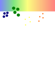
SDS gel during electrophoresis
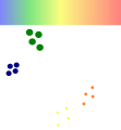
finished SDS gel


Mark and detect the proteins
Marking in the living cell ( in vivo )
Parameters such as the production of proteins in a certain period of time (protein synthesis rate) or the phosphorylation of proteins per unit of time are preferably determined by the incorporation of ( radioactive ) isotopes . For this purpose, the bacteria / cell culture is given a nutrient substrate with an unusual isotope, which is then incorporated into the proteins ( 35 S) or into the phosphate groups of phosphorylated ( 32/33 P) proteins. If the time span of the installation is chosen to be relatively short, the result is more likely to be a snapshot of what is happening in the cell; if the time span is longer, the cumulative image of many individual events can be captured. The protein extract of the marked cells is separated and the proportion of marked proteins is determined using autoradiography or mass spectrometric methods in a 2D pattern. A metabolic labeling can also be done with chemically modified amino acids which were equipped for subsequent labeling with fluorescent dyes with a biologically inert linker ( bioorthogonal chemistry ). In addition to permanent metabolic or isotope labeling, one of the methods explained below can also be used to determine the amount of protein that has accumulated.
Labeling prior to gel electrophoretic separation
Covalently binding fluorescent dyes are used especially to separate several samples on a 2D gel . To do this, a maximum of three different protein extracts are marked with a dye each, mixed and separated together on the same gel . Since the dyes can be detected separately from one another, the generation and differential comparison of three sample-specific protein patterns is possible ( Difference Gel Electrophoresis , DIGE). The mass influencing of the proteins by the bound dye is problematic. In addition, the dyes are relatively unstable and expensive.
Be used:
- Cy2; Cy3; Cy5
- Flashpro dyes
- G-Dyes
Marking or coloring after separation
The classic protein staining takes place after the electrophoretic separation. With the non-linear staining method, the quantity can only be determined approximately by adding proteins of known concentrations to the sample (e.g. a comigration standard ). Depending on the required specificity and sensitivity, the following are used:
Adsorbent dyes
- Coomassie brilliant blue - amount of protein / non-linear
- colloidal Coomassie brilliant blue - amount of protein / non-linear
- Silver staining - amount of protein / non-linear / highly sensitive
- negative zinc staining - amount of protein / not linear / not very sensitive
Fluorescent dyes
- SYPRO Ruby - amount of protein / linear / sensitive
- Krypton stain - amount of protein
- Flamingo staining - amount of protein / linear / highly sensitive
- DeepPurple staining - amount of protein
- Diamond ProQ - phosphorylated proteins (see also post-translational modification )
- Emerald ProQ - glycosylated proteins (see also post-translational modification )
By means of two-dimensional gel electrophoresis, far more than a thousand different protein species can often be detected in bacterial extracts after staining the proteins. Approximately 10,000 spots could be displayed in mouse embryos.
Analyze and interpret 2D gels
Digitize 2D gels
When digitizing 2D gels, the 2D patterns are broken down into pixels with different gray values. The resolution determines the accuracy in the x and y directions, the color depth the amount of gray levels that is available for mapping the amount of protein per pixel.
Imaging devices
- For all visible absorption dyes, higher quality white light scanners with high sample penetration capacity (transmitted light scans with an OD up to 4.0) are used. Without a corresponding calibration of the protein concentrations, dye signals and gray values, quantitative statements are only possible to a limited extent later. Camera-based systems are now also available for digitizing the gels with visible dyes. Color filters can u. U. help improve the utilization of the dynamic range.
- For the detection of fluorescent dyes, high-intensity laser scanners are mainly used, in which the wavelength of the excitation light and the filter optics for the emitted light of the fluorescent dyes can be flexibly adjusted. Here, too, the established systems are now being complemented by camera-based devices.
- So-called phosphor imagers are used to detect radioactive isotopes that have been built into proteins . Here a so-called imaging plate is exposed to the dried 2D gel for several hours. The released radioactive radiation is stored in the imaging plate and read out by the phosphor imager. The stored energy released from the imaging plate is translated linearly into gray levels of the digitized image (see autoradiogram above).
- Systems for the detection of the self-fluorescence of proteins under UV light by stimulating aromatic amino acids such as tyrosine , phenylalanine and tryptophan have so far not been able to establish themselves, but promise an interesting alternative, since no expensive fluorescent dyes have to be used. This would lead to massive time savings and, moreover, to a reduction in diffusion and other effects caused by dyeing and washing steps.
For more information on scanning gels, download a manual.
Single channel techniques
With the single channel techniques, a series of gels is usually acquired, which has been completely stained in the same way.
Multiplexing
With gel multiplexing, several images are generated independently of one another from the same gel. This can be possible under different circumstances:
- Different dyes or labels are used that represent different properties of the separated proteins.
- For example, the amount of protein by the flamingo dye and the protein phosphorylation by the ProQ-Diamond dye can be detected in the same gel. Both fluorescent dyes are detected separately by the scanner and saved in two gray-scale images. These can be combined to form a false color image using the appropriate imaging software.
- Another possibility is the combination of an autoradiogram (this shows the protein synthesis determined by a radioactive pulse marking ) and a protein quantity image (detection of the proteins with the silver color). In the false color image shown, the newly synthesized proteins are visible in red and the accumulated proteins in green.
- Combinations of Emerald ProQ images (coloring of proteins with sugar molecules as side chains), Diamond ProQ images (coloring of the phosphate residues on the protein) and SyproRuby (coloring of the amount of protein) in three color channels at the same time can detect phosphorylation and glycosylation of proteins at the same time .
- As an alternative to the independent detection of different protein properties using different specific dyes or labels, the DIGE can detect up to three samples simultaneously on one gel. For this purpose, the proteins of the three samples are covalently linked with the cyanine dyes Cy2, Cy3 and Cy5 separately before the electrophoretic run . Then the samples are combined and simultaneously separated on the same gel. The gels are detected with three lasers of different wavelengths and with appropriate detection optics.

Evaluate 2D protein samples
Classically, 2D gels are evaluated visually on the light or, in the case of fluorescent dyes, on the UV table. Due to the complexity of protein patterns, software-based analysis leads to more reliable results. For an overview of the current status of the procedure, see
Prepare gel images

In order to analyze gel images quantitatively, they should be freed from the inhomogeneous background typical of 2D gels and cleaned of artificial signals. The picture opposite shows, for example, the breakdown of a gel image into a background component, a component with artificial signals and the spot component used for further quantitative analysis.
Correct the position of the gel images
A problem that had not been resolved for a long time was the difficult positional reproducibility of the protein patterns in the 2D gel. One possible solution was to avoid independently produced gels.
- DIGE, with the simultaneous separation of up to three samples per gel, is a viable option, but with more than three samples it is confronted with the reproducibility problem again, since several gels have to be compared with each other. Since there is still no satisfactory experimental solution to avoid running differences, a solution had to be found at the software analysis level.
- In the first step of spot pattern matching, software is used to search for protein spots in the raw 2D gel image. With the help of the computer-generated spot information, an attempt is made to find partners belonging to an expression profile (spot matching). However, weaknesses in spot detection lead to spot assignment errors in these methods.
- The so-called image warping (image distortion or rectification) was introduced into 2D gel analysis in 2000. Automatic image warping processes use the entire pixel information present in the 2D gel images to calculate an image transformation that leads to the best possible positional correspondence between the gel images to be compared. Because of the positional correction, complex spot matching is no longer necessary, since spots belonging to one another are already at the same positions in the gels to be compared. Positionally transformed gels correspond, so to speak, to ideal gels that no longer show any system-related distortions.


Reference gels and proteome maps
For a scientifically sound interpretation of the 2D gels, the identity of the proteins behind the protein spots is determined. This can be done using various technologies such as Edman degradation or mass spectrometric methods such as MALDI-TOF mass spectrometry . While in the 1990s the protein spots were still cut out manually from the gels, robots have now found their way into laboratories to cut out the spots and for pipetting . With the help of robot technology, several hundred proteins can be identified almost overnight.

Due to the introduction of computer-aided positional correction of protein patterns, it became possible to easily transfer protein spot identifications from one gel to another without having to identify the spots again. A 2D gel, which shows the protein extract from a cell culture, only shows a subset of all possible cell proteins. Only gel series from cell cultures grown under different growth conditions can show the total number of all possible proteins. The reason is the differential gene expression, which only allows the production of currently important proteins and prevents the synthesis of proteins that are currently not needed. For the construction of comprehensive proteome maps that contain a large number of all possible proteins, individual gel images are positionally aligned and combined into a composite image using image fusion algorithms. The composite image is combined with the data from the protein identifications and can then be used as a reference for the interpretation of further experiments.
Detect and quantify protein spots
For a (semi) quantitative evaluation of the 2D gels, the total absorption (absorption dyes), the total radio signal (radioactively labeled proteins, autoradiogram) or the total fluorescence signal (fluorescence dyes) of a protein spot is determined over all image points. In the first spot detection step the coordinates of the protein spots and in the second the corresponding spot shapes are determined. The spot outlines can be determined close to the pixel information or using mathematical models. Disturbance information, such as B. Background, artifacts and image noise (see image preparation) are excluded before quantification. If necessary, the gray values of the pixels are corrected with device and dye-dependent calibration curves and then added up to a raw quantity within the spot outlines found. The raw quantities are standardized and assigned to the corresponding protein spots. Since, as already mentioned above, spot detection does not provide absolutely reproducible results from gel to gel, errors can occur when assigning proteins to their expression profiles, which sometimes cannot be resolved with the established methods. That is why a new method for spot matching was introduced in 2003. This is based on the definition of a spot consensus from all the gels to be analyzed in an experiment on the basis of a composite image. Because the composite image contains all the spot information from the overall experiment, the spot consensus can be used at least on all of those gels for spot quantification from which the composite image was created. Spot allocation errors can be ruled out using this method.
Visualize data

The application of proteome maps and composite image-based spot detection opens up completely new possibilities for the visualization of protein spots that were conspicuous in the analyzed experiment. For example, the adjacent picture shows a summary of four different protein synthesis patterns. The colors indicate under which environmental conditions which protein is increased at least twice in its synthesis. With the help of such color coding, it is now possible for the first time to visualize regulatory data in addition to the positional data of the protein spots. Biomarkers can thus be identified quickly and reliably.
Modifications of the classic 2D gels
Separation system chemistry
While the classic 2D gels separate proteins in the 1st dimension according to their isoelectric point and in the 2nd dimension according to their size, in practice other separation methods are also combined in two dimensions for proteins. Examples are:
- the combination of two different detergents in the two dimensions.
- "Blue native" electrophoresis in the first dimension and SDS gel electrophoresis in the second dimension
- Separation under oxic (first dimension) and reducing (second dimension) conditions
Geometry of the separation system
- The separation of the proteins according to the isoelectric point usually takes place horizontally in gel strips or vertically in long cylindrical gels which were produced in tubes.
- The second separation is carried out in sandwiches made of 2 glass plates with a gel in between. These glass-plate-gel sandwiches are hung vertically in stacks of 1, 2, 5, 6, 10 or 12 gels in tanks that contain the running buffer and are cooled by a cryostat during the separation.
- Commercially available ready-made gels are often used in horizontal systems on cooling plates for protein separation. Depending on the dimensions, 1 to 4 or more gels can also be processed in parallel here.
- For example, new is the use of radially symmetrical devices, which cut the IEF strips in the 2nd dimension from inside to outside into circular gel discs. Depending on the length of the IEF gels, up to six batches can be separated on a gel disk. It is intended to stack several such discs in one system. Since the electric field runs from the inside out, the field lines move away from each other the further the proteins migrate outwards. This prevents small proteins in particular from moving too quickly and leaving the gel again before the large, slow proteins have been sufficiently separated.
- In order to push the parallel separation of samples to the extreme, geometrically three-dimensional gels were developed (www.3d-gel.com), which can simultaneously separate 36 IEF gels on a gel block. As with traditional DNA sequencers, the proteins are detected in a gel level illuminated by a laser. Emission light is detected during the passage of the proteins marked with fluorescent dyes before separation. The measured signals are saved. The gel images are reconstructed from the stored image stack by software and conventionally evaluated.
Benefits of 2D gels
- The visualization of the protein pattern allows a qualitative assessment of the results without further analysis
- Different protein species of one and the same primary protein are immediately accessible to analysis without any preconditions, provided they have sufficiently different masses and / or isoelectric points.
- The method is highly parallelizable. Depending on the 2D gel electrophoresis device, up to 12 gels can be produced simultaneously.
- The necessary investments are relatively manageable compared to gel-free methods.
- The separated gel can be stored for a long time and further processed later.
- In the meantime, analysis systems are available that can compare a large number of samples in a short time and without advanced computer knowledge.
Problems of 2D gel electrophoresis
Like any other technique in protein biochemistry, 2D gel electrophoresis has some problems:
- Due to the separation system in the aqueous environment, mainly hydrophilic proteins with a GRAVY index smaller and close to 1 are separated.
- Despite the great separation potential, strong protein spots can certainly overlap or completely mask weak spots. These masked spots are no longer accessible to a quantitative analysis.
- Due to the limited sensitivity of Coomassie or silver staining on the one hand and the limited capacity of a 2D gel on the other hand, the sensitivity of the method is limited. 2D gels can therefore only represent the most abundant proteins of a cell (e.g. cytoskeletal proteins, metabolic enzymes), while low-expression proteins such as (e.g. transcription factors) mostly remain undetected.
- Too large total protein amounts lead to discrimination phenomena when different protein species enter the IEF gel and thus to a distortion of the protein species ratios.
- The more basic a protein, the more difficult it is to separate it. Furthermore, the 2D gel system is mainly efficient in a pH range of 3 to 10.
- The 2D gels have a limited reproducibility , which depends on the manufacturer and user as well as on the gel components and electrophoresis buffer compositions used .
See also
- Agarose gel electrophoresis
- Discontinuous electrophoresis
- Gel electrophoresis
- Native PAGE
- Polyacrylamide gel electrophoresis
Individual evidence
- ^ PH O'Farrell: High resolution two-dimensional electrophoresis of proteins. In: J. Biol. Chem. 250, 1975, pp. 4007-4021. PMID 236308 ; PDF (free full text access)
- ^ J. Klose: Protein mapping by combined isoelectric focusing and electrophoresis in mouse tissues. A novel approach to testing for induced point mutations in mammals. In: Human Genetics. 26, 1975, pp. 231-243. PMID 1093965
- ↑ Thierry Rabilloud: Two-Dimensional Electrophoresis Protocols. Humana Springer, 2009, ISBN 978-1-58829-937-6 . Chapter 2: Solubilization of proteins in 2DE: An outline (PDF)
- ↑ A. Görg, C. Obermaier, G. Boguth, A. Harder, B. Scheibe, R. Wildgruber, W. Weiss: The current state of two-dimensional electrophoresis with immobilized pH gradients. In: Electrophoresis. 21 (6), Apr 2000, pp. 1037-1053. PMID 10786879
- ^ R. Westermeier, W. Postel, J. Weser, A. Görg: High-resolution two-dimensional electrophoresis with isoelectric focusing in immobilized pH gradients. In: J Biochem Biophys Methods. 8 (4), Dec 1983, pp. 321-330. PMID 6663005
- ↑ Joachim Hartinger, Katinka Stenius, Dagmar Högemann, Reinhard Jahn : 16-BAC / SDS-PAGE: a two-dimensional gel electrophoresis system suitable for the separation of integral membrane proteins. In: Analytical Biochemistry. Volume 240, No. 1, 1996, pp. 126-133. PMID 8811889 , doi: 10.1006 / abio.1996.0339 .
- ^ RP Zahedi, J. Moebius, A. Sickmann: Two-dimensional BAC / SDS-PAGE for membrane proteomics. In: Sub-cellular biochemistry. Volume 43, 2007, pp. 13-20. PMID 17953388 .
- ↑ decodon.com: Imaging Guide (English)
- ↑ M. Berth, FM Moser, M. Kolbe et al: The state of the art in the analysis of two-dimensional gel electrophoresis images. In: Appl Microbiol Biotechnol. 76 (6), 2007, pp. 1223–1243, PMC 2279157 (free full text)
- ↑ JE Bandow, JD Baker, M. Berth, C. Painter, OJ Sepulveda, KA Clark, I. Kilty, RA VanBogelen: Improved image analysis workflow for 2-D gels enables large-scale 2-D gel-based proteomics studies– COPD biomarker discovery study. In: Proteomics. Volume 8, Number 15, August 2008, pp. 3030-3041, ISSN 1615-9861 . doi: 10.1002 / pmic.200701184 . PMID 18618493 .
literature
- RC Allen, B. Budowle: Gel Electrophoresis of Proteins and Nucleic Acids: Selected Techniques . Walter de Gruyter, 1994, ISBN 3-11-013896-4 .
- JE Bandow, JD Baker, M. Berth, C. Painter, OJ Sepulveda, KA Clark, I. Kilty, RA VanBogelen: Improved image analysis workflow for 2-D gels enables large-scale 2-D gel-based proteomics studies – COPD biomarker discovery study. In: Proteomics. Volume 8, Number 15, August 2008, pp. 3030-3041, ISSN 1615-9861 . doi: 10.1002 / pmic.200701184 . PMID 18618493 .
- M. Berth, FM Moser, M. Kolbe et al .: The state of the art in the analysis of two-dimensional gel electrophoresis images. In: Appl Microbiol Biotechnol. 76 (6), 2007, pp. 1223–1243, PMC 2279157 (free full text)
- MH Hamdan, PG Righetti: Proteomics Today: Protein Assessment and Biomarkers Using Mass Spectrometry, 2D Electrophoresis, and Microarray Technology . Wiley-Interscience, 2005, ISBN 0-471-64817-5 .
- T. Rabilloud (Ed.): Proteome Research: Two-Dimensional Gel Electrophoresis and Identification Methods (Principles and Practice) . Springer, 1999, ISBN 3-540-65792-4 .
- Sabine Schmitz: The experimenter: cell culture . 1st edition. Spectrum Academic Publishing House, 2007, ISBN 978-3-8274-1564-6 .