Pressure ulcer
| Classification according to ICD-10 | |
|---|---|
| L89 | Decubitus ulcer |
| ICD-10 online (WHO version 2019) | |
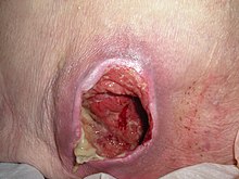
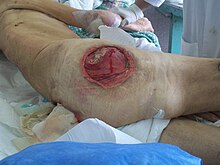
A pressure ulcer (from Latin decumbere , `` to lie down '' ) is local damage to the skin and the underlying tissue due to prolonged pressure that disrupts the blood circulation in the skin. Other names are decubitus ulcer , pressure ulcer , decubitus ulcer (or each - ulcer ).
Pressure ulcers are chronic wounds that occur primarily in patients with reduced mobility , especially when they are bedridden or dependent on a wheelchair . They can indicate care errors and are therefore also rated as an indicator of care quality .
Open pressure ulcers can become the gateway for pathogens that not only cause local infections. A decubital lesion can therefore lead to serious and possibly fatal secondary diseases such as pneumonia or blood poisoning (sepsis) due to the spread of pus through the bloodstream .
Pressure ulcer classification
The classification of a pressure ulcer can - depending on the classification used - be very different; therefore, it should be clear from the care documentation which system was used. Some classifications have grading that can only indicate worsening of a disease. Most classifications divide the pressure ulcer into stages so that an improvement in the disease can also be shown.
Phillips finger test
With the Phillips finger test , a first-degree pressure ulcer can be differentiated from reddening of the skin from another cause: Reddening can be pushed away if a white outline appears when you press your finger on a reddened skin region and the fingerprint appears white for a brief moment when you let go. This means that there is no pressure ulcer; it is then possibly an allergic or inflammatory rash. If, on the other hand, the reddening persists after letting go, there is pressure-related skin damage.
By degree (Shea, 1975)
Pressure ulcer ulcers are divided into four grades according to JD Shea.
- Grade 1: circumscribed reddening of the skin that cannot be pushed away on intact skin. Further clinical signs can be edema formation, induration and local overheating.
- Grade 2: partial loss of skin; The epidermis and parts of the corium are damaged. The pressure damage is superficial and can clinically appear as a blister, abrasion, or shallow ulcer.
- Grade 3: Loss of all skin layers including damage or necrosis of the subcutaneous tissue that may extend up to, but not below, the underlying fascia . The pressure ulcer shows up clinically as a deep, open ulcer.
- Grade 4: Loss of all skin layers with extensive destruction, tissue necrosis or damage to muscles , bones or supporting structures such as tendons or joint capsules , with or without loss of all skin layers.
A pressure ulcer grade cannot improve; Even after healing, the damage remains, which is filled in by scar tissue. A grade 3 pressure ulcer that has healed is referred to as a grade 3 pressure ulcer - closed . Further classifications that use grading are the pressure ulcer classification according to Campbell and the severity classification according to Daniel.
Degree / stage (Seiler, 1979)
The Swiss physician Walter O. Seiler differentiates four degrees of pressure ulcer with regard to the depth of infiltration and three stages of the wound condition:
Infiltration depth:
- Grade 1: redness
- Grade 2: redness, blistering, defects in the top layers of the skin
- Grade 3: Defect of the subcutaneous tissue and underlying tissue (muscles, tendons, ligaments) up to the periosteum (periosteum)
- Grade 4: like Grade 3, with inflammation of the bone (osteomyelitis)
Wound condition:
- Stage A: clean wound, granulation tissue present, no necrosis
- Stage B: necrotic wound, no infiltration of the surrounding area
- Stage C: like B, but infiltration of the wound environment, wound infection
A division into stages describes the course of a disease based on the appearance and can also show an improvement in the pressure ulcer. Other classifications that use staging are the division of pressure ulcers according to Torrance (five stages), Surrey (four stages) and Stirling (twelve stages).
Category (EPUAP and NPUAP, 2009 and 2014)
The traditional division by severity or stage, e.g. B. according to Shea or Seiler, implies a progressive development of the disease: The pressure ulcer progresses from degree to degree, or changes its stage. Since a pressure ulcer does not automatically move from one degree or stage to the next, a classification into categories was developed. The European Pressure Ulcer Advisory Panel (EPUAP) developed a classification in 2009 that comprises four categories.
- Category I: Reddening that can not be pushed away on intact skin
- Category II: Superficial pressure ulcer with partial loss of the skin with damaged epidermis and / or dermis
- Category III: Loss of skin layers and damage (or necrosis) to subcutaneous tissue
- Category IV: Complete loss of skin or tissue
Nevertheless, the term “degree” or “decubitus degree” is still in use, among other things because the ICD coding also uses this term.
In 2014 two new classifications were added:
- Cannot be assigned to any category / stage: Depth unknown : This is a complete loss of tissue in which the wound bed is covered by plaque or scabs , so that the actual depth and therefore the category can only be determined after the plaque has been removed. Intact, dry, adherent eschar without erythema and fluid on the heels should not be removed as it acts as "natural (biological) protection for the body".
- Suspected deep tissue damage: Depth unknown : This is an area of discolored but intact skin or a blood-filled blister that has formed due to damage to the underlying soft tissue from pressure or shear forces. Such tissue damage can be difficult to detect in people with dark skin. A thin blister or scab may appear over a dark wound bed. The wound can continue to change underneath, and even with optimal treatment it can progress rapidly, exposing additional layers of tissue.
Graduation for ICD coding
The degrees of the ICD coding for the "decubitus ulcer and pressure zone" (L89.-) are based on the Shea classification:
- Decubitus 1st degree: pressure zone with reddening that cannot be pushed away with intact skin
- 2nd degree pressure ulcer : pressure ulcer with abrasion, blister, (partial) loss of skin
- 3rd degree pressure ulcer : pressure ulcer with loss of all skin layers
- Grade 4 bedsores: Pressure ulcer with necrosis of muscles, bones, or supporting structures
- Decubitus, grade unspecified: Pressure ulcer without indication of a grade
Emergence
The term pressure ulcer refers to the local pressure load as a decisive development factor. The load can be evaluated using the formula: pressure × time. If external pressure on the vessels exceeds the capillary pressure of the vessels, trophic disturbances occur. This limit value is often referred to in the literature as the physiological capillary pressure. As a rule, the dead weight of the respective (immobile) body part is sufficient to exceed the capillary pressure. Various studies to determine it (by E. M. Landis, K.-D. Neander, Yamada and Burton, among others) yielded values between 32 and 70 mmHg for an interruption of the blood supply.
If a pressure load above the capillary pressure threshold lasts longer, the cells are insufficiently supplied with oxygen ( hypoxia ) and nutrients . The oxygen partial pressure sinks to 0 mmHg ( ischemia ) and toxic (acidic) metabolic products accumulate. The tissue becomes necrotic and nerve cells suffer irreversible damage. In healthy people, the increase in acidic metabolic products triggers a reflex to relocate and thus relieve stressed areas of the skin before permanent damage occurs. In elderly and sick people, these reflexes are often only limited or nonexistent, and the necessary relief of the tissue is not provided. As a result of the acidification of the tissue, the body expands the vessels ( vascular dilatation ), so that these areas of the skin are supplied with more blood - the result is reddening of the skin, even with pressure - decubitus grade I. Areas with low soft tissue coverage ( muscles or fatty tissue ) and outwardly curved (convex) bony abutments are particularly at risk : the sacrum , the spinous processes of the spine, the heels , the rolling hills of the thigh bones and the ankles . The contact pressure here hits small areas and is therefore higher.
Risk factors
The development of a pressure ulcer must be seen as a multifactorial event as a result of intrinsic and extrinsic risk factors . The intrinsic factors lie “in the patient himself” (including reduced mobility , old age / old age , dehydration , underweight , infections , diabetes mellitus , urinary or fecal incontinence , sensory disorders ). The extrinsic factors are determined by the patient's environment. In the best case, they can be positively influenced by mobilization , suitable aids and correct repositioning (see also pressure ulcer mattress ) as well as consistently planned care of the person concerned.
Other extrinsic factors that favor the development of a pressure ulcer are:
- Shear forces lead to twisting of the blood vessels; trophic disorders are the result. Especially in older people, in whom a decrease in the water content of the skin leads to a loss of elasticity, shear forces can also lead to a separation of entire skin layers from one another;
- Friction leads to injuries on the skin surface;
- Temperature in the non-physiological area caused by fever or from outside (hot water bottle, heating pad, cooling pack) can affect the blood circulation
In addition, the following factors favor the development of a pressure ulcer or have a detrimental effect on healing:
- Long-term moisture (for example, a damp environment in protective trousers, heavy sweating ) softens the upper skin layer ( maceration ), making it more prone to injury.
- Obesity , due to a lack of exercise or limited mobility; higher pressure due to more weight and delayed wound healing in the presence of pressure ulcers
- Cachexia , due to a lack of padding due to the lack of subcutaneous fat, especially on protruding bones
- Paraplegia , as possible pressure points (especially on the buttocks) are not noticed in time due to a lack of sensitivity
- Diseases, syndromes , drugs or procedures such as heart failure , diabetes mellitus, immune deficiency , broken bones , coma , contractures , anesthesia , neurological disorders , peripheral arterial disease , mental illness, strong painkillers , psychotropic drugs , sedatives and sleeping pills
Measurability of the risk
In professional nursing and nursing science , various assessment instruments have been developed within the nursing assessment . These scoring systems have proven to be beneficial for assessing the pressure ulcer risk based on intrinsic factors and making it measurable. For this purpose, points are awarded for different categories (for example mental state, physical state, flexibility, ...). Patients below a certain number of points are then considered to be at risk.
Doreen Norton developed the Norton scale back in the 1950s . The initially inadequate and sometimes vaguely formulated scale was expanded in 1985 to the modified Norton scale . In addition to the Medley and Waterlow scales , which tend to be based on specific patient ideas or care areas, the Braden scale is primarily used today in the USA , which introduces the categories “ friction and shear forces ” and “sensory sensitivity”.
The first national expert standard pressure ulcer prophylaxis in nursing in Germany recommended the use of standardized risk scales in 2004, but deleted this recommendation in the first update in 2010 because none of the scales took into account all possible risk factors and a clear effect of the benefit was not proven. An experienced nurse can use their specialist expertise to assess the overall condition of the patient and thus their individual pressure ulcer risk, even without assessment instruments, and derive the necessary measures from them. It was found, however, that risk scales can be helpful for nurses with little professional experience in making an assessment.
treatment
The treatment consists primarily in the complete pressure relief of the affected part of the body through regular changes in position , the frequency of which must be determined individually. As with decubitus prophylaxis, movement, positioning and transfer techniques are used that are gentle on the skin and tissue. In addition, the decubitus is treated according to the principles of wound treatment . If pressure relief is not possible or only inadequately possible, wound healing is delayed or excluded. Aids for pressure reduction and skin care support wound healing, but cannot replace complete pressure relief or (instructions for) self-movement.
prevention
All measures to prevent pressure ulcers are called pressure ulcer prophylaxis . For professional care in Germany, the national expert standard for pressure ulcer prophylaxis applies .
For professional prophylaxis , the individual degree of risk is first determined ("initial screening") and corresponding risk factors are identified and, if possible, eliminated. The patient at risk and the caregivers (including caring relatives) are included in the information, planning and implementation of the necessary measures; in addition, the patient or his or her legal representative must agree to the procedure.
The decisive practical measure is the pressure relief of endangered parts of the body by regular mobilization , changes of position or positioning of the patient with restricted mobility while avoiding shear forces and with the targeted use of aids. Appropriate skin care and appropriate nutrition and hydration complement an effective prophylaxis, but cannot replace pressure relief.
The psychological situation of those affected is not to be neglected. You are to be stimulated by a holistic care concept for your own motivation, nutrition, mobilization, prophylaxis etc. Patients and their relatives receive the necessary information and practical guidance from the relevant nursing staff .
Individual exercise plan
In order to plan individual mobilization measures, the actual condition, the movement resources and movement impairments of the patient are first recorded, which result from the care history and are observed when carrying out care measures. This includes a risk assessment for contractures and falls, information on the condition of the skin, the nutritional situation and any continence problems, the resources relevant for effective prophylaxis (e.g. enjoyment of movement, adherence), a description of existing skills ("sits on the edge of the bed without help "," turns independently when lying down on its side ") and the acceptance or rejection of certain positions, positions and other measures. Certain situations or times of the day can lead to the acceptance or rejection of measures, for example the desire for an undisturbed night's sleep. Possible active or passive movement exercises as well as standing and balance training are included in the care , if necessary with the help of physiotherapy . The care plan contains a comprehensible description of the transfer and lists the aids required (e.g. slide board, sliding mat, lifter), shoes or non-slip socks. Several caregivers are required for certain measures, depending on the patient's body weight and their ability to cooperate.
Avoidance of pressure points
- Promotion of self-movement, early mobilization
- Exposure or padding of predilection sites such as protruding bone points
- Smoothing out wrinkles in clothing or documents
- Keep contact surfaces of inlet and outlet lines (e.g. urinary catheter tube, infusion lines) and small parts free (e.g. sealing plugs, cannula sleeves)
- Avoid tight clothing and shoes
- Padding of tight-fitting orthoses and prosthetic arms and legs
Tools
Aids for pressure ulcer prophylaxis should have proven pressure distributing and relieving properties. These include alternating pressure or soft mattresses and micro-stimulation systems . In addition, numerous other aids are offered, including pillows of various shapes with different fillings, sliding mats, synthetic skins or natural sheepskins made of virgin wool , which, in addition to relieving pressure, should reduce the shear forces on the skin and wick away moisture well. However, the 2010 national expert standard for pressure ulcer prophylaxis does not assess the study situation as sufficient to recommend the use of special sheepskins as a suitable aid. In contrast, the international guideline gives a restricted recommendation for natural sheepskins. Aids such as sliding boards or sliding mats avoid shear forces when changing positions. A lifter is helpful when transferring (moving) immobile and heavy patients.
In order to find suitable aids, it must be considered beforehand which care and therapy goals are to be achieved for the patient, and which of these are to be achieved first: For a patient with regular self-movement, a super soft position is unsuitable, for a weakened, dying patient, for example Pain reduction can be more important than promoting one's own movement. The general and nutritional condition plays a role, as well as the presence of breathing problems, contractures , paralysis, spasticity and perception disorders. In addition, the aid should be easy to use and hygienic to prepare.
Skin care
The aim of skin care is to keep the skin of endangered body regions clean, dry and supple. Suitable incontinence aids are particularly important for patients with urinary or stool incontinence . Endangered parts of the body that are often exposed to moisture can be protected from moisture with special products such as barrier cream or transparent skin protection film. Excretions should be removed immediately. A pH-neutral skin cleanser is recommended for cleaning . Strong rubbing should be avoided; massages are also not recommended.
nutrition
Both malnutrition and underweight are associated with the incidence of pressure ulcers. However, a balanced, protein and vitamin-rich diet and fluid intake alone will not help prevent pressure sores. But it lowers the risk of dehydration , malnutrition and malnutrition . Food supplements are not necessary with a balanced mixed diet. If the patient has difficulty eating or intolerance, they can be used to compensate for deficiencies.
literature
- Jennifer Anders et al .: Pressure ulcers - Pathophysiology and primary prevention. In: Deutsches Ärzteblatt. Volume 107, 2010, pp. 371-382.
- German network for quality development in nursing (DNQP, ed.): Expert standard pressure ulcer prophylaxis in nursing. 2. Update 2017 including commentary and literature review. Series of publications by the German Network for Quality Development in Nursing. Osnabrück ISBN 978-3-00-009033-2
- Waltraud Steigele: Movement, mobilization and positioning in nursing. Practical tips for movement exercises and changing positions. Springer-Verlag Berlin Heidelberg, 2016 ISBN 978-3-662-47270-5
Web links
- Abridged version of the pressure ulcer guideline of the National Pressure Ulcer Advisory Panel, European Pressure Ulcer Advisory Panel and Pan Pacific Pressure Injury Alliance (2014)
- PflegeWiki.de - decubitus
- Expert standard pressure ulcer prophylaxis in nursing, second update 2017 (content and 1st chapter)
- Decubitus ulcer - information at Gesundheitsinformation.de (online offer of the Institute for Quality and Efficiency in Healthcare )
Individual evidence
- ↑ http://dictionary.reference.com/search?q=decubitus (English)
- ↑ a b c Eva-Maria Panfil, Gerhard Schröder (Ed.): Care of people with chronic wounds. 3. Edition. Verlag Hans Huber, Bern 2013, ISBN 978-3-456-85194-5 , p. 201.
- ^ Jenny Phillips: Pressure Sores (= Access to Clinical Education ). 1st edition. Churchill Livingston, New York 1997, ISBN 978-0-443-05532-4 .
- ↑ p.17 Project group 32 of the MDS eV: Policy statement decubitus. On mds-ev.de, June 2001 ; accessed on December 24, 2018
- ↑ J. Darrell Shea: Pressure sores: classification and management. In: Clinical Orthopedics and Related Research 112, pp. 89-100.
- ↑ Dirk J. Schaefer: Decubitus diagnosis and classification. 56th Basel Decubitus Seminar 2014 ; accessed on December 17, 2018
- ↑ a b I care care. Georg Thieme Verlag, Stuttgart 2015, ISBN 978-3-13-165651-3 , p. 401.
- ^ Kerstin Protz: Modern wound care , Urban & Fischer, Munich, 7th edition 2014, p. 76.
- ↑ ICD coding of Dekubutalgeschwürs
- ^ Angelika Bischoff: Obesity - serious problems in the case of an operation. In: Deutsches Ärzteblatt 2007; 104 (22): A-1560 / B-1382 / C-1322
- ↑ Kerstin Protz: Modern wound care. Practical knowledge, standards and documentation. 6th edition, Urban & Fischer, Munich 2011, p. 73. ISBN 978-3-437-27883-9
- ↑ Pressure ulcer (decubitus) - treatment on informedhealth.org ; accessed on December 20, 2018
- ↑ Kerstin Protz: Modern wound care. Practical knowledge, standards and documentation. 6th edition, Urban & Fischer, Munich 2011, p. 80. ISBN 978-3-437-27883-9
- ^ German network for quality development in nursing: Expert standard pressure ulcer prophylaxis in nursing; 2nd update 2017. (PDF) DNQP, June 2017, accessed on December 17, 2018 .
- ↑ National Pressure Ulcer Advisory Panel, European Pressure Ulcer Advisory Panel and Pan Pacific Pressure Injury Alliance. Prevention and Treatment of Pressure Ulcers: Quick Reference Guide. Emily Haesler (Ed.). Cambridge Media: Osborne Park, Australia 2014; German translation Prevention and treatment of pressure ulcers: short version of the guideline. P.32 ; accessed on December 18, 2018
- ↑ Elke Kuno: Skin protection for incontinence: Sour keeps you healthy. On bibliomed-pflege.de from October 28, 2016 ; accessed on December 18, 2018
- ↑ Short version of the guideline NPUAP, EPUAP, PPPIA 2014; P.21 ; accessed on December 18, 2018
- ↑ S. Eberhardt, A. Heinemann, W. Kulp, W. Greiner, C. Leffmann, M. Leutenegger, J. Anders, F. Pröfener, U. Balmaceda, O. Cordes, U. Zimmermann, J.-M. Graf von der Schulenburg: Decubitus prophylaxis and therapy. Editor: German Agency for Health Technology Assessment of the German Institute for Medical Documentation and Information, Cologne 2005, p. 133 ; accessed on December 18, 2018


