Dendrite (biology)
| Parent |
| Neuron / cell |
| Subordinate |
|
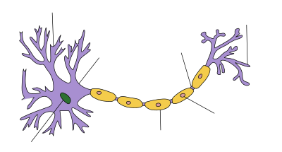
Thorn process terminus shaft branching dendr. cytoplasm |
| Gene Ontology |
|---|
| QuickGO |
| Structure of a nerve cell |
|---|
Dendrites ( old Gr. Δgrνδρον dendron 'tree' or dendrites 'belonging to the tree') are called in biology cell extensions of nerve cells that arise from the cell body and are primarily used to absorb stimuli.
A nerve cell typically consists of three parts: the cell body, called soma or perikaryon , and cell processes, the dendrites on the one hand and the neurite - the axon in the glial envelope - on the other. There are also specialized neurons that have no axon (e.g. the amacrine cells of the retina) or that have no dendrites (e.g. the rods and cones of the retina) or those in which the cell body is no longer between the dendritic stem and axon and the processes merge into one another ( pseudounipolar nerve cells like the sensitive dorsal root ganglion cells ).
Dendrites as parts of a cell should not be confused with dendritic cells of the immune system.
Dendrite growth
Despite the importance of dendrites for neurons, little is known about how dendrites grow and how they orient and branch in vivo . One assumption about these formation processes is the synaptotrophic hypothesis , according to which the formation of synapses plays a special role in the growth of dendrites.
Otherwise, the dendrite growth is similar to that of neurites explains, through so-called growth cones (English growth cones ). According to this, both neurites and dendrites have conical or bulb-like swellings at their tips, which are in an exploratory interaction with the immediate surroundings and decide on progress or continuation - the extent and direction of the outgrowth of processes - and essentially determine the further behavior of the neurons. Cell culture techniques with time-lapse recordings can give a clear view of how nerve cell processes sprout out searching their surroundings. In the body there are different signals and different process paths that can be used to regulate the start, direction and speed as well as pauses in dendrite growth.
Most of the growth of dendrites in the human brain occurs during late embryonic and early childhood brain development. In this phase, dendrites with a total length of many hundreds of kilometers grow out of the 100 billion nerve cells in our brain. The enzyme Nedd4-1 is considered to be an important protein for the growth of the cell skeleton during dendrite development and is said to be essential for normal dendrite growth .
Neurites or axons and dendrites differ in their growth and growth phases. From a cellular point of view, roughly speaking, a stabilized supporting skeleton made of microtubules is required for the growth of the process to advance the tip of the growth. But then for the back and forth-playing growth in this region - in an unstable equilibrium - build-up and breakdown processes are needed, with which individual actin molecules (spherical, globular G-actin) form chains (thread-like, filamentary F- Actin) strung together - and can disintegrate again. No growth is possible with unstable microtubules and / or stable actin filaments . In the early phases of development, the dendrite growth can be temporarily stopped via inst / stabilization on the one hand, in favor of the growth in length of the neurite on the other. In principle, these relationships also apply later, for example during regeneration processes after lesions.
Anatomy of dendrites
shape

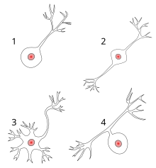
1 unipolar nerve cell
2 bipolar nerve cell
3 multipolar nerve cell
4 pseudounipolar nerve cell
The variety of forms and functions of neurons is essentially determined by the different characteristics of the dendrites. The figure shows the morphological differentiation of nerve cells, which u. a. then it is determined whether a nerve cell has no, one or more dendrites. Some neurons have real dendrite trees , in others the relationship between soma surface and dendrite surface is more balanced. Finally, there are neurons that do not have dendrites. Following this morphological classification, one can say that dendrites only occur in bipolar nerve cells and multipolar nerve cells . In pseudounipolar nerve cells, the distal end of the peripheral process is typically dendritic in character.
The number and shape of the dendrites contribute significantly to the enlargement of the receptive surface of the nerve cells. It has been estimated that up to 200,000 axons terminate at the dendrites of a single Purkinje cell . As a rule, the dendrites are branched, branched processes of the perikaryon .
Cell components

The structure of the dendrite is closer to the cell body than the neurite. In some respects, dendrites and perikaryon can even be understood as a functional unit and are also referred to as a somatodendritic compartment . The composition of the dendritic cytoplasm essentially corresponds to that of the perikaryon. It is therefore impossible to draw a sharp line between the parts of the nerve cell.
Knowledge of the cytoplasm , organelles and cytoskeleton allows a well-founded approach to differentiating the processes (axon / dendrites).
The following morphological peculiarities can be found:
- In contrast to the axon, dendrites are unmyelinated .
- In the larger Stammdendriten to find similar organelles as in the soma. Especially in the broad-based origin (close to the perikaryon) z. Sometimes even Nissl clods (rough endoplasmic reticulum ) can be found. In addition to the smooth and rough endoplasmic reticulum, there are free ribosomes , microfilaments (actin) and also bundles of parallel microtubules . However, their functional orientation is not uniform (as with the axon), but their polarity is variable, i.e. H. its plus end can point either to the periphery or to the perikaryon.
While fibrils and Nissl clods can still be seen under a light microscope , the other components are only visible under an electron microscope . - With each branching, the diameter of the dendrites becomes smaller. Mitochondria are absent in very thin dendrites . The end sections of the dendrites contain few organelles, and the cytoskeleton is also poorly developed.
- Compared to the axons (which in humans can sometimes be over 1 m long), dendrites are very small and only reach lengths of a few hundred micrometers (µm). Neurites that grow into the periphery can, however, reach a length of 1 to 1.20 m with a diameter of only 2–16 µm.
Distinctions of dendrites

Various distinguishing features of dendrites can be found in the literature.
If you look at pyramidal cells (a fairly large nerve cell), two types of dendrites can be distinguished: apical dendrites and basal dendrites . Both arise at the top of the pyramidal cells, but apical dendrites are longer than basal dendrites. The apical dendrites point in the opposite direction to the axon and extend transversely vertically through the layers of the cerebral cortex . Both apical and basal dendrites have thorns . While there are many basal dendrites, only a long, strong apical dendrite rises to the surface of the cortex .
Sometimes the apical dendrites are divided into distal and proximal dendrites . The distal apical dendrites are longer and project in the opposite direction from the axon. Because of their length, they form non-local synapses that are far away from the nerve cell. Proximal apical dendrites are shorter and receive impulses from nearby neurons, such as interneurons .
Furthermore, one can differentiate between dendrites according to whether they have dendritic thorns or not. Accordingly, one speaks of smooth ("smooth dendrites") or thorny ("spiny dendrites") dendrites. With smooth dendrites, the nerve impulse is picked up directly. In the case of thorny dendrites, both the dendrite stem and the thorns absorb the impulse.
As a rule, the thorny dendrites receive excitatory signals, while inhibitory synapses are more likely to be found on smooth dendrites (sections).
Dendritic thorns
The small thorn- like projections on the surfaces of branched dendritic trees are called dendritic thorns (English spines , Latin spinula dendritica or gemmula dendritica ) or thorny processes. Most of the synaptic contacts are often located here.
As a rule, a thorn process receives input from exactly one synapse of an axon. These fine processes (on a dendrite as nerve cell process) support the afferent transmission of electrical signals to the cell body of the neuron. The thorns can take different shapes, clearly formed often have a bulbous head and a thin neck, which connects the head with the dendritic trunk. The dendrites of a single neuron can have hundreds or thousands of thorns. In addition to their function as a postsynaptic region (postsynapse) - some with a thorn apparatus as a calcium store - and the ability to increase synaptic transmission ( long-term potentiation LTP), dendrites can also serve to increase the possible number of contacts between neurons.
Thorn processes represent a kind of sub-compartmentalization of the dendritic membrane. The possible fine-tuning of the individual thorn process through its special ionic environment or its specific cAMP level can be important for the selectivity and storage of information.
Functions
The glial cells , a kind of supporting tissue, take over the largest part of the supply of the neurons . But the dendrites are also involved in nourishing the nerve cells. Their main task, however, is to receive stimuli or signals, mostly from other nerve cells, and to forward the impulses formed as a result to the perikaryon (nerve cell body) ( afferent or cellulipetal ) - in contrast to the neurite or the axon , via the signals from this neuron on the axon hill beginning on ( efferent ) and directed to other cells.
Signal recording
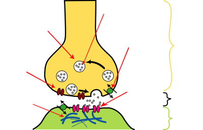
| Construction of a chemical synapse |
|---|
A nerve cell can be a sensory cell - such as olfactory cells ( olfactory receptors ) or visual cells ( photoreceptors ) - or it can receive signals from upstream cells - for example from other nerve cells, in that neurotransmitters dock with specific receptors in the postsynaptic membrane regions of this nerve cell. These postsynapses are usually not located in the area of the axon, axon mound or soma (body) of the nerve cell, but on its dendrites. Contact points between neurons are called interneuronal synapses , with several types being distinguished (see also Classifications of Synapses ). Dendrites are involved in the following types:
-
Dendro-dendritic synapses: They connect different dendrites with one another.
Some dendrites show presynaptic specializations through which they come into contact with other (postsynaptic) dendrites and thus can form dendrodendritic synapses . As chemical synapses, these can be formed with presynaptic vesicles and postsynaptic membrane regions. Sometimes synapses, even as gap junctions, have neither vesicles nor the usual membrane densities and can transmit signals not only in one direction (unidirectional), but bidirectionally. Chemical dendrodendritic synapses with mirror-image synaptic vesicles and membrane attachments also occur as so-called reciprocal synapses , in which an exciting transmitter (e.g. glycine ) is released in one direction and an inhibiting transmitter (e.g. GABA ) is released in the other .
Examples of dendrodendritic synapse contact in the animal world are bidirectional synapses in the stomatogastric ganglion of the lobster, which connects the oral cavity with the stomach, or the Reichardt motion detectors in the fly's eye. In humans, for example, reciprocal synapses occur in the olfactory bulb of the olfactory tract . - Axodendritic synapses: Usually axons or their branches (axon collaterals) within the nervous system end as a presynaptic end at a dendrite and thus form axodendritic synapses.
- Axospinal dendritic synapses: In this special case of axodendritic synapses, the axon encompasses the thorny process of a dendrite.
The ( afferent ) impulse arriving at one of the many different synapses of a nerve cell changes the membrane potential in this region (postsynaptic potential). This change in potential spreads rapidly over the neighboring membrane areas, becoming weaker with increasing distance, and can either be depolarizing ( EPSP ) or hyperpolarizing ( IPSP ). The transmission of depolarizing potentials can be canceled by hyperpolarized regions. If sufficiently strong depolarizations converge on the axon hill at a certain point in time, so that a certain threshold value is exceeded - then an action potential is triggered and the neuron is excited . Almost simultaneously incoming stimuli can add up in their effect and build up an excitation potential on the axon hill through summation . In general, the closer a synapse is to the axon mound, the stronger its influence on the excitation of this nerve cell, the formation of action potentials - because the more postsynaptic potential changes (electrotonically) spread, the more they are weakened. Investigations into the dendrite potential were made very early on.
Individual evidence
- ^ Dendrite - definition in the Roche Lexicon for Medicine
- ↑ Dendrites - definition in the compact dictionary of biology .
- ↑ a b c d L.CU Junqueira, José Carneirohofer: Histology . Springer Berlin Heidelberg (September 15, 2004). ISBN 978-3-540-21965-1 . Pp. 109-112.
- ↑ 34.3.4.1 Exploratory growth cones looking the axon the best way - Deed at zum.de .
- ↑ a b How nerve cells grow - press release from the Max Planck Institute, February 16, 2010.
- ↑ Release the growth brakes in the spinal cord - press release at the German Center for Neurodegenerative Diseases (DZNE) , Bonn, February 8, 2012.
- ↑ Niels Birbaumer, Robert F. Schmidt: Biological Psychology . Jumper; Edition: 7th, completely revised. u. supplemented edition (July 21, 2010). ISBN 978-3-540-95937-3 . P. 23.
- ^ A b c Karl Zilles, Bernhard Tillmann : Anatomie . Springer Berlin Heidelberg; Edition: 1st edition (August 10, 2010). ISBN 978-3-540-69481-6 . P. 47.
- ↑ Werner Linß, Jochen Fanghänel: Histology . Gruyter; Edition: 1 (November 4, 1998). ISBN 978-3-11-014032-3 . P. 81.
- ^ Theodor H. Schiebler, Horst-W. Korf: Anatomy: histology, history of development, macroscopic and microscopic anatomy, topography . Steinkopff; Edition: 10th, completely revised. Edition (September 21, 2007). ISBN 978-3-7985-1770-7 . P. 72.
- ↑ Johannes W. Rohen : Functional anatomy of the nervous system. Textbook and atlas. 5th edition, p. 61. Schattauer, Stuttgart 1994, ISBN 3-7945-1573-0 .
- ↑ Clemens Cherry: Biopsychologie from A to Z . Springer Berlin Heidelberg; Edition: 1 (February 2008). ISBN 978-3-540-39603-1 . Pp. 20/34
- ^ Nervous system - cerebrum ( Memento from October 24, 2013 in the Internet Archive ) - Article at the University of Freiburg
- ↑ Werner Kahle, Michael Frotscher: Pocket Atlas of Anatomy. Volume 3 . Thieme, Stuttgart; Edition: 10th, revised edition. (August 26, 2009). ISBN 978-3-13-492210-3 . P. 242.
- ↑ a b What Are the Different Types of Dendrites? - Article at wisegeek.com .
- ↑ Michaela Hartmann, Maria Anna Pabst, Gottfried Dohr: Cytology, histology and microscopic anatomy: light and electron microscopic image atlas . Facultas; Edition: 5th, revised edition. (December 2010). ISBN 978-3-7089-0682-9 . P. 51.
- ↑ Lüllmann-Rauch - Pocket Textbook of Histology, Chap. 9 nerve tissue.
- ↑ Pschyrembel, 257th edition, 1994, p. 308; Roche Lexicon Medicine, 5th ed. 2003, p. 406.
- ^ Roger Eckert, David Randall, Warren Burggren, Kathleen French; et alii: Tierphysiologie , 4. A, Thieme Verlag, 2002, ISBN 3-13-664004-7 , p.256 .
- ^ Katharina Munk: Pocket textbook Biology: Zoologie : Thieme, Stuttgart; Edition: 1 (November 10, 2010). ISBN 978-3-13-144841-5 . P. 461.
- ↑ Scientific journal of the Karl Marx University: Mathematisch-Naturwissenschaftliche Reihe, Volume 33, 1984, p. 479 .
- ↑ About the relationship between dendrite potential and direct voltage on the cerebral cortex - Article by HEINZ CASPERS (1959), published by Springer Verlag.
literature
- Oliver Arendt: Investigations into the diffusible mobility of calcium-binding proteins in dendrites of nerve cells . Leipziger Universitätsvlg; Edition: 1 (November 30, 2009). ISBN 978-3-86583-393-8
- Arne Blichenberg: Dendritic localization of neuronal mRNAs: characterization of cis-acting elements in transcripts of microtubule-associated protein 2 and Ca2 + / calmodulin-dependent protein kinase II . The other publishing house; Edition: 1st edition (2000). ISBN 978-3-934366-98-5
- Jan Eschrich: On signal propagation and convergence in the dendrite system using the example of the electrosensory afference of the cluster-forming catfish Schilbe mystis . 2003. ISBN 3-933508-21-5
- Greg Stuart, Nelson Spruston, Michael Hausser: Dendrites . Oxford University Press; Edition: 2nd Revised edition (REV). (September 27, 2007). ISBN 978-0-19-856656-4
- Rafael Yuste: Dendritic Spines . With Pr (September 24, 2010). ISBN 978-0-262-01350-5
Web links
- Entry on Dendrit in the Flexikon , a Wiki of the DocCheck company
- Dendro-dendritic synapse - Article about dendrodendritic synapses at scholarpedia.org


