Flow cytometry
The term flow cytometry (cytometry = cell measurement) describes a measurement method that is used in biology and medicine. It allows the analysis of cells that are flowing individually at high speed past an electrical voltage or a beam of light. Depending on the shape, structure and / or color of the cells, different effects are generated from which the properties of the cell can be derived.
In one form of flow cytometry, fluorescence- marked cells are sorted into different reagent vessels depending on their color. Corresponding devices are as a flow sorter (in German: Flow sorter) or FACS (= f luorescence- a ctivated C ell S denotes orting). However, FACS is a registered trademark of a device manufacturer. Sometimes the term FACS (= is f luorescence- a ctivated C ell S canning) is used with this meaning for devices that do not perform sorting of the cells, but only an analysis of their properties.
history
Fluorescence-based flow cytometry was developed by Wolfgang Göhde at the Westphalian Wilhelms University in Münster in 1968 and a patent was applied for on December 18, 1968. At the time, absorption methods were still largely favored and considered to be far superior to fluorescence-based methods, especially in the USA.
The world's first commercially available fluorescence-based flow cytometer was the ICP 11 from the German developer and manufacturer Partec (licensed to Phywe AG Göttingen ), followed by the cytofluorograph (Bio / Physics Systems 1971, later Ortho Diagnostics), the PAS 8000 (1973) from Partec, the first generation of FACS devices from Becton Dickinson (1974, note: "FACS" is a registered trademark of the company BD), the ICP 22 (1975) from Partec / Phywe and ICP 22A (1977) from Partec / Ortho Instruments and the EPICS from Coulter Electronics (1977/78).
This technique was first used for eukaryotic cells. But it can also be used for the analysis of bacteria.
The term “flow cytometry” was defined as the standard term for the technology at the “5th American Engineering Foundation Conference on Automated Cytology” in Pensacola (Florida) in 1976.
principle
The principle of the investigation is based on the emission of optical signals by the cell when it passes a laser beam . Focused by a sheath current, the sample enters the microchannel of a high-precision cuvette made of glass or quartz, so that each cell is guided through the measuring range of a laser beam one after the other. The resulting scattered light or fluorescence signal is evaluated by a detector. The result is quantitative information about each individual cell analyzed . By analyzing a large number of cells within a very short time interval (> 1000 cells / sec), representative information about cell populations is quickly obtained. The amount of light scattered correlates with the size of the cell and with its complexity. Granulocytes , for example , which have a rough surface and many vesicles inside, scatter significantly more light than the very smooth T cells . Forward scatter (FSC ) is a measure of the diffraction of light at a flat angle and depends on the volume of the cell. The side scatter (SSC ) is a measure of the refraction of light at right angles , which is influenced by the granularity of the cell, the size and structure of its nucleus and the number of vesicles in a cell. With these two parameters, for example, the cells of the blood can already be distinguished quite well.
Fluorescence measurements
At the same time as the scattered light, fluorescent colors can be measured in the flow cytometer . Only a few cells emit fluorescent light per se. Therefore, one uses dyes that bind to certain components of the cells. If you put z. B. the dyes DAPI and propidium iodide , which bind to the DNA of a cell (DAPI) or intercalate into it (propidium iodide) - d. H. intercalate between the bases - the brightness of the cell can be used to examine how much DNA it contains. Also, antibodies that are labeled with fluorescent dyes can be used. The antibodies are mostly directed against certain surface proteins (e.g. proteins of the CD classification; CD = cluster of differentiation ). After marking, the sorting can then also take place according to these characteristics. By using different colored lasers and, above all, filters, the number of usable dyes and thus the information density can be increased.
Structure of a flow cytometer
The flow cytometer consists of
- the flow cell: the cell suspension is passed through this microchannel cuvette made of glass and quartz in a very thin stream. This is where the measurement takes place.
- the light source: Usually several lasers, but xenon or argon lamps can also be used.
- the filters for separating the fluorescence signals on different detectors.
- The detectors: Usually, photomultipliers are used to amplify the incoming signals. The measurement can be linear or logarithmic.
- the computer.
Fluorescence-activated Cell Sorting (FACS)
After the fluorescence detectors, a FACS device also has a vibrator for dividing the liquid flow into small droplets (hydrodynamic focusing) and an electrostatic sorting mechanism. The droplet size is chosen so that only a little more than one cell fits into it, which leads to isolation. At the electrode of the sorting mechanism, the drop in a cell to be sorted is polarized reversely and, due to an electric field , falls into a vessel other than the cells that are not to be sorted. The term FACS is a registered trademark of Becton Dickinson , but is used as a generic term for all cell sorting based on fluorescence. The forerunner of the FACS sorted on the basis of certain changes in impedance and was developed in 1965 by Mack Jett Fulwyler at Beckman Coulter . The FACS was developed in 1969 by Leonard A. Herzenberg , who received the Kyoto Prize for it in 2006 .
High throughput screening
The optical principle of the flow cytometer is very similar to that of the ( fluorescence ) microscope . In contrast to the microscope, up to 10,000 cells per second can be typed in the flow cytometer. The measurement can be checked in real time.
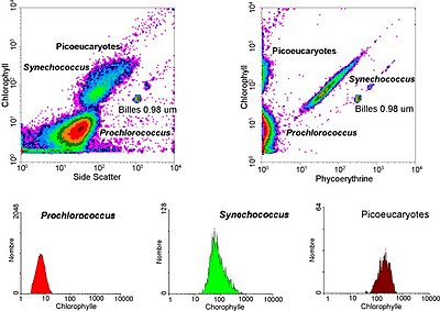
The data collected in the flow cytometer is presented in graphs in which one or two parameters are viewed at the same time. A subset of the cells that lie within a freely selectable region can be selected for further analysis with the help of a so-called "gate". Detailed analyzes can be made through sequences of sequential gates.
Presentation of flow cytometry data
Flow cytometry data is typically presented in two ways: histograms , which measure or compare only a single parameter, and dotplots , which compare two or three parameters simultaneously on a two- or three-dimensional scatter plot. A histogram typically records the intensity sensed in a single channel along one axis. The number of events recorded at this intensity is on a separate axis. A large number of events captured at a certain intensity will show up as a peak on the histogram. If two or three different parameters are recorded during a measurement, a histogram is not sufficient. A multi-dimensional representation is used - the point diagram or dot plot - in order to be able to show the correlation distribution of the parameters. Unlike the histogram, the dot plot shows each event as a single point on a scatter plot. The intensity of two different channels (or three different channels in a three-dimensional representation) is shown along the different axes.
application
Flow cytometry is used in the clinic for routine diagnostics in areas such as hematology , infectious diseases and immunology . Another large area of application is basic research in medicine and cell biology. This method is also used in biotechnology, e.g. B. to separate sperm cells with the sex chromosome X and those with the chromosome Y from each other (whereby density gradient centrifugation is also suitable). Thus, one can determine the sex of an embryo created by in vitro fertilization by separating the sperm with an X and a Y chromosome prior to in vitro fertilization .
Another application in biology is the quantitative examination of cells. In addition to the simple determination of the number of cells, the cells can usually be stained with the fluorescent DNA dye SYBR Green I in the course of a cell viability determination to differentiate between living and dead cells. Propidium iodide only gets into cells with a no longer intact cell membrane and thus only stains dead cells. In combination with a DNA dye, the proportion of dead cells can be determined from the total number of cells. In addition, cell functions (such as phagocytosis , reactive oxygen species, LE cell test) can be analyzed using flow cytometry.
At the end of 2012, flow cytometry was included in the Swiss Food Register as a recommended method for determining the total number of cells in fresh water . .
Food
In food analysis, flow cytometry can be used to determine the bacterial count in raw milk . In many companies, the cytometrically determined number of live bacteria is also used for end product control. This applies in particular to the approval of modern variants such as UHT milk or ESL milk, where shelf life is a particularly important quality feature .
literature
- Bacterial Detection and Live / Dead Discrimination by Flow Cytometry. Application Note from BD Biosciences (April 2002)
- G. Nebe-von-Caron, PJ Stephens et al. a .: Analysis of bacterial function by multi-color fluorescence flow cytometry and single cell sorting. In: Journal of microbiological methods. Volume 42, Number 1, September 2000, pp. 97-114. PMID 11000436 (Review).
Individual evidence
- ↑ Patent DE1815352 : FLOW-THROUGH CHAMBER FOR PHOTOMETERS TO MEASURE AND COUNT PARTICLES IN A DISPERSION MEDIUM. Registered on December 18, 1968 , inventors: Wolfgang Dittrich, Wolfgang Göhde.
- ↑ L. Kamentsky: In: DMD Evans (Ed.): Proceedings of the 1968 Conference "Cytology Automation". 1970.
- ↑ U. Sack et al.: Cellular diagnostics. Karger Publishers, 2006.
- ^ G. Valet: Past and present concepts in flow cytometry: A European perspective. In: Journal of Biological Regulators and Homeostatic Agents . Important Editore, 2003.
- ↑ MJ Fulwyler: Electronic separation of biological cells by volume. In: Science. Volume 150, No. 3698, 1965, pp. 910-911. PMID 5891056 .
- ↑ HR Hulett, WA Bonner, J. Barrett, LA Herzenberg: Cell sorting: automated separation of mammalian cells as a function of intracellular fluorescence. In: Science. Volume 166, No. 3906, 1969, pp. 747-749. PMID 4898615 .
- ↑ I. Böhm: Flow cytometric analysis of the LE cell phenomenon. In: Autoimmunity. 37, 2004, pp. 37-44.
- ↑ Much more lives in drinking water than previously assumed. Media releases from the Federal Council , Dübendorf, January 24, 2013.
- ↑ Official collection of test methods according to § 64 LFGB : L 01.01-7: 2002-05 Analysis of foodstuffs - Determination of the germ count in raw milk - Flow cytometric counting of microorganisms (routine method) , Beuth Verlag , May 2002.
- ↑ Burkhard Schütze: Microbiological results in a few minutes , LVT Lebensmittel Industrie , March 2016.
- ↑ So that the milk is in order - high-tech germ count determination with flow cytometry , LABO online, May 26, 2016.


