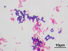
Gram stain


The Gram staining (or Gram stain ) is the Danish bacteriologist Hans Christian Gram developed method (1853-1938) for differentiating coloring of bacteria for microscopic examination. It enables bacteria to be divided into two large groups that differ in the structure of their cell walls. A distinction is made between gram-positive and gram-negative bacteria. However, not all species of bacteria by this technique classifies be, so there are Gram variable and Gram-indeterminate species.
meaning
The Gram stain is a valuable diagnostic tool in scientific and medical microbiology : With its help, bacteria can be easily differentiated according to the structure of their cell wall , since the different colorability of bacteria is based on their chemical and physical properties. The difference in the structure of the cell wall is an important systematic distinguishing feature in bacteria, so the differentiability using Gram staining serves as a taxonomic feature.
Important Gram staining is in the diagnosis of infectious diseases . Gram-positive and gram-negative bacteria can often only be combated with different antibiotics . After the bacterial smear has dried (about 5–15 minutes, depending on the type of material) and fixed (usually heat fixation by briefly pulling it three times over a strong flame (Bunsen burner)), the “grief behavior” is determined in about five minutes. This means that the doctor can start antibiotic therapy immediately before the result of the culture of pathogen cultivation lasting at least 24 hours with the following determination is available.
Staining method
In the Gram-staining of the bacteria with a basic dye (usually after staining crystal violet ) by treatment with Lugol's solution (containing an iodine - Potassium iodide - complex ) a dye-iodine complex formed in the bacteria. In contrast to the primary dye, this color complex is insoluble in water. In contrast, it is soluble in ethanol and is therefore extracted from gram-negative bacteria by treatment with ethanol. Because of the thicker murein layer , on the other hand, it is not extracted from Gram-positive bacteria under the conditions of Gram staining by ethanol.
The staining process consists of three steps:
- Dyeing : The first step is to dye with a solution of crystal violet ( gentian violet ) to which 15 g / l phenol ( carbolic acid ) is added, the so-called "carbolic gentian violet". All bacteria, gram-positive as well as gram-negative, are colored. During the subsequent treatment with Lugol's solution , larger dye complexes are formed and all bacteria appear dark blue.
- Decolorization ("differentiation"): The second step is treatment with 96% ethanol . Gram-positive and Gram- negative bacteria behave differently: Gram- negative bacteria are quickly decolorized again, while the blue dye complexes from Gram-positive bacteria can only be washed out after much longer treatment with ethanol.
- Counterstaining : To show the gram-negative bacteria, they can then be counterstained with dilute fuchsine solution (a solution of fuchsine with phenol in about 1/10 of the usual concentrations of "carbol fuchsin") or saffranine solution, whereupon they appear red or red-orange.
The treatments with Lugol's solution and with alcohol are the crucial steps in Gram staining.
Causes of the different coloring behavior
The difference in the Gram staining is due to the structure of the cell wall:

1. Gram- positive cell wall
2. Gram- negative cell wall
3. Peptidoglycan (Murein)
4. Plasma membrane
5. Cytoplasm
6. Periplasmic space , in grampos. Bacteria the inner wall zone (IWZ)
7. outer membrane
-
Gram-positive bacteria have a thick, multi-layered "murein shell" (consisting of peptidoglycans or "murein") deposited on the membrane . This can account for up to 50% of the dry shell mass. In addition, the cell wall contains between 20% and 40% lipoteichoic acids . Lugol's solution collects in the interstices of the Murein envelope. Here the alcohol has a dehydrating effect and reduces the distance between the molecules so that the dye complexes cannot be washed out by the alcohol. The dark blue color is thus retained.
Examples of gram-positive bacteria are all species of the Actinobacteria strain , such as those of the genera Actinomyces and Streptomyces , and almost all species of the Firmicutes strain , such as those of the genera Streptococcus , Enterococcus , Staphylococcus , Listeria , Bacillus , Clostridium , Lactobacillus , and the species Erysipelothrix rhusiopathiae .
-
Gram-negative bacteria, on the other hand, only have a thin, single-layer murein shell. This makes up only about 10% of the dry mass of the bacterial shell and does not contain any teichonic acids. In addition, a second it is also lipid - membrane superimposed. The alcohol has a lipid-dissolving effect, so that the deposited lipid membrane is dissolved and the thin murein shell is exposed. The dye complexes are washed out by the alcohol - the bacterium is decolorized again . The gram-negative bacteria are then stained red by counterstaining with fuchsin.
Examples of gram-negative bacteria are all species of the Proteobacteria department , such as the enterobacteria ( Escherichia coli , Salmonella , Shigella , Klebsiella , Proteus , Enterobacter ) as well as the genera Pseudomonas , Legionella , Neisseria , Rickettsia and the species Pasteurella multocida ; Representatives of other departments, such as Streptobacillus moniliformis (a species of the Fusobacteria tribe ), Meningococcus , Chlamydophila , Chlamydia , the spirochetes , all species of the Bacteroidetes and cyanobacteria . In addition, the species of the genus Veillonella of the family Acidaminococcaceae (strain Firmicutes ) are gram-negative, although all other species of the strain Firmicutes are gram-positive.
Alternatives to Gram stain
The following short tests can be used to distinguish bacteria based on the same cell wall characteristics as with Gram stain:
KOH test
A small amount of bacterial mass (from an agar culture) is suspended in a drop of 3% potassium hydroxide solution . In the case of gram-positive ones , this lye is too weak to lyse the cell wall . If you pull a needle or toothpick through the mixture, it behaves like a liquid with a viscosity like water (no thread formation can be seen). The cell wall of gram-negative bacteria, on the other hand, is much thinner and is lysed by the potassium hydroxide solution. The cells break open and the DNA is released. If the needle is pulled through this solution, thread formation can be observed due to the increased viscosity caused by the released DNA. It should be emphasized that this is a quick test that is only partially reliable. It is often difficult for beginners to see thread formation. The use of an incorrect alkali concentration can also be a source of error here. If this is too strong, gram-positive bacteria will also be lysed . On the other hand, if it is too weak, gram-negative bacteria are not lysed either.
Aminopeptidase test
The aminopeptidase test is based on the fact that the enzyme L-alanine aminopeptidase , with a few exceptions, can only be detected in gram-negative bacteria. A small amount of the bacteria to be examined is suspended in a test tube or micro-reaction vessel in a little sterile , distilled water . For detection, L -alanine-4-nitroanilide is used, which is cleaved by the enzyme with cleavage of an amide bond into L -alanine and the yellow-colored 4-nitroaniline. Thus, a yellow coloration of the suspension indicates the presence of a gram-negative bacterium. Industrially manufactured test strips are available for this reaction. It is advisable to always include a negative control in order to have a comparison. In addition, the test can only be used for bacterial colonies without a strong inherent color.
history
The Danish physician Hans Christian Gram developed the staining method as an employee at Carl Friedländer in Berlin. He was looking for a staining method with which bacteria can be represented in animal tissues, i.e. stained in a way that contrasts them with the tissue cells. The found staining method, published in 1884, was only successful with some bacteria, the gram-positive ones. Émile Roux applied the method to differentiate gram-positive and gram-negative bacteria by dyeing, in particular to determine gonococci (gram-negative in contrast to many other cocci) (published in 1886).
Further development
Scientists at Harvard University in Boston (USA) have developed the Gram stain into a magnetic detection method. After coloring with modified crystal violet , magnetic nanoparticles are attached to the dye. The bacteria can then be detected using NMR devices and separated magnetically. The advantage of magnetic detection is its high sensitivity. With the help of miniaturized micro-NMR devices, a rapid and sensitive on-site diagnosis is conceivable.
literature
- C. Gram: About the isolated coloration of the schizomycetes in section and dry specimens. In: Advances in Medicin . Vol. 2, 1884, pp. 185-189.
- Steve K. Alexander, Dennis Strete: Basic microbiological internship - a color atlas. Pearson, Munich 2006, ISBN 978-3-8273-7201-7 .
Web links
Individual evidence
- ^ Carl Roth GmbH + Co. KG: Gram dyeing instructions. Accessed April 15, 2020 (German).
- ↑ Benoît Zuber et al. : Granular Layer in the Periplasmic Space of Gram-Positive Bacteria and Fine Structures of Enterococcus gallinarum and Streptococcus gordonii Septa Revealed by Cryo-Electron Microscopy of Vitreous Sections In: J Bacteriol 2006, 188 (18): 6652-6660. PMC 1595480 (free full text).
- ↑ Wissenschaft-Online-Lexika: Entry on Murein in the Lexikon der Biologie, accessed on November 22, 2008.
- ↑ Ghyslain Budin, Hyun Jung Chung, Hakho Lee, Ralph Weissleder: A Magnetic Gram Stain for Bacterial Detection . In: Angewandte Chemie . tape 124 , no. 24 , 2012, doi : 10.1002 / anie.201202982 .

