Diatoms
| Diatoms | ||||||||
|---|---|---|---|---|---|---|---|---|

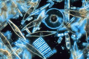
These marine diatoms live as " ice algae " in the interior of the sea ice of the Antarctic McMurdo Sound . |
||||||||
| Systematics | ||||||||
|
||||||||
| Scientific name | ||||||||
| Bacillariophyta | ||||||||
| Haeckel |
The diatoms or diatoms (Bacillariophyta) form a taxon of photosynthesis operated protists (Protista) and in the group of Stramenopilen belongs (Stramenopiles). The group is often referred to by the synonymous name Diatomea Dumortier, alternatively the synonymous names Fragilariophyceae, Diatomophyceae are also used. Some authors call the diatoms Bacillariophyceae, so they place them as a class in the photosynthetic representatives of the stramenopiles (Ochrophyta), which are then understood as phylum, this view is represented in the databases DiatomBase and WoRMS . This use is misleading, however, since other taxonomists list a more narrowly defined class Bacillariophyceae, as one of three classes within the diatoms. When using this name, the respective opinion must be checked, otherwise misunderstandings will arise.
Today there are around 6,000 species . However, it is believed that there are up to 100,000 species in total.
features
German their trivial names owe the diatom of the cell cases (frustule), which consists predominantly of silicon dioxide ( empirical formula : SiO 2 is, dem) anhydride of silica (simplified empirical formula: SiO 2 · n H 2 O). In the German-speaking world, however, silica is often incorrectly equated with its anhydride. The organism gets the silicon dioxide from the monosilicic acid Si (OH) 4 .

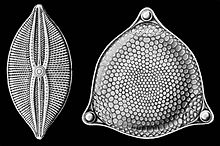
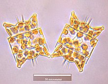
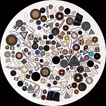
The Frustel is box-shaped and consists of two bowl-shaped parts of different sizes, one of which ("Epitheka") reaches with its opening over the opening of the other ("Hypotheka"). The bowls are structured in characteristic patterns. Due to the shell geometry, two types of diatoms are distinguished: centric diatoms (Centrales) mostly have round, sometimes also triangular shells, while pennate diatoms (Pennales) form rod-shaped or boat-shaped, sometimes also arched or S-shaped curved shells. The shells are created using special peptides called silaffins . The silaffins enable the precipitation of silicon dioxide in small globular silicon dioxide aggregates, the so-called "nanospheres". These have a diameter of 30 to 50 nm and in their entirety form the actual shells. Many pennate diatoms can crawl on a firm surface with the help of a raphe . The speed is up to 20 μm / s.
Diatoms are unicellular and almost always not flagellated . Only in some species do the male gametes have a flagellum, a forward-pointing cilia flagellum . The plastids created by secondary endosymbiosis with a red alga are colored brown, as the xanthophyll fucoxanthin covers the color of the chlorophylls (chlorophyll a and c). Chrysolaminarin serves as a reserve substance .
As a rule, diatoms are microscopic in size. For example, the Achnanthes reaches a length of 40 micrometers. However, some species can grow up to 2 millimeters long.
Multiplication

The diatoms are diploid and reproduce mainly asexually by cell division. The daughter cells each receive a shell part and form the other part anew; the term “diatom” ( ancient Greek διατέμνειν (diatemnein) = to split) is derived from this. The new shell part is always the smaller mortgage, so that the cell size of almost all offspring continuously shrinks over the course of the generations, only the daughter cell line of the original epitheka retains the original maximum size. If the size falls below a minimum, the individual dies. However, before a minimum size is reached, sexual processes can take place. Haploid gametes are formed from the cells through meiosis . Oogamy has been proven in centric diatoms : the gametes become free, after a female and a male gamete merge, the zygote forms a permanent form, a so-called auxospore, with growth in size. Conjugation has been observed in pennate diatoms : two partners lie next to each other and form a common cytoplasmic bridge (“conjugation channel”) into which a haploid nucleus and a chloroplast of the two partners immigrate. The zygote formed in this way forms an auxospore in which nuclear fusion ( karyogamy ) takes place. A larger new diatom with a new two-part shell is formed from the auxospores of the centric and pennate diatoms.
Occurrence
Diatoms are mainly found in the sea and in freshwater in planktonic or benthic form , or they are found on stones or aquatic plants ( epiphytes ). Some species need pure and hardly polluted water and for this reason are also pointer organisms for unpolluted waters. Other types, in turn, Also referred to as the agricultural guild , are typical of bodies of water that are particularly polluted by agricultural inputs, e.g. by overfertilization. These are u. a. Navicula radiosa , Melosira varians , Nitzschia palea , Diatoma vulgare or Amphora perpusilla are counted. Even terrestrial species are found among the diatoms; these colonize soils and, in tropical areas, also leaves from trees.
Systematics
The diatoms were traditionally divided into the radially symmetrical Centrales and the bilaterally symmetrical Pennales. The Centrales, however, are paraphyletic , a stable system based on molecular genetics has not yet been established. According to the taxonomic database Diatombase, there is currently no generally accepted sub-taxa classification of diatoms.
A widespread structure, but not recognized by all taxonomists, could look like this; the higher structure was adopted in a comparable form in the widely used classification of eukaryotes by Sina Adl and colleagues.
- Coscinodiscophytina subdivision
- Class Coscinodiscophyceae
- Order Asterolamprales
- Order Arachnoidiscales
- Order Aulacoseirales
- Genus Aulacoseira
- Order Chrysanthemodiscales
- Order Corethrales
- Order Coscinodiscales
- Order ethmodiscales
- Order Melosirales
- Genus Melosira
- Order Orthoseirales
- Order Rhizosoleniales
- Genus Rhizosolenia
- Order Stictocyclales
- Order stictodiscales
- Order Leptocylinderales
- Class Coscinodiscophyceae
- Bacillariophytina subdivision
- Class Mediophyceae
- Order anaulales
- Order Biddulphiales
- Order Chaetocerotales
- Order Cymatosirales
- Order Eupodiscales (Tricertiales)
- Genus Pleurosira
- Order Hemiaulales
- Order Lithodesmiales
- Order thalassiosirales
- Genus Cyclotella
- Order Toxariales
- Class Bacillariophyceae (largely corresponds to the earlier Pennales)
- Order Raphoneidales
- Order Rhabdonematales
- Order Cocconeidales
- Genus algae lice ( Cocconeis )
- Order fragile (presumably paraphyletic)
- Genus Asterionella
- Genus Diatoma
- Genus Fragilaria
- Genus Meridion
- Genus Synedra
- Order Tabellariales
- Genus Tabellaria
- Order Licmophorales
- Order Thalassionematales
- Order Eunotiales
- Genus Eunotia
- Order Lyrellales
- Order Mastogloiales
- Order Dictyoneidales
- Order Cymbellales
- Genus Cymbella
- Genus Didymosphenia (including Didymosphenia geminata )
- Genus Gomphonema
- Genus Rhoicosphenia
- Order Achnanthales
- Genus Achnanthes
- Order Naviculales
- Genus Diploneis
- Genus Gyrosigma
- Genus Navicula
- Genus Pinnularia
- Genus Stauroneis
- Order Thalassiophysales
- Genus amphora
- Order Bacillariales
- Genus Bacillaria
- Genus Nitzschia
- Order Rhopalodiales
- Genus Epithemia
- Order surirellales
- Genus Cymatopleura
- Genus Surirella
- Class Mediophyceae
meaning
The diatoms are the main component of the marine phytoplankton and are the main primary producers of organic matter, so they form an essential part of the base of the food pyramid . As oxygen- producing (oxygenic) phototrophs , they also generate a large part of the oxygen in the earth's atmosphere .
From the relative species composition of the diatom population of a water body, its degree of trophism can be derived quite precisely ( diatom index ), as well as other water parameters such as pH value , salinity , saprobism, etc. These methods can also be applied to sediments or oil deposits and then provide information the previously prevailing living conditions.
To identify the species, a specimen of diatoms is treated with sulfuric acid , hydrogen peroxide , potassium dichromate or another oxidizing agent and all organic components of the specimen are dissolved in this way. Only the pure silicon dioxide shells remain. These are embedded in an inclusion medium with a high optical refractive index (e.g. Naphrax ™) and identified with a light microscope using the phase contrast method at approx . 1000x magnification.
If the cells die, they sink to the bottom of the water, the organic components are broken down and the silicon dioxide shells form a deposit, the so-called kieselguhr (diatomaceous earth). This process, especially in the marine environment, is only efficient enough below the CCD ( Calcite Compensation Depth ) to form large deposits. The resulting diatomaceous earth is used in technology and medicine. Diatom shells are used, among other things, as a filter, for the production of dynamite , in toothpaste as a cleaning body, as a non-toxic insect control agent in the form of vermin powder , and as a reflective material in the color used for road markings in road construction. Diatoms are also used in forensic medicine ( diatom detection ). Their informative value for the proof of a drowning death is, however, controversial.
In the 19th century in particular, diatoms were used to make aesthetic microscopic specimens, and viewing them together in salons was a popular pastime in higher social circles. Johann Diedrich Möller from Wedel near Hamburg achieved the highest level of perfection in the extremely difficult craftsmanship of such preparations .
Marine diatoms can lead to poisoning in humans and animals, as some species, in particular Pseudo-nitzschia , Nitzschia or amphora, produce domoic acid . In filtering seawater organisms such. B. mussels , or species that feed on these animals, such as. B. fishing , these diatoms can accumulate. If such organisms enriched with domoic acid are consumed by humans, symptoms of poisoning occur, which are known as amnesic shellfish poisoning (ASP). Symptoms include memory loss , nausea , cramps , diarrhea , headache, and difficulty breathing .
literature

- Friedrich Hustedt: Bacillariophyta (Diatomeae). 2nd Edition. Fischer, Jena 1930.
- Friedrich Hustedt: Diatoms (diatoms). 3. Edition. Franckh, Stuttgart 1965.
- Kurt Krammer: Diatoms. Franck, Stuttgart 1986.
- Kurt Krammer, Horst Lange-Bertalot: Bacillariophyceae in freshwater flora of Central Europe 1986–2000. Fischer, Stuttgart and Spektrum, Academic Publishing House, Heidelberg.
- Volume 1: Naviculaceae. 1986.
- Volume 2: Bacillariaceae, Epithemiaceae, Surirellaceae. 1988.
- Volume 3: Centrales, Fragilariaceae, Eunotiaceae. 1991.
- Volume 4: Achnanthaceae. 1991.
- Volume 5: English and French translations and additions. 2000.
-
Diatoms of Europe: diatoms of the European inland waters and comparable habitats. Gantner, Rugell.
- Volume 1: Kurt Krammer: The genus Pinnularia. 2000.
- Volume 2: Horst Lange-Bertalot: Navicula sensu stricto. 2001.
- Volume 3: Kurt Krammer: The genus Cymbella. 2002.
- Volume 4: Kurt Krammer: The genera Cymbopleura, Delicata, Navicymbula, Gomphocymbellopsis and Afrocymbella. 2003.
- FE Ross, RM Crawford, DG Mann: The Diatom. Biology and Morphology of The Genera. Cambridge Univ. Press, 1990.
Individual evidence
- ↑ a b Class Bacillariophyceae. in Kociolek, JP; Balasubramanian, K .; Blanco, S .; Coste, M .; Ector, L .; Liu, Y .; Kulikovskiy, M .; Lundholm, N .; Ludwig, T .; Potapova, M .; Rimet, F .; Sabbe, K .; Sala, S .; Sar, E .; Taylor, J .; Van de Vijver, B .; Wetzel, CE; Williams, DM; Witkowski, A .; Witkowski, J. (2017). DiatomBase. Retrieved December 4, 2017
- ^ TA Norton, M. Melkonian, RA Andersen: Algal biodiversity. In: Phycologia. 35, pp. 308-326.
- ^ N. Kroger: The sweetness of diatom molecular engineering. In: Journal of Phycology. Volume 37, 2001, pp. 93-112.
- ↑ N. Kroger, M. Sumper: The molecular basis of diatom biosilica formation. In: E. Baeuerlein (Ed.): Biomineralization: Progress in Biology, Molecular Biology and Application. 2004, pp. 137-158, Wiley-VCH, Weinheim
- ^ Secondary Endosymbiosis Exposed. In: The Scientist. June 2005, accessed June 1, 2016 .
- ↑ JL Richardson, NS Mody, ME Stacey: Diatoms and water quality in Lancaster County (PA) streams: a 45 year perspective. In: Jornal - Pennsylvania Academy of Sciences. Volume 70, 1996, pp. 30-39.
- ↑ Linda K. Metlin (2016): Evolution of the diatoms: major steps in their evolution and a review of the supporting molecular and morphological evidence. Phycologia 55 (1): 79-103. doi: 10.2216 / 15-105.1
- ↑ Adl, SM, Simpson, AGB, Lane, CE, Lukeš, J., Bass, D., Bowser, SS, Brown, MW, Burki, F., Dunthorn, M., Hampl, V., Heiss, A. , Hoppenrath, M., Lara, E., le Gall, L., Lynn, DH, McManus, H., Mitchell, EAD, Mozley-Stanridge, SE, Parfrey, LW, Pawlowski, J., Rueckert, S., Shadwick, L., Schoch, CL, Smirnov, A. and Spiegel, FW (2012): The Revised Classification of Eukaryotes. Journal of Eukaryotic Microbiology 59: 429-514. doi: 10.1111 / j.1550-7408.2012.00644.x
- ↑ B. Madea: Practice forensic medicine: assessment, reconstruction, assessment. 2nd updated edition. Springer, Berlin / Heidelberg / New York 2007.
- ↑ P. Lunetta, JH Modell: Macropathological, Microscopical, and Laboratory Findings in Drowning Victims. In: M. Tsokos (Ed.): Forensic Pathology Reviews. Vol. 3, Humana Press, Totowa, NJ 2005, pp. 4-77.
- ↑ SS Bates: Domoic-acid-producing diatoms: another genus added. In: Journal of Phycology. Volume 36, 2000, pp. 978-985.
- ↑ Health consequences of toxic algal blooms ( Memento of the original dated February 9, 2007 in the Internet Archive ) Info: The archive link was inserted automatically and has not yet been checked. Please check the original and archive link according to the instructions and then remove this notice.
Web links
- Dark breathing with nitrate
- Urea as an anabolic steroid for diatoms
- Videos on diatoms , published by the Institute for Scientific Film . Provided in the AV portal of the technical information library


