Canine Malassezia Dermatitis

The Malassezia dermatitis of the dog (syn. Pityrosporie) is in domestic dogs occur through saprobische fungus ( Malassezia spp., Ustilagomycotina ) caused inflammation of the skin . Occasionally, only the skin of the external auditory canal is affected, in which case one speaks of Malassezia otitis ( inflammation of the outer ear caused by Malassezia). The pathogen belongs to the natural skin flora and only develops its pathogenic effect when the balance of this flora is disturbed . The disease is characterized by reddening of the skin and severe itching , and in the chronic course by severe skin changes.
Cause and occurrence

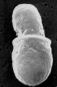
Of the ten Malassezia species identified so far, Malassezia pachydermatis (outdated: Pityrosporum pachydermatis ) is of greater clinical importance in dogs. The pathogen is lipophilic but independent of lipids, which means that it does not necessarily need fats as a carbon source, but grows better when they are present. In contrast, most other malassezia are lipid dependent. Malassezia pachydermatis is not mycelium-forming , about 2–5 µm in size and, under the microscope, has a typical peanut-like shape, which comes about through the multiplication of the fungus in the form of one-sided budding . As a further pathogen, Malassezia sympodialis can also trigger a disease with mostly slight itching.
Malassezia also occur in healthy dogs and other mammals as part of the natural skin flora, the detection rate in healthy dogs is up to 40% and varies depending on the dog breed and the body region. They mainly colonize the skin of the area between the toes , the lips and the ear canal as well as the mucous membranes of the vagina , anal pouch and anus . The spread on the animal takes place mostly by licking. It is assumed that Malassezia as commensals keep other more pathogenic fungi in check and are kept under control by various defense mechanisms of the skin ( exfoliation of the epithelium , fatty film, immunoglobulin A ).
Other mammals are much less affected by Malassezia dermatitis. In humans, infections with Malassezia furfur play a major role. Malassezia pachydermatis can be transmitted by hospital staff who own dogs and occasionally cause disease in newborns and immunocompromised adults ( nosocomial infection ). An individual case was also described in an otherwise healthy dog owner. In domestic cats , the disease is rare, above all breeds are affected with genetic Haarabnormitäten: Sphynx and Devon Rex . Basic diseases such as " cat aids" , feline leukemia , diabetes and herpes infections are favorable factors in cats.
Disease emergence
Clinically, malassezia usually only appear secondarily with the presence of another skin disease ( idiopathic seborrhea , epidermal dysplasia , allergies , hormonal disorders ) or other predisposing factors (strong skin wrinkling, warmth, increased skin moisture, immunosuppression , prolonged use of antibiotics ) that lead to a disruption of the Balance the skin flora. In some dog breeds, however, primary Malassezia dermatitis cannot be ruled out. West Highland White Terriers (predisposing to epidermal dysplasia), poodles , basset hounds (predisposing to idiopathic seborrhea), Shih Tzu , Pekingese , Cocker Spaniels (predisposing to idiopathic seborrhea), Labrador Retrievers , German Shepherds and Jack Russell Terriers are particularly susceptible .
The pathogenic effect of Malassezia is mediated by two enzymes . A lipase breaks down fatty acids in the sebum film . The resulting short-chain fatty acids have a skin-irritating effect and cause the typical rancid odor and may also trigger allergies . In addition, zymogen activates the complement system .
Clinical picture
Malassezia dermatitis has severe, often glucocorticoid- resistant itching ( pruritus ) and severe reddening of the skin ( erythema ). The yeast breakdown of the fatty acids often results in a rancid skin odor. The increased sebum production ( seborrhea ) often leads to yellowish, greasy deposits, but "dry" seborrhea is also possible. The changes usually start in the abdomen and then spread to other parts of the body. If the claws are also affected, the base of the claws shows brownish discoloration. Malassezia dermatitis can also manifest itself in the wrinkles of the lower lip or as a chronic, recurrent inflammation of the anal glands . In Malassezia otitis, dark ear wax is typical, which can lead to confusion with an infestation by ear mites .
Scratching the itchy area can develop various secondary skin changes such as abrasions, excoriations, and crusts. It is not uncommon for malassezia dermatitis to be aggravated by the multiplication of skin-dwelling bacteria (especially Staphylococcus intermedius ), so that a purulent skin inflammation ( pyoderma ) can develop.
With a chronic course, hair loss ( hypotrichosis , alopecia ), skin thickening ( lichenification ) and increased pigment storage in the skin ( hyperpigmentation ) can occur.
Diagnosis
Malassezia can be detected on impression specimens using adhesive tape, microscope slides or swabs or a superficial skin scraper . The specimens are briefly heat-set, stained with routine stains and then examined microscopically . However, it is problematic to distinguish it from a natural skin colonization, as there are large differences depending on the breed. As a guideline for a pathological colonization, at least 10 Malassezia per field of view are given at 400x magnification in the microscope. For microscopic findings, predisposing factors, localization and any previous illnesses must always be included. The specificity is very high at 95%, but the sensitivity is low at around 30%.
It is also possible to create a Malassezia culture with growing the pathogens on a nutrient medium , but this involves more effort and is expensive. The sensitivity is 82%. Since Malassezia also occur in healthy dogs, quantification must be carried out using colony-forming units .
In the differential diagnosis , allergies ( atopic dermatitis , food or flea allergy ), superficial pyoderma (in particular bacterial overgrowth of the skin and bacterial folliculitis ), zinc-reactive dermatosis , cornification disorders of the skin and sarcoptic mange are to be excluded, although these diseases should also be excluded as a pre-existing condition can favor malassezia dermatitis.
therapy
There are several options for treating Malassezia dermatitis:
- Local treatment (topical therapy): It is done with shampoos or creams with malassezia-effective ingredients such as chlorhexidine , dichlorfen , selenium disulfide or polyvidon-iodine , the combination of chlorhexidine and miconazole showing the best effect. Topical treatment is indicated for smaller herds, otitis and supportive also for systemic treatment.
- Whole-body treatment (systemic therapy): Here malassecia-effective drugs such as ketoconazole , more rarely itraconazole or fluconazole, are used for 3–6 weeks.
Since Malassezia dermatitis often occurs as a secondary disease, the primary disease must also be diagnosed and treated. In animals with a non-treatable underlying disease (e.g. seborrhoea oleosa ) or a very advanced malassezia dermatitis in the chronic stage, lifelong continuous therapy is usually required.
To support therapy, a change in feed or the addition of unsaturated fatty acids to the feed can be carried out.
literature
- B. Bigler: Mycotic Skin Diseases. In: PF Suter, B. Kohn (Hrsg.): Internship at the dog clinic. 10th edition. Parey, 2006, ISBN 3-8304-4141-X , pp. 366-367.
- Richard G. Harvey et al: Ear diseases in dogs and cats . Schattauer, 2003, ISBN 3-7945-2235-4 .
- Ch. Noli, F. Scarampella: Malassezia dermatitis. In: Practical Dermatology in Dogs and Cats. 2nd Edition. Schlütersche Verlagsanstalt, 2005, ISBN 3-87706-713-1 , pp. 210-214.
- George T. Wilkinson, Richard G. Harvey: Color Atlas of Skin Diseases in Small Pets. 2nd Edition. Schlütersche, 1999, ISBN 3-87706-554-6 .
Individual evidence
- ^ A b Daniel O. Morris et al: Malassezia pachydermatis Carriage in Dog Owners. In: Emerging Infectious Diseases . 11 (2005). Full text
- ↑ a b c C. Cafarchia et al .: Frequency, body distribution, and population size of Malassezia species in healthy dogs and in dogs with localized cutaneous lesions. In: J Vet Diagn Invest. 17 (2005), pp. 316-322. PMID 16130988
- ↑ YM Fan ua: Granulomatous skin infection caused by Malassezia pachydermatis in a dog owner. In: Arch Dermatol. 142 (2006), pp. 1181-1184. PMID 16983005
- ^ PJ McKeever, HW Richardson: Otitis externa. Part 2:. Clinical appearance and diagnostic methods. In: Comp. Anim. Pract. 2, pp. 25-31 (1988).
- ↑ R. Bond et al .: Comparison of two shampoos for treatment of Malassezia pachydermatis-associated seborrhoeic dermatitis in basset hounds. In: J Small Anim Pract. 36: 99-104 (1995). PMID 7783442
