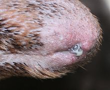
Penis of dogs

The penis of male dogs (the males ) has in comparison to the general structure of the penis of the mammals a variety of anatomical and functional peculiarities associated with the mating behavior of dogs. The basic anatomy of the penis is identical in all types of dogs: The glans is divided into two sections and swells after being inserted into the vagina of the bitch due to the pronounced erectile tissue of the penis , which in connection with the vaginal muscles of the bitch causes the dog to "hang" up to 30 minutes after penetration. The penis is largely transformed into the penis bone in dogs .
In domestic dogs , the most common dysfunction of the penis is preputial catarrh, an increase in secretion from glands in the foreskin.
Anatomy and physiology
Erectile tissue
As in all mammals, the penis has three erectile tissue in dogs , namely the two paired erectile tissue ( Corpora cavernosa penis ) and the unpaired urethral erectile tissue ( Corpus spongiosum penis) . The latter continues into the glans as the corpus spongiosum glandis . In contrast to humans, the part of the penis that is visible during erection consists exclusively of the glans; the penis shaft with the penile cavernous bodies remains hidden under the skin of the interlegs between the thighs and the penile cavernous bodies hardly swell during erection .
The retractor penis muscle attaches to the penis shaft , a pair of smooth muscles that the male can use to retract his penis into the foreskin.
Acorn
The glans ( glans penis ) in dogs is divided into two parts: Behind the long part ( pars longa glandis ) lies the "node" ( bulb glandis ). This swells only after it has penetrated the vagina and ensures that the male dog remains connected to the bitch for some time (between 15 and 30 minutes) after ejaculation ("hanging"). The swelling is caused by the filling of the corpus spongiosum glandis ( corpus spongiosum glandis ), which in dogs - in contrast to most other mammals - is strongly erect due to the restriction of the blood flow through the vena dorsalis penis by the muscle ischiourethralis . This increases the chances of fertilization and prevents, at least in the short term, other pack members from being able to mate with the bitch.
Behind the knot, the penis is very flexible in the horizontal plane, even when erect, which enables the male to get off while hanging.
shaft
The shaft of the dog's penis ( corpus penis ) is also not visible during erection. However, its course can easily be felt, starting right behind the knot between the hind legs and up to the anus.
Penis bones and urethra

The penis bone ( os penis ) is located in the corpus spongiosum . This enables the male to enter the bitch's vagina before the penis has swollen. The urethra runs in a downwardly open groove in the penis bone and ends at the outermost "tip" of the penis (urethral process , processus urethrae ), which is sometimes also referred to as "glans tickler" because of its appearance and its extraordinary sensitivity.
A little above the urethral process, a small indentation forms in the penis during erection. This is due to the fact that the front end of the penis bone is connected to the skin at the tip of the penis inside via a cartilaginous structure. If the penis swells during erection , the bones and cartilage remain the same size and therefore pull the skin slightly inwards at its "attachment point".
foreskin
The penis foreskin (the preputium penis ) completely surrounds the glans when it is not erect. The back of the foreskin is fused with the skin of the abdomen; the front part, reaching almost to the navel, is free. The inner foreskin sheet ( lamina interna ) is covered with cutaneous mucous membrane like the glans , the outer sheet ( lamina externa ) with normal, hairy epidermis . The opening of the foreskin at the transition between the internal lamina and the external lamina is called the ostium preputiale , the transition between the prepuce and the penis is called the fundus preputialis or, more rarely, the fornix ("vault"). In the non-erect state, the foreskin cavity ( cavum preputiale ) is located between the lamina interna and the penis .
The paired foreskin muscle ( Musculus preputialis cranialis ) attaches in front of the prepuce , a smooth muscle that pulls the foreskin over the glans.
Frenulum
In contrast to humans, the foreskin ligament ( frenulum praeputii ), which connects the glans and foreskin, tears in dogs before they reach sexual maturity . The former connection point remains visible as a " seam " ( raphe ) down the entire length of the glans. In individual cases, the frenulum can also persist into sexually mature age and then prevent the male from digging. This condition is rare and can easily be treated by the veterinarian.
Blood supply, lymph drainage and innervation
The penile blood supply is ensured by the penile artery and vein ( arteria and vena penis ). The penile artery is the terminal branch of the internal pubic artery ( internal pudendal artery ). At the base of the penis, it divides into three branches: the arteria bulbi penis feeds the urethral erectile tissue, the deep artery penis the penile erectile tissue. The arteria dorsalis penis is mostly unpaired, runs on the back side of the penis up to the penis tip and supplies the glans, the foreskin and the skin around the penis shaft. In dogs, it has an additional inflow via the anterior penile artery ( arteria penis cranialis ), which arises from the external pubic artery ( arteria pudenda externa ). The blood flow to the foreskin is also ensured by branches of the arteria and vena epigastrica caudalis superficialis , which also originate from the external pubic artery.
The lymph vessels of the penis lead to the superficial inguinal lymph nodes ( Lymphonodi inguinales superficiales s. Scrotales ).
The penis is innervated by the dorsal nerve penis , the terminal branch of the pudendal nerve . This nerve receives sympathetic and parasympathetic nerve fibers in addition to sensitive ones . The parasympathetic fibers control the erection, the sympathetic fibers the ejaculation. In addition, the genitofemoral nerve , and in part also the first two lumbar nerves ( iliohypogastric nerve and ilioinguinal nerve ), contribute to the innervation of the foreskin.
ejaculation
In dogs - in contrast to humans - ejaculation takes place in three phases. The first, sperm- poor fraction is released during penetration until the penis is fully erected. The second, sperm-rich fraction is released shortly after a full erection has been achieved. The third, again sperm-poor fraction is released during the entire remaining phase of the hanging and has by far the largest volume of the three fractions.
Diseases
The following diseases, which are described for domestic dogs , also apply at least in part to other dogs.
Preputial catarrh

A preputial catarrh is caused by an increased secretion activity of the glands of the inner foreskin and manifests itself by the drainage of a slimy, cloudy, white-yellowish secretion from the foreskin opening. It is the most common disease of the dog penis. The disorder can be differentiated from balanoposthitis on the basis of the absence of symptoms of inflammation (warmth, redness, swelling, painfulness). The cause of the disease has not yet been clarified.
Mild preputial catarrh is present in many adult males and is usually clinically insignificant. The administration of antibiotics does not improve symptoms. The usual therapeutic procedure is repeated rinsing of the prepuce with mild disinfectants such as chlorhexidine solution or care substances such as diluted chamomile extract . A castration often leads to a decrease in the secretion.
As with balanoposthitis (see below), the presence of other possible causes, such as foreign bodies, must be excluded before symptomatic treatment of preputial catarrh is started.
Balanoposthitis
In contrast to preputial catarrh, inflammation of the glans and foreskin (balanoposthitis) is associated with clear symptoms of inflammation. The penile and foreskin mucous membrane appears irregularly reddened; As with preputial catarrh, a purulent-mucous (mucopurulent) secretion, whitish, yellowish or greenish depending on the bacteria involved , is secreted, which in severe cases can also become purulent to pus-like. In more severe cases, the mucous membrane of the genitals can take on a bumpy surface structure caused by the swelling of lymph follicles .
Mild balanoposthitis is treated like preputial catarrh. If the symptoms are worse, local (ointments) or systemic administration of antibiotics may be necessary. Before symptomatic treatment, the dog must be examined for underlying problems such as foreign bodies or the like.
foreign body
Occasionally, foreign bodies are found in the preputial cavity. Often these are awns , which penetrate deeper and deeper due to their surface structure and lead to balanoposthitis. From time to time hair penetrates the preputial cavity, which also causes balanoposthitis, but in the worst case can also lead to constriction and death of parts of the penis.
Urinary stones
Due to the fact that in dogs the urethra runs in a furrow of the penis bone and is therefore not very flexible, urinary stones washed away from the bladder often get stuck at the narrowing that has arisen, which leads to an occlusion of the urethra. If the outflow of urine is completely prevented by this, the dog can no longer excrete the toxic metabolic products contained in the urine, so that severe symptoms of postrenal uremia occur very quickly .
Complete occlusion of the urethra is a urological emergency. Affected males repeatedly try to urinate without success, are restless and lick their penis. As the disease progresses, there are uremic symptoms as well as changes in the electrolyte balance, which can lead to cardiac arrhythmias and ultimately death. Treatment consists of surgically removing the urinary stones and treating their cause.
Phimosis
A phimosis is expressed in an abnormally small opening of the foreskin, which prevents excavation of the penis and in severe cases leads to problems in urination. It can be congenital, but it can also be acquired through scarring, chronic inflammatory processes or neoplasms . The phimosis usually goes unnoticed until the first attempt at mating. If necessary, the foreskin opening can be surgically enlarged. The most important differential diagnosis is the persistent foreskin ligament (see above).
Paraphimosis
In paraphimosis , the erect penis can no longer be pulled back into the foreskin. This leads to a constriction and further edematous swelling of the exposed part of the penis, which can die as a result. Paraphimosis is an andrological emergency and should be corrected as soon as possible.
Paraphimosis can be caused by a slight phimosis on the one hand, but on the other hand it can also be an inversion of the outer foreskin sheet into the foreskin cavity, whereby the retraction of the penis into the foreskin is blocked. The treatment consists in the careful manual correction of this inversion with generous use of lubricant. In advanced cases, however, surgical intervention may be necessary.
Tumor diseases
Tumors of the penis and prepuce in domestic dogs are rare in Central Europe. The so-called sticker sarcoma , a sexually transmitted malignant tumor, occurs more frequently in tropical and subtropical regions in particular and is also the most frequently diagnosed tumor type on dog penis.
Besides occur squamous , adenocarcinoma , malignant Hemangioendotheliomas , fibrosarcoma , papilloma , lymphoma , fibroma and hemangiomas in the penis mucosa. The prepuce can be affected by all tumors on the skin. Melanomas , mast cell tumors and squamous cell carcinomas are most frequently found here .
Fracture of the penile bone
A fracture of the penis bone is rare, but it can lead to problems with urination through to retention of urine due to a blockage or rupture of the urethra and, in the long term, problems with mating due to the curvature of the penis. Possible causes are traffic accidents, but also mistreatment (especially kicks). The fracture is diagnosed by x-rays . Unsuspended fractures are usually not operated on because the penis provides sufficient support; however, an indwelling catheter may be used to stabilize and ensure the flow of urine . Fractures with displaced ends are surgically reduced and fixed with orthopedic wire. Urethral tears must also be corrected surgically.
During healing, the callus that forms can lead to an obstruction of the urethra. As an alternative to surgical reduction and fixation, penile amputation can also be considered.
Herpes
The canine herpes virus can affect the mucous membrane of the penis and foreskin in males. This is a mostly inapparent, persistent infection , so clinical symptoms only occur rarely and irregularly in an infected dog. Outbreaks can be caused by stress, but also by treatment with corticosteroids and lead to a vesicular inflammation of the genital mucous membrane (" cold sores "). Viruses are excreted at irregular intervals even without the appearance of lesions on the penis, so that affected males can be contagious despite a clinically normal-looking penis. Males infected with herpes should therefore only be used for breeding with caution, as herpes infection can lead to puppy deaths .
Hypospadias
The Hypospadias is a congenital malformation , in which the urethra does not end at the tip of the penis, but later on the shaft. This condition is rare and usually does not require treatment. The libido is not affected by this; however, depending on the severity, fertility may be limited. However, since there is a hereditary component in hypospadias , affected males should be excluded from breeding anyway. In severe cases, surgery may be necessary.
Other diseases
In addition to the hypospadias and the persistent foreskin ligament, other family-related diseases have been described: a deformation of the penis bone can lead from the inability to mate to necrosis of the glans. Congenital shortenings of the foreskin are also described as having a family history and can cause penile injuries and even necrosis due to the unprotected glans.
literature
- Anonymous: Reproductive Diseases of the Male Small Animal . In: Cynthia M. Kahn (Ed.): The Merck Veterinary Manual . 9th ed. Merck & Co., Whitehouse Station, NJ 2005, ISBN 0-911910-50-6 , pp. 1158 f.
- Klaus-Dieter Budras et al. (Ed.): Atlas of the anatomy of the dog (textbook for veterinarians and students). 7th edition Schlütersche, Hannover 2004, ISBN 3-89993-012-6 .
- Uwe Gille: Male sexual organs . In: Franz-Viktor Salomon et al. (Hrsg.): Anatomie für die Tiermedizin . Enke, Stuttgart 2004, pp. 389-403. ISBN 3-8304-1007-7 .
- Anne-Rose Günzel-Apel: Fertility control and semen transmission in dogs. Gustav Fischer Verlag, Jena 1994, ISBN 3-334-60512-4 .
- Richard W. Nelson (Ed.): Diseases of the penis, prepuce and testicles . In the S. u. a .: Internal medicine of small animals (Small animal internal medicine). Elsevier, Urban & Fischer, Munich 2006, ISBN 3-437-57040-4 .
Individual evidence
- ↑ Leland E. Carmichael and Craig E. Greene: Canine Herpesvirus Infection. In: Craig E. Greene (Ed.): Infectious Diseases of the Dog and Cat. Saunders, Philadelphia 1998, pp. 28-32, ISBN 0-7216-2737-4 .
- ^ John D. Hoskins: Congenital defects of the dog. In: Stephen J. Ettinger, Edward C. Feldmann (Eds.): Textbook of veterinary internal medicine. Diseases of the dog and cat. Saunders, Philadelphia 2000: 1994, ISBN 0-7216-6797-X .

