Rabies virus
| Rabies virus | ||||||||||||||||||||
|---|---|---|---|---|---|---|---|---|---|---|---|---|---|---|---|---|---|---|---|---|

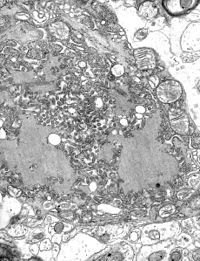
Rabies virus in a cell, electron microscope . |
||||||||||||||||||||
| Systematics | ||||||||||||||||||||
|
||||||||||||||||||||
| Taxonomic characteristics | ||||||||||||||||||||
|
||||||||||||||||||||
| Scientific name | ||||||||||||||||||||
| Rabies lyssavirus | ||||||||||||||||||||
| Short name | ||||||||||||||||||||
| RABV | ||||||||||||||||||||
| Left | ||||||||||||||||||||
|
The rabies virus , scientifically Rabies lyssavirus , commonly also called rabies virus , is a virus that attacks the nervous system and triggers rabies in animals and humans . The result is acute, life-threatening encephalitis (inflammation of the brain), which is usually fatal. It can be transmitted via the saliva of animals. The rabies virus is an enveloped virus of cylindrical shape.
It is a member of the genus Lyssavirus and belongs to the Rhabdoviridae family , the members of which have a single-stranded RNA with negative polarity as their genome . The genome is completely sequenced . The genetic information is packaged as a ribonucleoprotein complex in which the RNA is tightly bound to the viral nucleoprotein N. The RNA genome codes for five genes, the arrangement of which on the genome is highly conserved. These are the genes for nucleoprotein (N), phosphoprotein (P), matrix protein (M), glycoprotein (G) and the viral RNA polymerase (L).
The transcription and replication take place in the cytoplasm of the host cell within special "virus factories" (VF, also viroplasms ), which are referred to under light microscopy as so-called Negri bodies (named after Adelchi Negri ). They have a diameter of 2–10 µm and are typical of rabies infection, so that they serve as a pathognomonic feature.
Systematics
The lyssaviruses include the rabies virus, which is usually associated with rabies, various bat lyssaviruses and the mokola virus (English Mokola lyssavirus ). Together with the causative agent of vesicular stomatitis and others, they form the Rhabdoviridae family. Rhabdoviridae characteristically have a broad host spectrum, which can range from plants to insects to vertebrates.
structure
Lyssaviruses have a helical symmetry, the virions have a cylindrical shape. This is in contrast to other viruses that affect humans, which usually have cubic symmetry.
The rabies virus is elongated in shape with a length of about 180 nm and a diameter of about 75 nm. One end is rounded while the other is planar. The virus envelope contains so-called "spikes" (protuberances) that are formed by glycoprotein G. These protuberances are absent from the planar end of the virion. Below the shell is a layer of matrix protein M, which covers the core of the virion made of helical ribonucleoprotein , which is composed of RNA and protein N.
Genome
The genome consists of unsegmented, linear, single-stranded RNA with negative polarity . The genome has been completely sequenced and is 11,900 nucleotides in length . The genetic information is packaged as a ribonucleoprotein complex in which the RNA is tightly bound to the viral nucleoprotein. The RNA genome codes for five genes, namely the genes for nucleoprotein (N), phosphoprotein (P), matrix protein (M), glycoprotein (G) and the viral RNA polymerase (L). The 3'-NPMGL-5 'arrangement of these genes is highly conserved.
Replication
Rabies viruses attach themselves to the cell surface via specific receptors ( nicotinic acetylcholine receptor , neural cell adhesion molecule 1 ) and are absorbed through endocytosis by an endosome vesicle that forms . Inside the endosome, the acidic pH induces the fusion of the endosome membrane and the virus envelope. This causes the capsid to enter the cytosol , disintegrate, and release the genome. Both receptor binding and membrane fusion are catalyzed by glycoprotein G, which plays an important role in pathogenesis (mutants without G are not infectious).
After entering the host cell, transcription of the viral genome is initiated by the L polymerase to produce more viral proteins. P is an essential cofactor for polymerase L. The viral polymerase only recognizes ribonucleoprotein and cannot use free RNA as a template to produce mRNA. Transcription is regulated by cis elements on the viral genome and by protein M. The latter is not only essential for the budding (ger .: budding ) of the virus from the membrane, but also regulates the balance between mRNA production and replication of the viral genome.
Later, the polymerase produces full-length positive polarity RNA. These complementary RNA strands are used as templates to create new RNA genomes with negative polarity. These are packed into ribonucleoprotein with protein N and can form new viruses.
transmission
The virus is present in the saliva of a rabid animal and the route of infection is almost always via a bite. But even the smallest injuries to the skin and mucous membranes can allow the virus to penetrate via smear infection or contact infection . Transmission through mucous membranes has occurred in vitro . Transmission in this form may have occurred in humans who explored caves populated by bats . With the exception of organ transplants (one case with three deaths in the USA at the beginning of 2004 and one case with three deaths in Germany at the beginning of 2005), human-to-human transmission has not yet been observed.
At the point of entry, the virus infects peripheral nerve cells and migrates along the nerve cells into the central nervous system (CNS). Retrograde axonal transport is the most important step in natural rabies infection. The exact molecular basis of this transport is not yet clear, but it has been shown that the rabies virus phosphoprotein P interacts with the DYNLL1 (LC8) protein of the light chain of Dynein . P also acts as an interferon antagonist , thereby reducing the immune response.
From the CNS, the virus spreads to other organs; it occurs in the saliva of infected animals and can thus spread further. Often there is increased aggressiveness with increased biting behavior, which increases the likelihood of spreading the virus further.
Symptoms
The first symptoms of rabies virus infection are similar to symptoms of the flu, such as physical weakness, fever, and headache. A tingling or itching sensation may be felt at the site of the bite. Within a few days of the bite, humans develop symptoms of cerebral dysfunction, anxiety, confusion, and restlessness. Furthermore, the person may experience delirium, abnormal behavior, hallucinations, and insomnia.
treatment
The rabies virus vaccine is usually given after infection.
brain research
A non-pathogenic variant of the rabies virus has recently been used as a viral vector to map areas of the brain and their connections to one another. This research area makes it possible to color the interconnection of certain areas (e.g. the sensory, motor cortex with other areas) and then to create high-resolution maps. This modern form of neuroanatomy enables individual connections to be traced over long distances and their axonal connections to be reconstructed in detail.
Reporting requirement
In Germany, direct or indirect evidence of the rabies virus must be reported by name in accordance with Section 7 of the Infection Protection Act (IfSG) if the evidence indicates an acute infection. The obligation to notify primarily concerns the management of laboratories ( § 8 IfSG).
In Switzerland, the positive and negative laboratory analysis findings to be rabies laboratory reportable namely after the Epidemics Act (EpG) in connection with the epidemic Regulation and Annex 3 of the Regulation of EDI on the reporting of observations of communicable diseases of man .
Individual evidence
- ↑ ICTV Master Species List 2018b.v2 . MSL # 34, March 2019
- ↑ a b ICTV: ICTV Taxonomy history: Akabane orthobunyavirus , EC 51, Berlin, Germany, July 2019; Email ratification March 2020 (MSL # 35)
- ↑ a b c d Finke S, Conzelmann KK: Replication strategies of rabies virus . In: Virus Res. . 111, No. 2, August 2005, pp. 120-31. doi : 10.1016 / j.virusres.2005.04.004 . PMID 15885837 .
- ^ A b Albertini AA, Schoehn G, Weissenhorn W, Ruigrok RW: Structural aspects of rabies virus replication . In: Cell. Mol. Life Sci. . 65, No. 2, January 2008, pp. 282-94. doi : 10.1007 / s00018-007-7298-1 . PMID 17938861 .
- ↑ Monique Lafon, Rabies virus receptors., In: Journal of Neurovirology 11, No. 1, February 2005, pp. 82-7. doi: 10.1080 / 13550280590900427 . PMID 15804965
- ↑ Three patients died: organ donors spread rabies. In: Spiegel Online . July 1, 2004, accessed March 5, 2020 .
- ↑ Rabies through organ donation - Via medici online . Retrieved February 2, 2011.
- ^ H. Raux, A. Flamand, D. Blondel: Interaction of the rabies virus P protein with the LC8 dynein light chain. In: Journal of Virology . Volume 74, number 21, November 2000, pp. 10212-10216, doi : 10.1128 / jvi.74.21.10212-10216.2000 , PMID 11024151 , PMC 102061 (free full text).
- ↑ What are the signs and symptoms of rabies? In: cdc.gov. June 11, 2019, accessed March 5, 2020 .
- ↑ Mebatsion T, Konig M, Conzelmann KK: Budding of rabies virus particles in the absence of the spike glycoprotein. . In: Cell . 84, No. 6, March 1996, pp. 941-951. PMID 8601317 .
- ↑ a b Ginger M, Haberl M, Conzelmann KK, Schwarz MK, Frick A: Revealing the secrets of neuronal circuits with recombinant rabies virus technology. . In: Front Neural Circuits . 7, No. 2, 2013. doi : 10.3389 / fncir.2013.00002 . PMID 23355811 .
- ↑ Haberl MG, Viana da Silva S, Guest JM, Ginger M, Ghanem A, Mulle C, Oberlaender M, Conzelmann KK, Frick A: An anterograde rabies virus vector for high-resolution large-scale reconstruction of 3D neuron morphology. . In: Brain Struct Funct . April 2014. doi : 10.1007 / s00429-014-0730-z . PMID 24723034 .