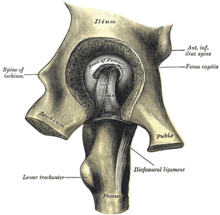
hip joint
The hip joint ( Latin Articulatio coxae , "joint of the hip", from early New High German hüffte from Old High German huffi , the plural for hoof , "hip", the lateral body part below the waist) is the second largest joint in mammals after the knee joint . The thigh bone ( femur ) and the pelvis ( pelvis ) or the hip bone form the bony joint partners. It is mainly involved in the (human) modes of locomotion of walking , in which, in contrast to running, there is no flight phase.
Bony structures and articular surfaces
The bone partners are in very close contact with one another. So that the hip joint can move painlessly and undisturbed on the contact surfaces, they (like all joint surfaces in the body) are coated with a very smooth, bluish-whitish layer of cartilage, the so-called hyaline cartilage .
Thigh bone (femur)
The upper end of the thigh bone forms a large spherical head ( caput femoris ). This represents a relatively regularly curved two-thirds sphere with an average radius of 2.5 centimeters. Only the near-joint ( proximal ) pole forms a flat surface around the retraction of the head of the femur ( fovea capitis femoris ) around the load to be transferred a point to move onto a ring. The head of the femur is completely covered by hyaline cartilage, but has a particularly pronounced layer of around 2.5 to 3.5 millimeters in its main stress zones. The thickness gradually decreases towards the thighbone neck ( collum femoris ).
Pelvis

The structure of the counterpart, the acetabular cup ( acetabulum ), all three pelvic bones are involved: the roof is from iliac ( ilium formed), the pubic bone ( pubic bone ) defines the front ( ventral ) and the seat leg ( ischium ) backwards and downwards ( dorsocaudal) the edge of the pelvis with its depression, the so-called acetabulum ("Essignäpfchen").
If you imagine the pan as a hollow hemisphere, its radius in humans is around 2.7 centimeters, depending on body size. However, an arched fiber cartilage lip ( labrum acetabuli or limbus acetabuli ) extends in places beyond the equator of the hemisphere and literally surrounds the head of the femur. This is why the hip joint is also referred to as a nut joint (special form of the ball joint ). On the other hand, radiating to the pubic bone hole ( foramen obturatum ), the lip of the cup is interrupted ( notch acetabuli ) so that it assumes a crescent-shaped shape. This interrupted part of the socket is bridged by a transverse ligament ( ligamentum transversum acetabuli ).
Dimensions
CCD angle
The angle at which the head of the femur is shifted more steeply upwards in relation to the bowl-shaped depression on the lateral edge of the pelvic bone is called the center - collum - diaphyseal angle (also called the CCD angle ). This is around 140 ° for children, around 126 ° for adults and around 120 ° for old people. Since the hip joint has to transfer large forces, the position of the joint socket, for example, must also be taken into account when assessing the axial relationships. Physiologically, people with steep joint sockets also have larger CCD angles. With pathologically high values for the CCD angle, the clinical picture of the bow leg ( coxa valga and genu varum ) is available (for example in the case of immobility after muscle paralysis), since the body load does not exert any mechanical pressure on the femoral neck. A knock-kneed leg ( coxa vara and genu valgum ) occurs when the neck of the femur is stressed proportionally too much . This can be the case with a reduced resistance and thus an increased resilience of the bone (for example in rickets ).
Antetorsion angle
The angle at which the femoral neck is shifted forward in relation to the bowl-shaped depression on the lateral edge of the pelvic bone is called the anteversion angle . This is approx. 30 ° for children and approx. 12 ° for adults. This results in peculiarities in the child's leg position (see: Naiadensitz ).
Hip value
The hip value is a measure of the formation of the socket of the hip joint, the utilization of the socket by the femoral head and its position in the hip joint. Normal values up to 15 apply to adults.
Joint capsule, fluid and space
The hip joint is enveloped by the strongest joint capsule in the human body, the tight hip joint capsule ( Capsula articularis coxae ). It is stabilized by straps located inside the capsule and strapped around the middle of the ring band.
Outer layer
The outer layer of the joint capsule ( membrana fibrosa capsulae ) arises at the limbus acetabuli and almost completely covers the femoral neck in a funnel shape, in order to insert in front on the line between the two femoral roll hillocks ( linea intertrochanterica ). At the back ( dorsal ) the line of attachment is about a finger's width above the edge ( crista intertrochanterica ) between the thigh knot ( trochanter major and trochanter lesser ).
Inner layer
The inner layer of the joint capsule is called the synovial membrane ( Membrana synovialis capsulae ).
It forms the joint fluid ( synovia ) , which is important for the nutrition of the cartilage . Movements of the hip joint mix the synovial fluid and thereby improve the absorption of nutrients by the cartilage cells ( chondrocytes ). The right amount and composition of the synovial fluid is also of crucial importance for the lubrication of the hip joint. They minimize the friction of the corresponding cartilage surfaces during the rolling-sliding movement.
Fat body
The hip joint has a body of fat ( corpus adiposum ), which is located in the hip socket.
Bursa
Between the capsule and the lumbar muscle there is a bursa ( bursa iliopectinea ), which is mostly connected to the hip joint.
Tapes
The hip joint is equipped with the strongest ligamentous apparatus in the human body, as it dispenses with a bone-accentuated rolling -sliding movement ( arthrokinematics and osteokinematics ), as is the case with the knee joint , due to the extensive range of motion .
The capsule reinforcing bands are so wound helically around the femur neck, that it is stationary and in stretching ( extension are excited), while they are at diffraction ( flexion handle). A ligament winds from each of the partial bones of the hip joint to the thigh bone near the joint. They stabilize by maintaining contact between the socket and the head. In addition, the bony socket has an additional ligament, which completes it. Another ligament leads into the thigh bone, which supplies the femoral head.
Ligaments outside the capsule
- The ring band ( zona orbicularis ) lies like a collar around the narrowest point of the thigh bone ( collum femoris ). On the inner surface of the joint capsule it can be seen as a clear annular bulge, while on the outside it is covered by the other ligaments that partially radiate into it.
- The iliofemorale ligament ("iliac thigh ligament ") is the strongest ligament in the body with a tensile strength of around 350 kg. It runs from the anterior lower pelvic tip ( spina iliaca anterior inferior ) and the edge of the acetabular roof to the line between the two thigh rolls. It is divided into a stronger rein that runs higher up and parallel to the axis of the femoral neck ( pars transversa ) and a weaker rein that runs a little further down and parallel to the longitudinal axis of the femur ( pars descendens ). Together, both reins prevent the pelvis from tilting backwards and thus stabilize the standing leg phase , i.e. the extension. The upper rein also inhibits outward rotation ( external rotation ), the lower rein inhibits inward rotation ( internal rotation ).
- The ischiofemoral ligament (" ischial thigh ligament ") runs from the ischium to the attachment of the upper rein of the iliac ligament . It also radiates into the ring band. The ischial thigh band inhibits inward rotation.
- The pubofemoral ligament ("pubic thigh ligament ") is the weakest of the hip ligaments . It runs from the pubic bone ( ramus superior ossis pubis , more precisely from the obturator crest and obturator membrane ) to the small roll hillock and radiates into the ring ligament and the joint capsule. The pubic thigh ligament restricts the splaying movement ( abduction ).
Ligaments within the capsule
- The thighbone head ligament ( ligamentum capitis femoris ) runs from the socket ( fossa acetabuli , more precisely incisura acetabuli ) in a small depression in the thighbone head ( fovea capitis femoris ). It has no mechanical function, but contains an artery that supplies the femoral head ( arteria capitis femoris ). Other arteries that supply the femoral head are the lateral circumflex artery and the medial circumflex artery . In horses , the femoral head ligament has a split-off ( ligamentum accessorium capitis femoris ) that attaches to the pubic bone.
- The transverse acetabular ligament ( ligamentum transversum acetabuli ) closes the gap in the acetabular socket ( Incisura acetabuli ) and thus completes the bony joint socket downwards.
Degrees of freedom
The hip joint is a nut joint, which, as a sub-form of the ball joint, allows all directions of movement, i.e. a total of three, as degrees of freedom :
- Flexion: up to approx. 140 °
- Extension: up to approx. 20 °
- Spreading movement: up to approx. 50 ° (with the hip joint extended)
- Approach movement: up to approx. 30 ° (with the hip joint extended)
- transverse splaying movement: up to approx. 80 ° (with 90 ° flexion in the hip joint)
- Transverse approach movement: up to approx. 20 ° (with 90 ° flexion in the hip joint)
- Inward rotation: up to approx. 40 ° (with the hip joint extended)
- Outward rotation: up to approx. 30 ° (with the hip joint extended)
- Inward rotation: up to approx. 40 ° (with the hip joint flexed 90 °)
- Outward rotation: up to approx. 50 ° (with hip joint flexed 90 °)
Musculature
Flexors
The inner lumbar muscles are two sub-muscles of the iliopsoas muscle (iliac muscle = iliacus muscle and large lumbar muscle = psoas major muscle ). They bend the hip joint, the iliac muscle also causes a slight approach movement and the large lumbar muscle a slight outward rotation. The small lumbar muscle ( Musculus psoas minor ), which occurs in only about 50% of people, supports the large one. In animals it has no effect on the hip joint as it attaches itself to the iliac bone.
Other flexors of the hip joint are the tailor's muscle ( sartorius muscle ), the comb muscle ( pectineus muscle ), the thigh ligament tensioner or sprinter muscle ( tensor fasciae latae muscle ) and the straight thigh muscle ( rectus femoris muscle ).
Extensors
The gluteus maximus muscle is the strongest extensor of the hip joint in humans, and the gluteus medius muscle in animals . The gluteal muscles also cause an outward rotation, the upper fibers a splaying motion, and the lower fibers an advancing motion. The long thigh muscles: the half-tendon muscle ( semitendinosus muscle ), the biceps ( biceps femoris muscle ) and the half-skinned muscle ( semimembranosus muscle ) mainly act as extensors on the hip joint.
Spreader (abductors)
The abductors of the hip include the gluteus medius muscle (with all fiber components) as well as the gluteus minimus muscle (with all fiber components) and the tensor fasciae latae muscle.
Pre-leader (adductor)
The adductors located on the inside of the hip joint : the comb muscle ( pectineus muscle ), the long ( adductor longus muscle ), short ( adductor brevis muscle ), large adductor ( adductor magnus muscle ) and the slender muscle ( gracilis muscle ) lead the thigh the body.
Away from home
The deep back layer of the rear, outer hip muscles is also known as the “small pelvic society”. It consists of the two hip-hole muscles ( obturator internus and extern obturator muscles ), the two twin muscles ( gemellus superior and gemellus inferior muscles) and the quadrangular thigh muscle ( quadratus femoris muscle ). All muscles cause an outward rotation, as does the pear muscle ( musculus piriformis ).
Inward turner
The middle gluteus muscle ( musculus gluteus medius ), the small gluteal muscle ( musculus gluteus minimus ) and the muscle tensor fasciae latae cause the thigh to twist inwards and spread apart. In other mammals, they act as hip extensors.
Diseases

The main diseases of the hip joint in humans are:
- congenital changes in the joint (more common in girls)
- flattened acetabular cups ( hip dysplasia , partly developmental in newborns)
- Flexion contracture
- Coxa vara
- Coxa valga
- Broken bones (fractures) of the pelvic bone ( acetabular fracture ) or the thigh bone
- Wear and tear on the joint surfaces ( hip arthrosis )
- Inflammation of the joint space ( arthritis )
- Femoral head necrosis
- Coxitis fugax , also called "hip runny nose"
- Adolescent femoral head solution (epiphyseolysis capitis femoris)
- Arching of the acetabulum and head ( protrusio acetabuli )
In domestic dogs inherited play malformations of the hip joint ( hip dysplasia of the dog ) a major role. Femoral head necrosis also occurs in dogs. Fractures can occur in any animal.
Changes in the context of syndromes
Typically, changes in the patella occur in the following diseases:
- Arthrogryposis multiplex congenita
- Trisomy 21 all possible dislocation
- Epiphyseal dysplasia
- Metaphyseal dysplasia possibly all deformation
- Kleidocranial Dysplasia
- rickets
- Spondyloepiphyseal dysplasia , tardaform, all coxa vara
- hemophilia
- Mucopolysaccharidosis all possibly femoral head necrosis
- Pseudoachondroplasia fragmented ossification nuclei
See also
Individual evidence
- ^ Friedrich Kluge , Alfred Götze : Etymological dictionary of the German language . 20th edition. Edited by Walther Mitzka . De Gruyter, Berlin / New York 1967; Reprint (“21st unchanged edition”) ibid 1975, ISBN 3-11-005709-3 , p. 319.
- ↑ F. Hefti: Pediatric Orthopedics in Practice. Springer 1998, p. 649, ISBN 3-540-61480-X .


