pupil
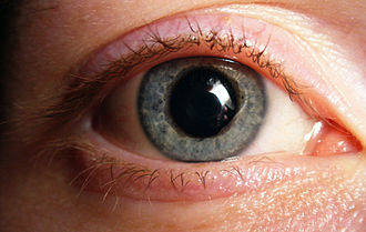
The pupil is the natural opening surrounded by the iris through which light can enter the inside of the eye . It is also called the eye hole . By reducing ( miosis ) or enlarging ( mydriasis ) the pupil with the aid of the sphincter pupillae or the dilatator pupillae muscle , the incidence of light on the retina can be adjusted.
The term pupil is derived from the “doll” (Latin pupilla ), which is reflected in the eye of the other person against a black background.
Pupillary shape and motor skills

The width and shape of the pupils are adjusted depending on the incidence of light via two smooth muscles in the iris. The pupil constrictor ( Musculus sphincter pupillae ) constricts the pupil, the pupil dilator ( Musculus dilatator pupillae ) widens it. This adaptation process ( adaptation ) is regulated unconsciously. A high intensity of the incident light is transmitted to the brain via the optic nerve ( nervus opticus ) and can trigger a pupillary constriction (miosis) starting from the Edinger-Westphal nucleus via the parasympathetic part of the nervus oculomotorius . When the incidence of light is low, the pupil is widened because of the lower parasympathetic effect (mydriasis), with the maximum pupil width depending on the sympathetically innervated dilatator pupillae muscle. An increase in the sympathetic tone (as in the case of fright) can also lead to mydriasis by activating the dilatator pupillae muscle.
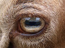
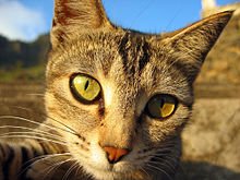
While a maximally dilated pupil is always round, the shape of the pupil can differ between the individual types if the pupil is narrowed. In some species (such as humans and dogs ) the pupil's sphincter is circular, so that the narrowed pupil is also round. In a number of other animals, on the other hand, this muscle runs like a scissor grid in such a way that, with narrowing, oval pupil shapes (e.g. in horses , cattle , marten-like ) or vertically slit-shaped (e.g. in wild / domestic cats , geckos or some snakes ) occur .
The pupil shapes of different animal species have developed in the course of evolution in such a way that they optimally complement the specific optical properties of the respective lens type . Slit-shaped pupils only occur in animals with multifocal lenses. These focus light of different wavelengths through different concentric (ring-shaped) zones of the lens. This creates a sharper image than is possible with eyes whose lenses focus incident light on a single point in the center. In the case of a multifocal lens, a round pupil would completely cover the outer circular regions of the lens, which, however, are needed to focus certain wavelengths of light. With slit-shaped pupils, however, light always also falls through a section of the concentric rings of the lens, so that optimal bundling of the different wavelengths is guaranteed.
The South American armored catfish have a completely different system for regulating the incidence of light , the so-called omega iris of which does not contract from the outside, but rather enlarges or shrinks as a kind of iris pendulum in the center of the pupil.
Physiological basics
The adjustment of the pupil size to the prevailing light conditions is ensured by a control circuit in which primarily regions in the midbrain and in the adjacent diencephalon are integrated as regulators ( control element ). The neural connections emanating from the photoreceptors in the retina as a sensor ( feeler element ) represent the afferent part of a reflex arc , the one going from there to the smooth muscles of the iris as an effector ( actuator ) its efferent part.
The afferents run from the retina via the optic nerve and the optic tract to nuclear areas in the area pretectalis . Connections take place between these, in particular from the nucleus pretectalis olivaris via the commissura epithalamica also to nuclei on the opposite side, which is why the contralateral pupil is consensually narrowed to the same extent when light falls on the retina of only one eye . From here there are connections on each side to the accessory nucleus nervi oculomotorii ( Edinger-Westphal nucleus ), the accessory autonomic subnucleus of the oculomotor nerve , which already belongs to the efferent part.
The parasympathetic efference runs, starting from the Edinger-Westphal nucleus, via the oculomotor nerve to the ciliary ganglion in the orbit. Postganglionic nerve fibers run to the sphincter pupillae muscle . The miosis of the close-up reaction probably takes place via the same nerve fibers as that of the light reaction.
The sympathetic efference that supplies the dilator pupillae muscle is not included in the light control circuit.
Psychological influence
Independently of one another, Israeli researchers and American psychologist Eckhard Hess discovered in the 1970s that the size of the pupil is also influenced by psychological processes. The reason for this is that the dilatator pupillae muscle, which dilates the pupil, is also indirectly connected to the limbic system in the brain via the sympathetic nervous system . The limbic system is also involved in the development of emotions , in learning processes and in the storage of what has been learned in long-term memory . When the limbic system is particularly active, the pupil is dilated.
Pupillary diagnostics
The inspection of the pupils and, at least since Caspar Stromayr , the examination of the pupillary reaction is part of a thorough physical examination ( pupillary light reflex ). It makes sense to check the efference first and only then the afference , since knowledge of the efferent functions is a prerequisite for an assessment of the afference. The so-called pupil comparison test ( swinging flashlight test ) can be used to test the affinity .
If the question is asked, the diameter (wide, normal and narrow), the reaction to light (speed, expression and uniformity) and whether both pupils are the same size ( isokor ) are assessed . Depending on the type of disturbance, there are indications of the location of the damage. In addition to checking the efference of light and close-up reactions, it is also advisable to examine the range of accommodation .
Certain drugs also affect pupil size and response. A mydriasis , i.e. dilated pupils, can be found e.g. B. in the treatment with some eye drops ( atropine , mydriaticum) or in the case of poisoning with hyoscyamine , as it can occur after ingestion of some plants such as thorn apple or deadly nightshade . In the case of poisoning or therapy with opioids , the pupils narrow ( miosis ).
For more in-depth differential diagnostic clarifications, some pharmaceuticals are therefore also used for so-called pharmacodynamic investigations, for example pilocarpine .
Pupil size
The diameter of the pupils determines the opening area for incident light and can be promptly adapted to changes in the ambient brightness, similar to the aperture in a photo camera . Rapid increases in the luminance of the observed environment are usually answered with a narrowing of the pupils ( miosis ), conversely, decreases with an enlargement ( mydriasis ), so that the amount of light irradiated on the retina fluctuates less strongly. A narrowed pupil not only reduces the light irradiation, but also reduces disruptive marginal rays, which enables a sharper image. In the physiological pupil play, the diameter in young people ranges between 1.5 mm (daytime vision) and 8 mm (night vision), which corresponds to a circular area of 1.8 mm 2 or 50 mm 2 . With age, the maximum opening width reduces to 4 to 5 mm.
The current pupil size is the result of the interplay of forces between two oppositely acting trains of smooth iris muscles. Since the pupil constriction is assigned to the parasympathetic nervous system, while the pupil dilator is assigned to the sympathetic nervous system, the relative weight of these vegetative innervations always plays a role. In a healthy person, both pupils are usually the same size, but side differences of up to a millimeter can occur without a pathological background. Two the condition differently on pupils called Anisocoria , one on either side of same diameter Isokorie .
The adaptation of the pupil size to the light environment is triggered by the joint activity of the five types of photoreceptors in the human eye: the rods , the three types of cones and the photosensitive ganglion cells . Depending on the nature of the light (wavelength, duration, intensity), these photoreceptors have varying degrees of influence on the control of the pupil size.
The measurement of the pupil diameter is called pupillometry ; For example, pupillometric methods can use an infrared camera to digitally photograph the pupil. The pupil width can then be determined with the aid of a computer.
Light reaction
With one-sided light irradiation, z. B. by means of a suitable lamp ( pupil light ), both the pupil of the illuminated eye ( direct reaction), and that of the opposite eye ( consensual or indirect light reaction) narrow.
Convergence miosis (convergence reaction)
When focusing on an object in the vicinity, the pupil constricts reflectively, and the depth of field is increased by reducing disturbing marginal rays . This takes place within the control loop described above, which is interconnected with convergence and accommodation and is called the close-up trias .
Lid closure reaction
The eyelid closure reaction (also: Westphal-Piltz phenomenon ) shows itself in the simultaneous narrowing of both pupils when trying to close the eyelids, possibly against resistance. A precise inspection of this phenomenon is not entirely unproblematic in practice, since in this situation the so-called Bell phenomenon , a reflex eye movement outwards and upwards, is triggered at the same time .
Sources of error in the assessment
Especially in emergency medicine with a medical history that is not yet available , illnesses, medication intake or the consequences of injuries can lure the examiner on the wrong track. It is understandable that a glass eye shows no light reactions. Treatment with drugs, e.g. B. pilocarpine to lower the intraocular pressure in glaucoma , cause a miosis , which in this case is wanted and necessary.
Pupil color
The pupil as the eye hole itself has no color. It usually appears black in humans because light entering the eye is absorbed by the inner eye skin - the light-sensitive retina with the darkening pigment epithelium - and not reflected back to the viewer . Supposed discoloration of the pupil can be caused by reflections on the underlying structures in the case of strong exposure, e.g. reflected in the healthy eye from the fundus of the eye as the " red-eye effect " in photographs, or in the case of disease-related changes, for example as the pupil appearing white ( leukocoria ) . In various (nocturnal) animals, the pupil can appear yellowish-green due to reflection on the tapetum lucidum .
Disorders of pupillary motor skills
Efferent pupillary disorder
The classic leading symptom of an efferent disorder is anisocoria . It must first be clarified which pupil is the diseased one, the narrower one or the wider one. As a rule, the pathological pupil changes less when there is a change in brightness than the healthy one, i.e. the light reaction has a smaller amplitude. If both pupils react equally quickly and extensively to light in an anisocoria, there may be a so-called central anisocoria (see below), or a weakening of the dilatator pupillae muscle, i.e. a Horner syndrome . If both pupils show a poor light reaction, there is probably a double-sided efferent disorder or, if the close-up reaction is intact, a reflex pupillary rigidity ( Argyll-Robertson sign )
Parasympathetic efferent disorders
These disorders always mean a paralysis of the sphincter pupillae muscle . The most common cause of this is the compression of the oculomotor nerve in its intracranial course, triggered for example by an aneurysm , a hematoma , tumors or massive brain edema . The pupil is then wide and reacts neither to the incidence of light nor to close focus ( absolute pupil rigidity ). Accommodation is also paralyzed. As a rule, the cases also show a simultaneous paralysis of the external ocular muscles innervated by the oculomotor nerve (see also Clivuskanten syndrome ).
Paralysis of the sphincter pupillae muscle and accommodation without involvement of the external eye muscles ( ophthalmoplegia interna ) suggests ciliary ganglionitis . There are various, usually harmless, causes for this common disease. After regeneration, nerve fibers, which are actually intended for the ciliary body , get lost in the sphincter pupillae muscle and thus result in the picture of pupillotonia , one of the most common parasympathetic innervation disorders. Symptomatic here is the wider pupil, even with strong lighting, which however becomes narrower than the healthy one in darkened rooms. There is also less pupillary excursion when the lighting conditions change. When focusing at close range, the pupils are narrowed in most cases, but the re-dilatation occurs tonicly slower when looking into the distance.
Pupillotonia almost always begins on one side, and in about 20% of cases the other side is affected later. Accommodation is also impaired in around 70%, and reflex disorders in the legs are found in around 50% ( Adie syndrome ).
Differential diagnosis
The frequent pupillotonia should not be confused with the much less common reflex pupillary rigidity ( Argyll-Robertson syndrome ). In both cases, the light reaction is greatly reduced or even eliminated, but in contrast to pupillotonia, both sides are usually affected and the close-up reaction is very prompt. In addition, in Argyll-Robertson syndrome, the pupil is usually very narrow and rounded.
A midbrain lesion, which affects both parts of the light control circuit and supranuclear inhibitor fibers acting on the Edinger-Westphal nucleus, is presumably decisive for reflex pupillary rigidity. The most common cause of reflex pupillary rigidity is syphilis .
Disorders of sympathetic efference
Disorders of the sympathetic efference show up in paralysis of the dilatator pupillae muscle . The sympathetic nervous system is not integrated into the lighting control circuit. Therefore, the light reaction goes out completely if the parasympathetic innervation is switched off, for example by administering atropine . The expression of the sympathetic innervation disorder is the Horner syndrome . Symptoms are the somewhat reduced amplitude of the light reaction with miosis, ptosis and the lower eyelid being slightly elevated.
Central anisocoria
A common and usually harmless disorder is what is known as central anisocoria . The difference in size of the pupil diameter often changes from hour to hour, can also be reversed and is no more than 1 mm. The light reaction of the smaller pupil also shows a smaller amplitude here. The reasons for this are unclear. Anomalous supranuclear inhibition of the Edinger-Westphal nucleus is assumed to be the mechanism. Apparently there is no connection to neurological diseases.
Afferent pupillary disorder
In contrast to efference disorders, there is no anisocoria in those of afferents . If the left optic nerve is completely interrupted, even intensive illumination of the left retina cannot trigger any pupillary constriction ( amaurotic pupillary rigidity ), while the pupils narrow normally when light falls on the right retina. However, minor optic nerve lesions cannot be detected with this method, since strong light stimuli can still lead to maximum miosis despite impaired afference.
For the examination, the pupil comparison test ( swinging flashlight test ) is recommended , a suitable method with which one-sided affections of the optic nerve and, if necessary, the optic nerve junction, optic chiasm can be recorded. On the affected side, the pupil narrows more slowly and expands more quickly ( Marcus-Gunn pupil sign , also: RAPD = relative afferent pupil defect ). Ocular damage, for example due to glaucoma or retinal detachment , can also be detected if necessary. This is also possible with significantly different impairments of the visual fields .
Local pupillary disorder
Local lesions of the iris or the middle skin of the eye ( uvea ) account for the majority of disorders of pupillary mobility and shape . These can u. a. have the following causes:
- congenital malformations ( e.g. persistent pupillary membrane , iris coloboma , aniridia )
- Inflammation (for example the many forms of iridocyclitis of various origins)
- traumatic injuries (for example, bruises)
- Age-related or metabolic degenerative changes ( e.g. iris atrophy )
- Tumors (for example iris cysts or melanomas )
The symptoms are very different depending on the cause and are expressed, for example, in sometimes massive deformities (e.g. clover-leaf pupil ), ruptures and perforations , injections ( ciliary injections ) or adhesions ( synechiae ). A glaucoma attack may cause a mydriatic pupillary immobility.
Hippus
With Hippus is called a partly physiological, partly pathological rhythmic restlessness of the pupils. The reasons for this are still unknown.
See also
literature
- Herbert Kaufmann (Ed.): Strabismus. With the collaboration of Wilfried de Decker u. a. Enke, Stuttgart 1986, ISBN 3-432-95391-7 .
- Theodor Axenfeld (founder), Hans Pau (ed.): Textbook and atlas of ophthalmology. With the collaboration of Rudolf Sachsenweger a . a. 12th, completely revised edition. Gustav Fischer, Stuttgart a. a. 1980, ISBN 3-437-00255-4 .
- Rudolf Sachsenweger: Neuroophthalmology. 3rd, revised edition. Thieme, Stuttgart a. a. 1982, ISBN 3-13-531003-5 , p. 307.
Web links
Individual evidence
- ↑ Duden: The dictionary of origin. An Etymology of the German Language. Bibliographical Institute. Mannheim, 1963. p. 539.
- ↑ Tim Malmström, Ronald HH Kröger: Pupil shapes and lens optics in the eyes of terrestrial vertebrates. In: The Journal of Experimental Biology. Vol. 209, No. 1, January 2006, ISSN 0366-0788 , pp. 18-25, doi : 10.1242 / jeb.01959 .
-
↑ Eckhard Hess : The Tell-Tale Eye: How Your Eyes Reveal Hidden Thoughts and Emotions. Van Nostrand Reinhold, New York 1975, ISBN 978-0-442-23390-7 .
German edition: The speaking eye. The pupil reveals hidden reactions. Kindler, Munich 1977, ISBN 978-3-463-00680-2 . - ↑ Caspar Stromayr: Practica copiosa from the right ground of Deß Bruch Schnidts (1559). Edited by Werner Friedrich Kümmel together with Gundolf Keil and Peter Proff, (Volume 1: Facsimile; Volume 2: Commentary) Munich 1978/1983.
- ↑ Gundolf Keil: "blutken - bloedekijn". Notes on the etiology of the hyposphagma genesis in the 'Pommersfeld Silesian Eye Booklet' (1st third of the 15th century). With an overview of the ophthalmological texts of the German Middle Ages. In: Specialized prose research - Crossing borders. Volume 8/9, 2012/2013, pp. 7–175, here: p. 29.
- ↑ See for example: Jay C. Bradley, Karl C. Bentley, Aleem I. Mughal, Hari Bodhireddy, Sandra M. Brown: Dark-adapted pupil diameter as a function of age measured with the NeurOptics pupillometer. In: Journal of Refractive Surgery . Vol. 27, No. 3, 2010, pp. 202-207, doi : 10.3928 / 1081597X-20100511-01 , examined on 263 people of different ages.
- ↑ Pablo A. Barrionuevo, Nathaniel Nicandro, J. Jason McAnany, Andrew J. Zele, Paul Gamlin: Assessing Rod, Cone, and Melanopsin Contributions to Human Pupil Flicker Responses . In: Investigative Opthalmology & Visual Science . tape 55 , no. 2 , February 4, 2014, ISSN 1552-5783 , p. 719 , doi : 10.1167 / iovs.13-13252 , PMID 24408974 , PMC 3915766 (free full text).
- ↑ Joshua J. Gooley, Ivan Ho Mien, Melissa A. St Hilaire, Sing-Chen Yeo, Eric Chern-Pin Chua: Melanopsin and rod-cone photoreceptors play different roles in mediating pupillary light responses during exposure to continuous light in humans . In: The Journal of Neuroscience: The Official Journal of the Society for Neuroscience . tape 32 , no. 41 , October 10, 2012, ISSN 1529-2401 , p. 14242–14253 , doi : 10.1523 / JNEUROSCI.1321-12.2012 , PMID 23055493 , PMC 3515688 (free full text).

