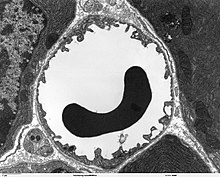
Capillary (anatomy)
In the anatomy ( histology ) of humans and animals, capillaries ( hair vessels ) are the smallest vessels . Instead of the three-layer wall structure made of tunica intima , T. media and T. adventitia , as found in larger vessels, capillaries only have an intima with a basement membrane .
In addition to the capillaries of the blood circulation, the initial lymph vessels are also referred to as lymph capillaries and the finest air routes in the bird's lungs are referred to as air capillaries .
Blood capillaries
Blood capillaries are about 0.5 millimeters long and have a lumen of 5 to 10 micrometers. In most of the organs and tissues of the body they form a fine network - the capillary network ( Latin: rete capillare ) - which is fed by arterioles and drained through venules . The capillary density is different in the individual tissues and corresponds roughly to the mean metabolic activity . Only the cornea , the lens of the eye , horn structures , hyaline cartilage and epithelia are capillary-free. Blood capillaries are not visible to the naked eye. The capillaries, postulated in the 13th century by the Syrian doctor Ibn al-Quff, were first discovered in 1661 by the Italian anatomist Marcello Malpighi in a frog's lung.
A constant exchange of substances takes place via the capillaries : nutrients are supplied to the tissue by means of microcirculation and the waste materials (metabolic end products ) are transported away. The thin capillary walls are semi-permeable (semi-permeable) and consist of a layer of endothelial cells that sit on a basal lamina , in which pericytes are mostly integrated .
A distinction is made between three types of capillary:
- Continuous capillaries have a closed endothelial layer and therefore only allow the passage ( permeation ) of very small molecules . They are typical of the skeletal muscles , the brain , the retina and the lungs .
- Fenestrated capillaries (“fenestrated capillaries”) have pores (“windows”) between the endothelial cells 5 to 12 nanometers, sometimes up to 60 nm , through which smaller proteins can pass. Such fenestrated capillaries are found in the organs in which substances are transported from the tissue into the interior of the vessels, for example in endocrine glands , in the bone marrow , in the intestine , in the pancreas and in the kidney . The largest pores are in the kidney corpuscles . The endothelial openings can be closed by a membrane approx. 4 nm thick or open-pored (kidney).
- Sinusoids ( discontinuous capillaries) are capillaries, enlarged to 30 to 40 µm, with a winding course, which are also fenestrated and have a fenestrated basal lamina. The pores of the sinusoids are around 70 to 80 nanometers in size and allow the permeation of larger proteins and blood cells . They are found in the liver (→ liver sinusoids ), in the spleen , in the bone marrow , in the lymph nodes and in the adrenal medulla . In addition to the gaps between the endothelial cells, there are also pores in the endothelial cells. In addition, there are cells in the vessel wall and the surrounding area that are capable of phagocytosis .
Lymph capillaries
Lymph capillaries represent the initial section of the lymphatic system in humans and in many vertebrates . They end blind and are embedded between the cells of the tissue with anchor filaments . Lymph capillaries consist of a simple layer of flat cells and an incomplete bass palm tree. The endothelial cells are connected to one another, but have gaps through which tissue fluid can flow. The endothelial cells overlap with oak leaf- like protuberances. The tile-like overlapping areas act as swinging tips as inlet valves. There are gaps between the endothelial cells that are so large that even cells (e.g. bacteria , leukocytes ), foreign bodies or proteins (e.g. albumins , antibodies , fibrin ) can penetrate the lymph capillaries through them . This is why the lumen of 30 to 50 micrometers is significantly larger than that of the blood capillaries ( see above ), which means that the lymphatic system is able to remove exudate from injuries, for example .
The lymph capillaries, which are filled with leaked tissue fluid - the lymph - open into larger lymph vessels , which ultimately lead the lymph to the lymph nodes .
Air capillaries
Air capillaries ( Pneumocapillares ) are the air exchange tissue of the bird's lungs . On the one hand, the air capillaries themselves form a network of tubes that usually communicate with one another ; on the other hand, this network of tubes is surrounded by dense blood capillary networks. In contrast to the alveoli of mammals are in the Pneumocapillares the birds not a blind-ended system , but an open (CRT) system.
literature
- Franz-Viktor Salomon, Hans Geyer, Uwe Gille (ed.): Anatomy for veterinary medicine . Among employees by Winnie Achilles. 3rd act. and exp. Edition. Enke , Stuttgart 2015, ISBN 978-3-8304-1288-5 , Chapter: 6 Heart, circulatory and immune systems, Angiologia. (U. Gille, F.-V. Salomon), p. 417-476 .
Individual evidence
- ^ Friedrun R. Hau: Ibn al-Quff (Abū l-Farağ ibn Yaʿqūb ibn al-Quff). In: Werner E. Gerabek , Bernhard D. Haage, Gundolf Keil , Wolfgang Wegner (eds.): Enzyklopädie Medizingeschichte. De Gruyter, Berlin / New York 2005, ISBN 3-11-015714-4 , pp. 1209 f.
- ↑ Renate Lüllmann-Rauch: Pocket textbook histology . Georg Thieme Verlag, Stuttgart 2006, ISBN 978-3-13-129242-1 , p. 246.
- ↑ a b c L.C. Junqueira, J. Carneiro: Histology: cytology, histology and microscopic human anatomy, taking into account the histophysiology . Springer-Verlag, 3rd edition 2013, ISBN 978-3-662-21994-2 , p. 287.
- ^ H. Sarin: Physiologic upper limits of pore size of different blood capillary types and another perspective on the dual pore theory of microvascular permeability. In: Journal of angiogenesis research. Volume 2, August 2010, p. 14, doi : 10.1186 / 2040-2384-2-14 , PMID 20701757 , PMC 2928191 (free full text).
- ↑ a b L.C. Junqueira, J. Carneiro: Histology: cytology, histology and microscopic human anatomy, taking into account the histophysiology . Springer-Verlag, 3rd edition 2013, ISBN 978-3-662-21994-2 , p. 288.
- ^ Margit Pavelka, Jürgen Roth: Fenestrated Capillary. In: Functional Ultrastructure. 2010, Springer, pp. 258-259. ISBN 978-3-211-99389-7
- ↑ Stefan Kubik, Ethel Földi, Michael Földi: Textbook Lymphology: for doctors, physiotherapists and masseurs / med. Lifeguard . Elsevier, Urban & FischerVerlag, 7th edition 2011, ISBN 978-3-437-59332-1 , p. 17.
- ↑ Bernd Vollmerhaus: Textbook of the anatomy of domestic animals . Volume 5, Georg Thieme, Stuttgart 2004, ISBN 978-3-8304-4153-3 , p. 171.

