Osteosynthesis
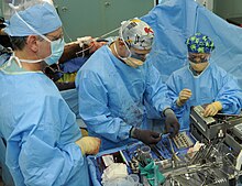
The osteosynthesis (from Greek: osteon , bone '; and synthesis , composition') is the surgical joining of two or more bones or bone fragments with the aim that they grow together.
A method that goes back in principle to the 19th century is understood to be the reduction and stabilization of a bone fracture by means of force carriers surgically attached to or in the bone .
General
An osteosynthesis is usually performed after bone fractures for stabilization , in stiffening operations on joints ( arthrodesis ) or on the spine ( spondylodesis ) and after osteotomies to correct misalignments. Rarer indications are stabilization of bone tumors or for the stabilization of bone at risk of fracture, such as B. in glass bone disease ( osteogenesis imperfecta ).
The goal of every osteosynthesis is the stable fixation of the related bone fragments in order to enable an early functional follow-up treatment with partial and in some cases even full loading of the fixed bones. Then no further immobilization z. B. necessary in a plaster cast and consequential damage is avoided.
In addition, the osteosynthesis should connect the bone fragments in a correct and corrected position in order to avoid misalignments, shortening and rotational errors. Especially in the case of bone fractures with joint involvement (“intra-articular fractures”), an anatomically exact and step-free restoration of the joint should take place in order to avoid post-traumatic joint wear and misalignment.
With an osteosynthesis an absolute or relative stability of the bone fragments is achieved. Absolute stability means that no micro-movements occur in the fracture gap under physiological stress after an osteosynthesis. This allows direct or primary fracture healing. In contrast to this, with a relative stability there is possible micro-movements in the fracture gap and thus indirect or secondary fracture healing by means of callus tissue . If the relative stability is insufficient and the movements are too great, hypertrophic pseudarthrosis can occur.
Types of osteosynthesis
A large number of different implants are available today . The choice of the procedure and the implant depends on many different factors. Depending on the shape, osteosynthesis can be divided into:
- Wire osteosynthesis (especially Kirschner wire or spike wire, also Cerclagen )
- Tension band osteosynthesis
- Screw osteosynthesis with compression screws inserted across the fracture or osteotomy gap
- Plate osteosynthesis , which includes screws for anchoring in the bone
- External fixator
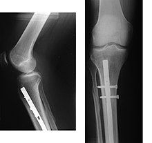
- Intramedullary nail osteosynthesis with long tubular bones inserted into the medullary canal
- Composite osteosynthesis using, for example, bone cement for augmentation of osteoporotic bone or with filling of bone defects
While the external fixator, some wire osteosynthesis and some modern plate osteosyntheses are introduced percutaneously without the need for surgical access to the bone fracture, the other osteosynthesis procedures involve a larger skin incision and opening of the bone fracture for "open reduction" with subsequent internal fixation, Called "ORIF" ( open reduction and internal fixation ).
Wire osteosynthesis
In spike wire osteosynthesis, the fragments (e.g. hand / elbow) are fixed after repositioning using so-called Kirschner wires. Drill wires are used particularly frequently in the treatment of fractures in children . Here the thin wires allow osteosynthesis across the growth plates without damaging them permanently. Therefore, after fracture treatment with drill wires in children there are usually no growth defects. However, the stability of an osteosynthesis with wires is often less than that of other methods. Therefore, the follow-up treatment is often carried out with an additional plaster splint. The advantage of an osteosynthesis with wires is that it can be done through relatively small incisions in the skin. After the fracture has healed, the wires can be removed again. Wire osteosyntheses are often introduced in addition to other procedures, e.g. B. for fracture treatment with external fixator .
Another technique uses wires ( wire cerclage ), which connect the two fragments as a loop and fix them against each other. Such cerclages are z. B. for osteosynthesis in fractures around endoprostheses (joint replacement) or after severing the sternum after heart surgery. This form of osteosynthesis offers little stability and is mainly used to fix fragments in a desired position.
Tension band osteosynthesis
Otherwise, wire osteosyntheses are used for tension strap osteosynthesis . The principle is derived from the Aachen engineer and orthopedic surgeon Friedrich Pauwels , the inventor of the process, from the engineering principle of prestressed concrete : In composite construction, compression-resistant and tensile-strength components are connected, which significantly increases their strength. With the same volume, a body is much more stable than bodies made from just one of the two materials. A concrete beam in which tensile steel bars are cast can withstand significantly higher loads than a pure concrete beam. If the tensile strength element is additionally prestressed, as in prestressed concrete, the load capacity increases and maximum strength can be achieved with a minimum of material.
In a tension band osteosynthesis with tension-resistant wire on pressure-stable bone, the pulling forces of the knee extensor tendon through force deflection and asymmetrical position of the wire cerclage (wire loop) lead to compression of the fragments and thus to possible bone healing. The tension belt principle is only implemented when the knee joint is in the flexed position. In the extended position, there is a gaping of the fracture surfaces near the joint. The desired dynamic compression of the fragments therefore requires early joint mobilization through physiotherapy . Immobilizing patellar fractures operated with tension belts is not effective. Tension band osteosynthesis is only used for fractures of the kneecap and at the olecranon (the end of the ulna on the elbow side ), where tendons pull on bone fragments, a deflecting part of the joint (femoral condyle or humerustrochlea) is present and the tension band principle can therefore be effective. The tension strap procedure is very cost-effective and, with perfect surgical technique, also very reliable. A detailed description of the procedure can be found for patellar fracture .
Screw fixation
Stabilization of the fragments in the shaft area of long tubular bones by screw osteosynthesis alone is not sufficiently stable during exercise. A good application is found in fractures close to the joint, such as fractures of the inner and outer ankle, the femoral condyle, the elbow joint or in the area of the wrist, fingers or foot.
As osteosynthesis usually is at Schraubenosteosynthesen Zugschraubenprinzip applied. For this purpose, the screw thread only engages in the fracture part distant from the head; the screw slides freely in the bone near the head. This attachment of the screw results in the desired compression of the fragments. To do this, a corresponding stepped hole must be drilled. For safe positioning of the screws, these and the instruments used are cannulated and can therefore be used over a drill wire. The drill wire serves as a guide rail, is introduced under the view of an X-ray fluoroscopy and is regularly removed after the osteosynthesis has been completed.
A typical screw fixation is the treatment of the scaphoid fracture of the hand with the Herbert screw named after its inventor.
Very often, osteotomies in the forefoot area are fixed with screws, this leads to an osteosynthesis that is stable during exercise, so that the forefoot must first be relieved until the bone heals. Typical osteosyntheses include a. the chevron osteotomy or the scarf osteotomy of the first metatarsal bone for hallux valgus or the shortening Weil osteotomy of the second to fifth metatarsal bones for claw-toe malposition.
Plate fixation
In plate fixation, fractures in the shaft area of a long tubular bone are stabilized with a metal plate. Osteosynthesis plates adapted to the anatomical conditions of the fracture are available for all types of fractures and for all regions of the body. They are usually combined with screw osteosynthesis for fractures close to the joint or for fractures affecting the joint. A distinction is made between force-fit and form- fit osteosynthesis.
Angular stable osteosynthesis
If a fracture zone has to be bridged without the bone fragments being anatomically reduced, angle-stable plates are required that act like an external fixator but are inserted under the skin surface. The plate is fastened with screws in the non-fractured part of the bone and thus bridges the fracture zone. The force acting on the bone (e.g. the body weight on the femur) is transferred from the bone to the plate via the screws on the proximal part of the bone, passed through the plate over the fracture zone and then back via the screws distal to the fracture passed into the bone. The fractured bone is therefore not involved in the flow of force.
Construction principle
Angular stable plates are called LC plates (or LCP). LCP stands for Locking Compression Plate. The plates are characterized by the fact that the head of the screw used for fastening is firmly anchored in the plate. This can be achieved in a number of ways:
- In most plates, the screw head provided with a thread enters into a force-locking screw connection with the thread in the plate hole. U. even cold-welded when screwing in. In this case, the screw must be placed at a fixed angle to the plate surface (usually 90 °).
- The threaded screw head cuts into the softer sheet material
- The screw is screwed into the plate hole. Then a cap is placed over the screw head. This cap has an external thread and is screwed tightly to the thread of the plate hole. The screw head and cap form a force-locking connection.
The last two methods mentioned allow the screw to be angled within certain limits; it then does not have to be inserted at a fixed predetermined angle. One speaks of polyaxial systems, while the first case is a monoaxial system.
advantages
The advantage of the angular stable implants is that the fracture does not have to be anatomically reduced. It is sufficient to align the broken bone in terms of length, rotation and axis. The debris zone does not have to be touched; the plate can be introduced in a minimally invasive manner using small incisions far away from the fracture zone. Since the bone is supplied with blood by the soft tissues attached to it (periosteum, muscles, tendons, etc.) and the endosteal blood supply via the periosteum has collapsed in the event of a comminuted fracture, the blood supply to the bone with this osteosynthesis procedure is practically no further than the trauma happen anyway, impaired. Bone healing is indirect, i.e. H. through the formation of callus clouds. Another advantage of this plate is the fact that it does not lie directly on the bone, but leaves a small distance between the bone and the plate. This in turn is important for the blood supply via the periosteum, which is not affected by the plate.
disadvantage
The use of an angle-stable plate is a technically very demanding construct, v. a. when rubble zones have to be bridged. The indirect bone healing of the fracture requires a certain degree of mobility of the individual bone parts. This so-called interfragmentary mobility must not exceed a minimum and a maximum dimension, and the maximum distance between the bones must not be too great. Otherwise, fracture healing is delayed, pseudarthrosis develops or, in the worst case, the plate breaks. Another disadvantage is that the screw heads in the plates are sometimes cold-welded. Metal removal is then very time-consuming because the screws have to be drilled out of the plate.
Fixed angle osteosynthesis
Conventional plates are friction-locked processes. They stabilize the bone by tightening the fragments firmly against the plate with screws. Their use assumes that the fracture is almost anatomically reduced. Debris fractures must therefore be reconstructed almost exactly like a puzzle, since the plate can only absorb forces through the force-fit connection between the bone and the plate. The fracture is stabilized by the fact that the plate is pressed firmly against the bone and frictional forces occur between the bone and the plate. By cleverly placing the screws (several screws in different directions), however, a form-fitting construct can be created that offers a certain resistance to the screw being torn out of the bone: the screws that are used to fix the plates can (in contrast to to angle-stable plates) can move freely in the plate hole. The movement is prevented by the fact that they are tightened firmly and that frictional forces act between the screw head and the plate. If, after the actual osteosynthesis, there is a secondary loss of reduction (i.e. the fragments that were captured with the screws change their position), the screw in the plate can loosen and the osteosynthesis construct can loosen until the osteosynthesis fails and the screws even out of the Tear out bones. In the case of comminuted fractures, it is therefore necessary to insert the fragments precisely. However, this would mean a detachment of all soft tissues from the fragments and thus a complete destruction of the important blood supply to the fragments. Healing disorders are promoted, which is why this osteosynthesis procedure was abandoned in favor of angle-stable implants in the case of comminuted fractures. Even in the osteoporotic bone, the screws are only slightly held or even tear off, which is why angle-stable plates are used in this case.
The friction-locked plates are often used for simple fractures with no or only one intermediate fragment. This is where the plates can show their design advantages: The plate is first attached to one side (A) of the broken bone. The fracture is then reduced and the plate is attached to the other side (B) of the fracture. Here the round screw head is turned eccentrically into an oval plate hole. The plate hole tapers towards the bone, causing the round screw head to slide from the edge of the hole to the center of the hole. Of course, the screw cannot slide perpendicularly to the drilled hole in the bone, rather it presses the plate along its longitudinal axis along the bone. This moves the plate a few millimeters and pulls the previously gripped fragment (A) in the direction of the fracture. The fracture gap closes, it is compressed. This dynamic compression plate (DCP) causes primary, direct bone healing. Since the plate only works when it is pressed firmly to the bone, it leads to disturbances in the blood supply. This circumstance is countered in that the underside of the plate is not flat, but is provided with recesses. It is therefore not on the bone with its entire lower surface. This limits the contact area between bone and plate and thus also the area in which the periosteal blood supply is impaired. These plates are called LC-DCP (Limited Contact Dynamic Compression Plate)
Various biomechanical constructs can be realized through a suitable combination of screw and plate and skillful filling of the oval screw holes (at the end of the hole facing the fracture, in its center or at the end of the hole facing away from the fracture). It is possible to compress the fracture as described, to attach individual fragments or to support fragments on the plate (support plate).
Plates are available in a multitude of shapes and thicknesses, so that there are now suitable plates for practically every fracture and every bone, which in their design take into account the shape and the biomechanical requirements of the bone region. There are also plates that combine the system of compression and angular stability, in which the plate is provided with longitudinally oval holes that only have a thread at one end to accommodate the appropriately configured screw head.
External fixator
In contrast to the other internal osteosynthesis procedures mentioned here, this is a concept that allows a fracture to be adequately stabilized by means of wires or screws attached to the respective fragments from the outside . The external fixator is mainly used for temporary stabilization in the case of severely injured persons ( multiple trauma ) or for severe soft tissue damage when a definitive restoration is not directly possible. The fixator is usually applied percutaneously using X-ray fluoroscopy , quickly and gently for the patient and allows intensive medical therapy to be continued quickly. Once the patient has stabilized, the fixator can be removed again and a definitive osteosynthesis, e.g. B. by means of a plate or intramedullary nail. In some cases, fracture healing can take place completely under the fixator. Particularly with special ring fixators ( Ilizarov fixators, hexapods ), complex fractures and malpositions can be corrected and healed.
Intramedullary nail osteosynthesis
Intramedullary or locking nails are particularly suitable for treating shaft fractures (e.g. upper and lower legs). These become minimally invasive , i.e. H. Only through small incisions, introduced into the bone cavity (medullary canal) along the axis of the bone and locked with transverse screws on both sides of the fracture. The advantages of intramedullary nail osteosynthesis lie primarily in the tissue-sparing surgical technique without large accesses, the mostly closed repositioning without showing the fracture and in most cases the primarily load-stable fixation. An early functional follow-up treatment under full load is often possible depending on the severity of the bone fracture.
The principle of angular stability is also used in locking nailing in order to further increase stability and to be able to treat fractures of long tubular bones near the joints. Intramedullary nails can also be used to lengthen bones, e.g. B. after healing of a fracture in shortening, can be used. One way of achieving this "angular stability" is to use a sleeve in combination with a special screw so that the screw "jams" in the nail hole.
Medullary canal splints can be made with so-called TENS nails (Titanium Elastic Nail). In children, however, TENS are often used to treat fractures of long tubular bones (forearm, upper and lower legs). In adults there are only a few indications for osteosynthesis using TENS, e.g. B. with simple fractures of the collarbone.
Composite osteosynthesis
Osteoporotic bone fractures are an increasing problem. Here the hold of the implants is clearly weakened due to the reduction in bone density. In these fractures, one principle for increasing the stability of a plate or intramedullary nail osteosynthesis is so-called augmentation . Special bone cement is placed around the screw tips in places with poor bone quality (e.g. humerus head, thigh near the hip joint) in order to prevent the osteosynthesis from failing due to the screws breaking out. This procedure is also being used increasingly on the spine.
Complications
The possible complications after almost all osteosynthesis procedures include the loss of the reduction (position) achieved, with consequences such as B. Axis deviation and shortening. In rare cases, the osteosynthesis may fail completely with loosening or breaking out of the implants. This often happens when the ends of the bones do not heal adequately.
It should be noted here that an osteosynthesis carried out does not accelerate the healing of the fracture, but primarily ensures that the fracture can heal together in the correct position.
Used material
The used implants are usually made of chromium-cobalt-molybdenum alloys , surgical steel or titanium - alloys such. B. Ti-6Al-4V , but also made of materials such as PEEK (polyetheretherketone), carbon, fiber composites . In rare cases, resorbable implants made of different polymers are used.
Removal of the osteosynthesis
The osteosynthesis material can be removed later (“metal removal”), but this is not mandatory and with some deep-seated osteosynthesis or fixations that have grown firmly into the bone, this is not possible at all or only with great difficulty. First of all, a reliable fusion of the bone fragments must be determined radiologically. Removal is particularly indicated if there is pain over the material, muscle or tendon irritation has formed, material extends into a joint, subsequent interventions must be made possible (such as the use of an endoprosthesis ), nerve or vascular damage occurs or pushes the material through the skin. Removal is also necessary if infection is suspected.
On radiographs are often found even compaction lines (Sklerosesäume) that trace the former implants after removal of an osteosynthesis supply. At the interface between the cancellous bone and the foreign material (e.g. screw) that has been introduced, the bone scleroses as an adaptation reaction to the locally increased load.
Dentistry and maxillofacial surgery
As internal osteosynthesis in the highly atrophic lower jaw, special jaw implants made of titanium correspond to the principle of intramedullary splinting and at the same time serve as anchoring for dental prostheses through posts that are placed at right angles and perforate the gingiva . Because of their microporous roughened surface (TPS) and when exposed to dentures at an early stage , they enter into an intensive, bacteria-proof connection with the bone ( osseointegration ) so that they can remain in the bone permanently. They are usually used to prevent fractures in the lower jaw bone.
History of osteosynthesis
For a long time, fractures were only treated with splinting and relieving the broken bone. The first osteosynthesis emerged in the 19th century; today's procedures were developed in the middle of the 20th century. The working group for osteosynthesis issues (AO) made a significant contribution to this.
Osteosynthesis pioneers were:
- Johann Friedrich Dieffenbach (1792–1847)
- Bernhard von Langenbeck (1810-1887)
- Ludwig Rehn (1849–1930)
- Carl Hansmann (1852-1917)
- Fritz König (1866–1952)
- Albin Lambotte (1866–1955)
- Martin Kirschner (1879–1942)
- Robert Danis (1880–1962)
- Heinrich Bürkle de la Camp (1895–1974)
- Friedrich Pauwels (1885–1980)
- Gerhard Küntscher (1900–1972)
- Alfred Nikolaus Witt (1914–1999)
- Martin Allgöwer (1917-2007)
- Maurice Edmond Müller (1918-2009)
- Gavriil Abramowitsch Ilisarov (1921–1992)
literature
- Carl Häbler : The osteosynthesis in the trade association treatment . Archive for orthopedic and trauma surgery 38 (1937), pp. 283–289.
- Dietmar Wolter, Walther Zimmer (ed.): The plate osteosynthesis and its competitive processes. From Hansmann to Ilisarow . Springer, Berlin / Heidelberg 1991, ISBN 3-540-53536-5 .
Web links
- The history of osteosynthesis (PDF; 277 kB)
Individual evidence
- ↑ Thomas Schlich: Osteosynthesis. In: Werner E. Gerabek , Bernhard D. Haage, Gundolf Keil , Wolfgang Wegner (eds.): Enzyklopädie Medizingeschichte. De Gruyter, Berlin / New York 2005, ISBN 3-11-015714-4 , p. 1083 f., Here: p. 1083.
- ↑ The fracture of the kneecap: Modern tension band osteosynthesis in a historical context I. Klute and NM Meenen 1998 booklets for the magazine "Der Unfallchirurg" 269 Springer. ISBN 978-3-540-63590-1
- ↑ SM Perren: The concept of biological plating using the limited contact-dynamic compression plate (LC-DCP). Scientific background, design and application. In: Injury. 22 (Suppl 1), 1991, pp. 1-41.
- ↑ F. Baumgaertel, L. Gotzen: The “biological” plate osteosynthesis in multi-fragment fractures of the para-articular femur. A prospective study. In: trauma surgeon. 97 (2), 1994, pp. 78-84.
- ^ John H Wilber, Friedrich Baumgaertel: Bridge plating. on: aofoundation.org
- ↑ Martin L. Hansis: fractures. In: Werner E. Gerabek , Bernhard D. Haage, Gundolf Keil , Wolfgang Wegner (eds.): Enzyklopädie Medizingeschichte. De Gruyter, Berlin / New York 2005, ISBN 3-11-015714-4 , p. 419.


