fin
A fin ( Latin pinna ) is a broad or hem-like drive, control and stabilization organ of animals or stages of development of animals that live permanently in water. In the narrower sense, fins are understood to mean the corresponding organs of the fish , and in a broader sense also the corresponding organs of terrestrial vertebrates such as penguins , sea turtles , whales and other marine mammals and various invertebrates .

construction
Fins consist of a structure connected to skin folds (fin skin), the fin rays . In bony fish these rays are ossified, cartilaginous fish have horn rays . The fin rays are anchored in the muscles with fin ray carriers. Some fish species also have skeletal fins ( adipose fin , see below).
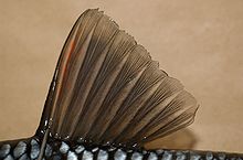
The fin rays of the bony fish are divided into hard (also spiked rays) and soft rays (also limb rays). Hard rays are undivided, mostly smooth pieces of bone, soft rays consist of two halves that have grown together. A distinction is made between different forms of soft rays:
- undivided, undivided, prickly
- undivided, structured
- Fan-like divided, structured
If a fin contains hard rays, these are always in front of the soft rays. The terms hard and soft rays are somewhat misleading. Hard rays can be flexible and soft, while undivided soft rays can be calcified and thorn-like. In case of doubt, the easiest way to distinguish between hard rays and non-structured soft rays is by looking from the front, which shows the two halves of the soft rays.
Real hard rays can only be found in the barbedfish . In some fish, some hard rays are provided with poison glands (e.g. the scorpion fish , weever , rabbit fish ) and a saw-shaped profile on the back is also possible.
Classification and arrangement
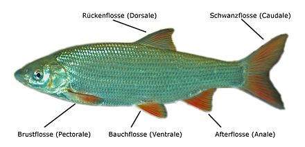
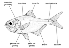
Most fish have seven fins. They are paired and unpaired (individual fins) on the fish body. The paired fins are homologous to the limbs of the terrestrial vertebrates (Tetrapoda), but have no connection with the spine , except for the meat fins .
Paired fins:
- Pectoral fin ( pectorale , pinna pectoralis )
- Ventral fin ( ventral , pinna ventralis )
Unpaired fins:
- Dorsal fin ( dorsal , pinna dorsalis )
- Caudal fin ( caudale , pinna caudalis )
- Anal fin ( anal , pinna analis )

Some species ( catfish , tetra , salmon-like ) also have a skin sac filled with fat, the adipose fin, between the dorsal and caudal fin .
In adaptation to the respective habitat, this basic configuration is partially significantly modified in many fish, so fins can be divided, grown together or greatly changed in shape and even completely missing. Functional adjustments could go so far that the respective fin can no longer be used as an organ of locomotion according to its original purpose.
Dorsal fin

The function of the dorsal fin or dorsal (abbreviation D ; short for Latin pinna dorsalis ) corresponds to that of a keel, that is, it stabilizes the vertical position of the fish in the water. It can be divided into several parts (e.g. perch ) or a whole series of small sections (e.g. Flössler ). The length of the dorsal fin and its position on the body is very variable. Usually there are hard rays in the front part of the dorsal fin or there is a completely hard-rayed front dorsal fin. The sticklebacks even have completely free and mobile spines in front of a rear dorsal fin. In some cases the hard rays have a locking mechanism that allows them to be held upright without muscle strength. The dorsal fin is only missing in very rare cases (e.g. electric eel ).
Special shapes:
- For pipefish this fin (along with the pectoral fin) is used to generate the propulsion.
- Most armfinches have developed baits from the dorsal fin rays to attract potential prey.
- The dorsal fin of the ship keepers is transformed into an adhesive organ.
Anal fin
The anal fin is similar in function and shape to the dorsal fin. It can also be divided and have hard rays in the front part.
Special shapes:
- Some families of the dentin have transformed the anal fin into a mating organ :
- The gonopodium in viviparous toothcarps .
- The andropodium in highland carp and half-beaked pike .
Pectoral fins
Corresponding to their function as height control, brake and stabilizing organ, a pectoral fin or pectoral (abbreviation P ; short for Latin pinna pectoralis ) is usually flexible, soft and transparent. Pectoral fins are connected to the skull via the skeleton and are therefore almost always located directly behind the gill covers. Occasionally, as is the case with many catfish , the leading edge is reinforced by hard soft rays. These fins can also be missing, for example with moray eels . Wrasse and surgeon fish swim through simultaneous beats of the pectoral fins (labriform), here the pectoral fins are the main driving organ.
Special shapes:
- A wing-like modification of the pectoral and pelvic fins allows the flying fish to glide longer in the air above the surface of the water.
- The mudskippers can move on land with the help of their stem-like pectoral fins.
Pelvic fins
These fins are usually relatively small, they take on control functions. The position on the fish's body varies between belly, chest and, in rare cases, still in front of the pectoral fins, throaty. Of all fin species, these fins are most often missing, including eels , sea wolves and most puffer fish relatives without ventral fins.
Special shapes:
- A suction disk formed from the ventral fins allows the gobies , the shield fish and the sea hares to find a better grip on stony ground.
- Gourami carry taste buds on their thread-like elongated pelvic fins.
- Gurnards have tactile organs formed from the first rays of the pelvic fins.
- In cartilaginous fish and plate membranes , parts of the pelvic fins in males are converted into clusters that serve as paired mating organs.
Caudal fin
Together with the caudal stalk, the caudal fin is the main driving organ in many fish. Fish usually generate propulsion by pushing their body forward through the water with powerful, side blows. The body of the fish performs strong, wave-like movements along its axis. The rays of the caudal fin are directly connected to the spine. Only in very few, highly specialized species, such as B. the seahorses , the caudal fin is missing.
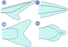
Tail fins are divided into six different types according to their anatomy (see graphic):
- Heterocerk (A): The end of the spine bends upwards and supports the larger upper part of the caudal fin, as is the case with most sharks and primeval bony fish such as the sturgeon (Acipenseriformes) and the bake (Lepisosteidae).
- Protocerk (B): The end of the spine is straight. The caudal fin forms a border around it, e.g. B. in the eel-like (Anguilliformes).
- Homocerk (C): The caudal fin is symmetrical, for example in most real bony fish (Teleostei). Even so, the end of the spine can still bend up a little with primitive shapes. It is no longer visible from the outside, but shows that the homocerke caudal fin has developed from the heterocerke.
- Diphycerk (D): The end of the spine is straight. The caudal fin consists of two parts above and below the spine, for example in the coelacanth ( Latimeria ). Sometimes the clavus of Mola mola is referred to as a diphycerker tail - which is not the case, because Mola completely loses its caudal fin as a larva and the clavus (also known as “nail”) is completely re-formed from the dorsal and anal fin. See below: gephyrocerk.
- Hypocerk: The end of the spine curves down and supports the lower part of the caudal fin, e.g. B. in the ichthyosaurs .
- Gephyrocerk: The caudal fin closes the blunt-ended body as a hem. This only occurs in the sunfish (Molidae).
Fin formula
The number and type of fin rays can be described with the help of the so-called fin formula .
The information on the individual fins is made up of three components: fin designation, number of hard rays, number of undivided and divided soft rays.
The fins are given by the Latin name, often only with the first letter: A for the anal fin (anal), C for the caudal fin (caudal), D for the dorsal fin (dorsal) and P for the pectoral fins (pectoral). If the same type of fin is present several times, an Arabic number is given immediately after the letter for counting.
Hard rays are indicated with Roman numerals , soft rays with Arabic numbers . Since hard rays and undivided soft rays are always at the beginning of the fin, the divided soft rays are always in the rear part of the fin, a clear representation can be created by separating them with a slash.
Examples:
- DI / 5: In the dorsal fin, a hard ray is followed by five soft rays.
- D2 3/9: In the second dorsal fin, three undivided soft rays are followed by nine divided soft rays.
- A II-III / 5-7: In the anal fin, two to three hard rays are followed by five to seven soft rays.
- C II (-III) / 7: In the caudal fin, two, in exceptional cases three, hard rays are followed by seven soft rays.
A fin formula using the example of largemouth bass : Dorsal X-XI / 12-13, Anale III / 10-11. Or shorter: D X-XI / 12-13, A III / 10-11.
Often not all fins are listed in the fin formula. In particular, the information on the caudal fin is often missing, as this is less significant and can be more difficult to count due to the pre-rays.
Notes on the evolution of the fins
Importance of the fins for the system
The shape, structure and number of fins are characteristic of a species and therefore play an important role in its description and determination.
Development of the caudal fin skeleton of bony fish
Despite its small size, the skeleton of the caudal fin - the rear end of the spine - has an astonishing variety of features, which has increasingly been recognized as being systematically important (T. Monod 1968). Within the ray fins we find a higher differentiation of the spinal column end, comparable to the evolution in birds from about the Archeopteryx stage. So here is an overview.
Since the tail fin of the real bony fish (Teleostei) is becoming more and more important for propulsion compared to the rest of the tail, the tail fin becomes larger and, above all, stiffer. It does this by fusing the bones involved ( synostoses ). The tuna are at the peak of development , but the Loricariidae also show a high degree of merging. Furthermore, Gonorynchus , for example , who has perfected the method of escaping his enemies by dipping into the sand at lightning speed - this requires strong acceleration through the caudal fin.
All the vertebrae that lie behind the bifurcation of the dorsal aorta (main artery) are called "caudal vertebrae" . In fish, the aorta runs under the spine in the hemal canal, protected by vertebral processes, from the head (gill basket) to the caudal fin. In the tail section of the body, these processes are united to form hemal arches. A distinction is made between prural vertebrae (PU) and ural vertebrae (U). Originally there were up to ten ural vertebrae ( acipenser ), at Hiodon eight are created.
The illustration shows the bent end of a spine, which here consists of three prural vertebrae. The rearmost one (PU 1) is a half-vortex (the PU are numbered from back to front). It is hollowed out like an hourglass at the front to accommodate an intervertebral chorda nucleus. The rear half, however, is narrowed and fused with two ural vertebrae (which can only be found outlined). The intergrowth complex PU1 + U1 + U2 is called urophore. It is a feature of the higher forms.
The long bone plates attached to the adhesions are also important. Usually there are four or five hypuralia (HU). The hypuralia are transformed (mostly widened) hemal appendages of former Ural vertebrae. Since they are bent up dorsally , the bone plates had space to widen into a fan to which the caudal fin rays attach at the rear.
In the picture we see the three lower hypuralia fused to PU 1 without a seam (it is unclear for HU 4). The parhypural (the hemal process of PU 2) and dorsally the stegural (descendant of the neural process of PU 1) are obviously also involved in the tail skeleton. The smaller lateral uroneuralia (descendants of the neural arches of ural vertebrae), of which there are usually two or three pairs, have already been lost . But uroneuralia can also fuse with the complex, as Gonorynchus shows.
Differentiation of the muscles and swimming techniques
The evolutionary problem with swimming has been that the large core muscles work inefficiently when the fish swims slowly or for short distances. Therefore, a lateral differentiation was initially created (near the sideline ): “Red” ( myoglobin-rich ) muscle fibers alone are responsible for swimming calmly. The large “white” part is then only used to escape or to pursue prey. The "white" muscles are responsible for oxygen and tire quickly. The trunk muscles of the anchovies are not divided into “red” and “white” - they are always nimble swimming animals.
When differentiating between “red” and “white”, the much larger “white” portion is inevitably moved with it even when swimming calmly, which costs energy. Many fish families have therefore produced even more "ingenious" solutions. They only use the smaller muscles of the fins for smooth movement. The white trunk muscle (which is divided by plates of connective tissue) is used solely to attack and flee.
Swimming ways of fish
Depending on the presence and the formation of the fins and their neural control, several types of locomotion have developed in the fish.
Movement with the core muscles
The following types use the core muscles to move. The list begins with winding the whole trunk, followed by types with a shorter trunk and with increasing importance of the caudal fin.
- The anguilliform type shows a body snaking (in "sine waves"), for example eels and moray eels (with or without a fin edge, therefore also useful in crevices or substrate!). This "original" mode of locomotion can already be seen in ginger and lampreys as well as the collar shark ( Chlamydoselachus anguineus ). The transition to the following type is shown by the sturgeon .
- The subcarangiform type is already shorter (fewer vertebrae), the torso stiffer: the "wave bellies" go back to 1. The majority of sharks and bony fish (approx. 85% of the species) belong to this type.
- In the carangiform type, the trunk is shorter, the tail and the caudal fin are becoming more and more important, the "sine wave" is only half present. The jacks (Carangidae) are "good", persistent swimmers. Herring sharks also belong here , e.g. B. Lamna - with transition to thunniform swimming style.
- The thunniform type only moves the caudal root and fin in a vigorous manner , the trunk muscle transmits its power to the very stiff fin blade by means of tendon plates. These fish also hardly “rest” (except possibly for “sunbathing”). Examples are thun (Thunnini) and probably also swordfish (Xiphiidae).
- The tail alone moves when fleeing, but also the boxfish (Ostraciidae), whose trunk is in a shell: ostraciiform type with largely dissolved trunk muscles; otherwise they swim tetraodontically.
Aphanopus carbo (Scombroidei) swims anguilliform, but when this pelagic predator spots prey, its trunk stiffens and it "sneaks up" ostraciiform (very small caudal fin). Theelectric rays (tail only!) And the elephant fish (Mormyridae) with stiffened tails (the muscles are transformed into an electrical organ)show echoes of ostraciiform swimming.
Smooth swimming thanks to the movement of the fins

In the following types of locomotion, no core muscles are used for slow swimming, and most of the time neither the caudal fin is used. Instead, other fins are used, for example with undulating movements (undulation). The caudal fin, if present at all, is usually only used for steering.
- Wavy fin movement
- Undulation of the dorsal fin: The bald pike ( Amia calva ) swims amiiformly , mainly due to undulation of the long dorsal fin. Similar numbers of Umberfishes (Sciaenidae), the Greater Nilpike ( Gymnarchus niloticus ), and also the Sea Cats (Chimaeriformes) (tail reduced) with the support of the pectoral fins. Under other conditions also the seahorses ( Hippocampus ) and the related pipefish (Syngnathidae).
- Undulation of the anal fin: Old World (Notopteridae) and New World knife fish (Gymnotiformes) swim gymnotiform , with anal fin undulation (the caudal fin is missing).
- Undulation of the dorsal and anal fin: The puffer fish (Tetraodontidae) have undulating, narrow dorsal and anal fins ( tetraodontiform swimming). They can use their tail fin to help them escape.
- Undulation of the pectoral fins: Porcupine fish (Diodontidae) swim diodontiformly , by means of undulation of the very broad pectoral fins. The rays (Batoidea) swim ( rajiform ), also with undulating, very widened pectoral fins (tail fin reduced or absent) - or the stingray-like (Myliobatiformes) more "flying".
- Undulation of the caudal fin: There are also fish that can move forward with undulations of the caudal fin (!), E.g. B. the groupers of the genus Epinephelus .
- More types
- Rowing pectoral fins: Wrasse (Labridae) swim labriform , mainly by means of the pectoral fins ("rowing"). The surgeon fish (Acanthuridae), the surf perch (Embiotocidae) u. a.
- Triggerfish (Balistidae) swim in the form of a balist , by “flapping” their dorsal and anal fins against each other. Similar to the sunfish (Molidae), which have no functional caudal fin and reduced trunk muscles; also with the support of "recoil" of water from the gill cavities.
Mammalian fins
The fins of the whales are called fluke for the caudal fin, flipper for the pectoral fin (pectoral fin ) and fin for the dorsal fin (dorsal fin).
Also as fins are designated with the like fins converted extremities sea cows , seals (Pinnipedia, which means "pinniped") and the platypus .
literature
- Horst Müller: Fish of Europe. 2nd Edition. Neumann Verlag, Leipzig / Radebeul 1983, ISBN 3-7402-0044-8 .
- Dietrich Starck (ed.): Textbook of special zoology. Volume 2: Vertebrates. Part 2: Kurt Fiedler: Fish. Fischer, Jena 1991, ISBN 3-334-00338-8 , pp. 16-21.
- Günther Sterba : Freshwater fish in the world. License issue. Weltbild-Verlag, Augsburg 1998, ISBN 3-89350-991-7 .
- Günther Sterba (Ed.): Lexicon of Aquaristics and Ichthyology. Edition Leipzig, Leipzig 1978.
- Gerhard KF Stinglwagner, Ronald Bachfischer: The great cosmos of fishing and fishing dictionary. Franckh-Kosmos, Stuttgart 2002, ISBN 3-440-09281-X .
Web link
Individual evidence
- ↑ Manfred Klinkhardt: D. , pinna dorsalis. In: Claus Schaefer, Torsten Schröer (Ed.) The large lexicon of aquaristics. 2 volumes, Eugen Ulmer, Stuttgart 2004, ISBN 3-8001-7497-9 , Volume 1, p. 318, and Volume 2, p. 778.
- ^ Manfred Klinkhardt: P. , pinna pectoralis. In: Claus Schaefer, Torsten Schröer (Ed.) The large lexicon of aquaristics. 2 volumes, Eugen Ulmer, Stuttgart 2004, ISBN 3-8001-7497-9 , volume 2, pp. 736 and 778.
- ↑ Théodore Monod : Le complexe urophore des téléostéens (= Mémoires de l'Institut Fondamental d'Afrique Noire 81, ISSN 0373-5338 ). Institut Fondamental d'Afrique Noire, Dakar 1968.
- ^ See Lance Grande, Terry Grande: Redescription of the type species for the genus † Notogoneus (Teleostei: Gonorynchidae) based on new, well-preserved material. In: Journal of Paleontology. 82, 2008, pp. 1-31. doi : 10.1666 / 0022-3360 (2008) 82 [1: ROTTSF] 2.0.CO; 2 , App. fig. 2.
- ↑ Spektrum.de . Batidoidimorpha . In: Compact Lexicon of Biology . Spectrum Academic Publishing House, Heidelberg. 2001


