Mouthparts
As mouthparts (also mouth limbs ) are generally structures of arthropods (Arthropoda) denotes serving of food intake. These are mainly specially trained extremities. The mouthparts of the various groups of arthropods vary greatly and can vary greatly within the individual taxa .
evolution
The mouthparts of the arthropods are based on transformed extremities of the front body segments that have fused to form the head . In fossil representatives from the Cambrian, unspecialized extremities predominate in the head area, which do not differ in structure and function from those of the trunk segments, so there was no difference between mouthparts and legs. Only the first pair of extremities, the later antennae , had a different shape. This is how the head appendages of the extinct trilobites are constructed . Unspecialized extremities in the trunk area, which serve not only for locomotion but also for breathing and feeding, still exist today in some arthropod groups such as the gill pods . However, there are no more recent arthropods with unspecialized head extremities, most of them died out in the Cambrian.
As is typical for arthropods, each of the original segments has at most one pair of limbs, but this can sometimes be completely regressed. How many segments formed the head in the various sub-tribes of the arthropods and how the various mouthparts can be homologated between them is still scientifically controversial.
Functionally, the mouthparts of the arthropods differ from the jaws of vertebrates and other animal phyla in that the mouthparts are always in front of the mouth opening. Within the individual groups, depending on the particularities of the acquisition of food, they are diversely transformed and modified. There are two basic types according to which (following today's prevailing hypothesis) the respective kinship groups are named. The chelicerae of the Chelicerata (arachnids and relatives) are tripartite in the basic plan that act as scissors . The mandibular animals, however, have chewing-biting mandibles as the first pair of mouth parts in the basic plan . The mouthparts have, depending on the group, additionally structured appendages that act as sensory organs and are called palps or buttons. In particular, in insects mouthparts but are completely modified in some groups, such as a leak organ or proboscis .
Columbus
The segmentation of the head by body appendages is clearly recognizable in the columbus (Onychophora). The jaw segment follows the antenna segment. It carries a pair of stubby short limbs that are trimmed with paired chitin at the top . The jaws cut from front to back in a motion that corresponds to the movement of the legs. In the segment behind the jaw segment, the extremities are transformed into stubby protuberances (oral papillae) into which the mucous glands open. The body parts with lip function (closing the mouth opening or opening and placing on the prey) surround the two jaws.
Claw bearers

With the jaw-claw carriers (Chelicerata) the head and the chest form a unit with six pairs of extremities. The last four take on the function of locomotion. The first two are known as the mouth limbs. The first pair of legs form the chelicerae (jaw feelers), the second pair of extremities is called the pedipalpus (jaw feeler).
be crazy
In the arachnids , the chelicerae consist of a powerful base member and the claw that can be struck against the base member. Poison glands almost always open near the tip of the claw. In primitive chelicerates, the two claws work parallel to the body axis (orthognathic position), otherwise against each other and perpendicular to the body axis (labidognathic position). The second pair of extremities is called the pedipalps (jaw probe). It has the function of a button, with the males it is also used to transfer the semen package. The first link is widened and covers the side of the mouth. A lower lip is separated from the abdominal shield of the exoskeleton towards the front and closes off the oral cavity towards the rear. At the front it is closed by the upper lip (epistome or rostrum). There are no chewing elements, as spiders only absorb liquefied food.
Scorpions
In the scorpions , the chelicerae are small, the pedipalps are structured similar to the striding legs. The last leg link carries a pair of pliers similar to Cancer's claws.
Mites
With the mites , the mouthparts sit on the front part of the front part of the body, the Gnathosoma (old Greek gnathos 'jaw' and soma 'body'). The gnathostoma is formed on the underside by the fused hip joints of the two mostly five-membered pedipalps, on the upper side by the tectum and tubularly encloses a space in front of the mouth. The chelicerae and pedipalps are transformed into a lancing device. The two- or three-part chelicerae work like scissors or knives or as needle-like stilettos.
Crustaceans

In the crustaceans (Crustacea) the first two head segments each carry a pair of antennae . The upper lip lies directly in front of the mouth opening. As with the other mandibular animals (to which the crustaceans and tracheal animals are grouped), the base of the extremity of the third head segment is transformed into a powerful chew ( mandible , upper jaw). The two following pairs of extremities also represent mouthparts, which are referred to as maxilla I and II (lower jaw and lower lip). They surround the mouth opening. They reveal the basic plan of the split foot, with an endopodit and an exopodit sitting on the base podid. In the mandibles and the two pairs of maxillae, serrated protrusions, the chewings, protrude from the trunk. Both, one or neither of the exopodit and endopodit can still be preserved as buttons. In various groups within Cancer, other extremities are used as mouthparts, which are then referred to as maxillipedes (jaw feet). In the case of crayfish, for example, three pairs of jaw feet are followed by the scissor foot. This can also grab food, chop it up and pass it on to the jaws.
Sackkiefler
The Sackkiefler (Entognatha altgr. Ento 'inside' and gnathos 'Kiefer') used to be placed next to the insects, today the hexapods are divided into Sackkiefler and insects. The sack pines include the double-tails , leg-teasers and springtails . With them, the mouthparts are inside a specially designed pocket. During the embryonic development, a mouth fold forms on each side, which deepens downwards. The two folds unite on or under the lower lip. The space in front of the actual mouth is thus directed forward and encloses a pair of mandibles and maxillae, which are also oriented forward. The mandibles sit in front of the maxillae and can be turned out forward.
- In springtails (Collembola), only the tips of the jaw protrude from the pocket. The upper and lower jaws are usually chewing, but can also be shaped like a pen and then used with a piercing and sucking technique. By changing the starting point of the muscles, the rotating movements when chewing become forward and backward movements when stabbing.
- The mouthparts of the leg tinkerers (Protura) are transformed into a bundle of needle-like stilettos with which they presumably drill into and suck out fungal hyphae.
- With the double tails (Diplura) the mouthworks are biting.
insects
The mouthparts of insects (sometimes called “jaws” by analogy with vertebrates) are formed by the appendages of four sections of the head capsule. Their job is to prepare the food for absorption in the digestive tract and to transport it into the mouth. With the exception of the upper lip, the mouthparts developed from three pairs of extremities of the head, which were constructed similarly to the split feet . The two basic types of mouthparts are the chewing-biting type for ingesting solid food and the sucking type with various sub-types for ingesting liquid food. Aristotle already made this distinction .
Biting mouthparts
| Mouthparts of different species of beetles | ||
|---|---|---|
|
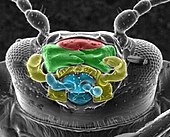
Fig. 2: Leaf beetle , head from below: red: upper lip green: upper jaw yellow: lower jaw blue: lower lip |
|
| Fig. 3: Mulmbock , upper lip, tinted blue on the right A: outside B: inside (epipharynx) |
||

|

|
|
| Fig. 4: Head from above, field tiger beetle, upper lip brown (natural), right lower jaw removed |
Fig. 5: Underside of the head Copper-colored sandpiper green: lip button blue: jaw button |
|
The biting or chewing mouthparts are classified as more primitive than the sucking mouthparts. The mouthparts chop up the food. Depending on the food source used, the focus is on tearing off or biting off, tearing or cutting up, gnawing, chewing or grinding the food or a combination thereof. The biting mouthparts are composed of four elements. These are called the upper lip (labrum), upper jaw (mandible), lower jaw (maxilla) and lower lip (labium), analogous to the naming of food preparation in humans. In insects, however, both in their structure and in their function they are completely different from the lips and jaws of mammals. The position of the four elements in a ladybug is shown in Fig. 2.
The upper lip (labrum)
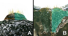
In front of the mouth is the unpaired upper lip. In the ancestors of the insects, the head was composed of its point (Akron) and probably six rings. The upper lip is considered to be the remnant of the last ring in front of the head. The upper lip is immobile, relatively stiff and prevents food from falling forward out of the mouth. From the outside you can only see a part, as the base of the upper lip is covered by the head shield (clypeus). The visible part can be semicircular (Fig. 3 A, right half tinted blue), rectangular, triangular, kidney-shaped or otherwise shaped. It is often filled with sensory bristles.
If you remove the rest of the mouthparts, you can see the side of the upper lip facing the mouth (Fig. 3 B, right half tinted blue). It closes the mouth at the front and is therefore called the epipharynx (epi, ancient Greek and pharynx, ancient Greek pharynx). The surface of the inside is not smooth, but structured in such a way that the food glides easily towards the mouth, but difficult in the opposite direction. Immediately in front of the oral cavity are sensory cells, with the help of which the final decision is made as to whether the food is suitable for consumption.
The upper jaw (mandibles)
| Deflection of the upper jaw into the head capsule using the example of the Mulmbock lower jaw and lower lip removed |
|
|---|---|

|
|
| Fig. 6: Lateral view of the upper jaw blue arrow: upper joint green arrow: lower joint brown arrow: joint cavity of the lower jaw |
|

|
|
| Fig. 8: lower joint and joint cavity A: upper jaw from below, green: joint head B, C, D: head capsule with upper jaw from below B green: position of the joint cavity in the head capsule D: brown: joint cavity of the lower jaw |
|
| Fig. 7: Upper joint A: Upper jaw from above B: Head capsule removed from the lower side of the head capsule |
|
Behind the upper lip lies a pair of jaws, the upper jaw. They developed from a pair of legs that lay next to the mouth. Each upper jaw consists of only one piece.
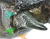
Only in the very primitive insect order of the rock jumpers (Archaeognatha) is the upper jaw connected to the head capsule by only one joint, similar to the antennae. In all other insects, the upper jaws are pivoted into the head capsule with two joints, comparable to the two pivots of a door. There is a joint above the upper jaw (blue arrow in Fig. 6) and a joint below the upper jaw (green arrow in Fig. 6). As a result, the jaw can now be moved very stably in just one plane perpendicular to the axis between the two joints. Only this enables powerful biting and grinding with the two upper jaws. The two deflections are further forward and further back on the head, so the jaws cannot be moved parallel to the head axis, but perpendicular to it. In Fig. 6–8, this turning is shown using the Mulmbock beetle as an example . On the underside of the upper jaw there is a strong, knob-shaped outgrowth (in Fig. 8 A tinted green). There is a corresponding cavity in the head capsule (in Fig. 8. B the location of the cavity is tinted green). Even in this joint, the upper jaw has little play when it rotates. On the upper side of the upper jaw there is a deep furrow in the form of an open ring (in Fig. 7. A the bottom of the annular recess is tinted blue). There is a corresponding ring wall-like elevation on the head capsule. With a natural constellation of the jaw, the blue-tinted surfaces grind against each other.
The upper jaw is moved by tendons of antagonistic muscles, the stronger adductors for closing and the weaker abductors for opening the upper jaw. In accordance with the food specialization, a specialization of the upper jaw also developed. In the basic type, an upper jaw is thick and curved on the outside, narrowing towards the inside to a cutting edge and towards the front to a point. These can be smooth or finely serrated or have individual blunt or pointed teeth. Often there are small bumps on the grinding surface of the cutting edge, which can be particularly hardened due to the inclusion of metals ( zinc and manganese , occasionally iron ). It is known that some beetle larvae are able to gnaw through iron fittings. The two upper jaws can meet like a pair of pliers or slide over one another like scissors with the cutting edges. The upper jaws in Figures six to eight are specialized in gnawing wood, the upper jaws in Figure 4 in securely grasping and tearing small flies. In the male stag beetle ( Lucanus cervus ), the upper jaws, also called antlers, are not used to acquire food, but are used in fights between the males to override the opponent.
The lower jaw (maxillae I)

|
|
| Chrysobothris affinis | Loricera pilicornis |

|

|
| Ovalisia rutilans | Trachys minutus |
| Fig. 9: Lower jaws of different beetle species | |
The lower jaw lies between the upper jaw and the lower lip. Each lower jaw has only one deflection with the head capsule (in Fig. 8. D the joint cavity of the lower jaw is tinted brown). Accordingly, the lower jaws have greater mobility than the upper jaws, but in principle they only move in one plane parallel to the upper jaws. In the case of the lower jaws, the origin of the legs is best understood. They are multi-part and have several attachments. The basal phalanx is divided into two parts, the base is called cardo , it is separated from the stipes by a flexion fold . The latter has two flat limbs, the inner ( Lacinia ) and the outer ( Galea ) chew, as well as a jaw probe, which consists of up to seven limbs. The maxillary palps and the galea carry sensory organs for finding and assessing possible food, the inner ark is mainly used for chewing and is therefore usually strongly structured on the inside. In Fig. 9, the lower jaws of four species of beetles show the wealth of shapes of the lower jaws. In Loricera pilicornis (Fig. 9) and Elaphrus cupreus (tinted light and dark blue on the right in Fig. 5), two palpitations can be seen. The lower jaw is responsible for chewing and transporting food.
The lower lip (labium, homologous maxilla II)

|

|

|
| Chrysobothris affinis | Ovalisia rutilans | Trachys minutus |
| Fig. 10: Lower lip of three species of beetle | ||
The rearmost or lowest element of the mouthparts is the lower lip or labium. It is unpaired, but also developed from a pair of head extremities. The lower lip does not begin in front of the chin, as the various sections of the head capsule grouped around the chin are also included in the lower lip. The back part, the postmentum (lat. Post 'after', 'behind' and mentum 'chin') is analogous to the cardo and is again divided into submentum (lat. Sub 'under') and mentum . The foremost part, the pre- mentum (Latin prae 'before'), is analogous to the stipes. The tongue ( glossa ) sits in the middle of the prementum . It is homologous to Lacinia. At the base of the tongue there is a side tongue (paraglossa) that corresponds to the galea. The paired origin of the lower lip is still partially recognizable (in Fig. 11 A, light blue). A pair of buttons, the lip buttons or labial palps, also arise on the lower lip. Fig. 10 shows the lower lips of three species of beetle.
Inner lip (hypopharynx)
Traditionally, the inner lip is not counted among the chewing-biting mouthparts ( hypopharynx from hypo, lower and pharynx, lower, throat). It divides the pre-oral space (prae- ( Latin ) in front of and oral (Latin) oral-) in front of the actual mouth and delimited by the mouthparts into the oral cavity ( cibarium ) and the salivary pocket ( salivarium ). The salivary glands empty into this. The inner lip plays an important role in the development of the sucking mouthparts.
Sucking mouthparts
| Fig. 11: Homology of mouthparts (A) grasshopper - (B) bee --- (C) butterfly - (D) mosquito ♀ |
||
|---|---|---|
|
c: composite eye ; a: antenna ; lr: labrum; md: mandible; mx: maxilla; lb: labium hp: hypopharynx |
||
| Upper lip (labrum) |
Upper jaw (mandible) |
Lower jaw (maxilla) |
| Lower lip (labium) |
Throat above (epipharynx) |
Throat below (hypopharynx) |
| Fig. 12 - Fig. 25 with the above color code c: food duct s: salivary duct |
|||||
|---|---|---|---|---|---|

|
Fig. 12: Honeybee detail of the mouthparts, lower jaw folded away on the right A: frontal B: on the left, partially tinted yellow GA: Galea yellow St: Stipes blue G: Glossa blue P: Lip button |
Fig. 13: Section, explanation in the text |
|||
|
Fig. 14: Butterfly trunk, rolled up |
Fig. 15: Section, explanation in the text |
||||
 Fig. 16: Real fly's trunk Fig. 19: Schnake head with trunk  |
 Fig. 17: Real fly |
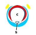 Fig. 18: Tsetse fly |
|||
 Fig. 20: Brake |
 Fig. 21: Mosquito |
||||
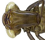 Fig. 22: Peat damsel larva, mask from below |
Fig. 23: Fire beetle , larva |
||||
 Fig. 24: Spotted back swimmer |
Fig. 26: Hemiptera |
||||
 Fig. 27: Fringed winged winged wing |
Fig. 28: Head flea |
Fig. 29: Flea |
|||
Insects with sucking mouthparts ingests liquid or liquefied food. For example, in butterflies and bees, this is flowers nectar, in leaf bugs , cicadas and aphids, plant sap , in fleas and animal lice, blood. The spectrum of food in flies is particularly rich (blood, liquefied food, liquids resulting from putrefaction). In the course of evolution, the mouthparts were transformed into a proboscis as a feeding tube for absorption . From a certain length of the trunk, a pump system was also required for the transport within the trunk. If saliva is to be transported in the opposite direction, this is most effective when the proboscis contains two separate ducts, a saliva tube and a feeding tube. The saliva of some stinging insects is not used to liquefy the food, but it hardens when it escapes, isolating the stab wound from the outside and thus enabling the food source to be used more effectively.
Not only the upper and lower lip and the upper and lower jaw work together in the formation of the trunk, but also the ceiling and floor of the oral cavity ( epipharynx and the inner lip of the hypopharynx ). This is also the prementum , mentum and Postmentum may be involved in the trunk education, further evidence is that they are homologous to portions of the head extremities.
The development of a proboscis occurred several times and in different groups of insects independently of one another. The share of the body parts just mentioned in the structure and function of the trunk is different. Of course, the jaws still lie within the lips, but they do not necessarily lie within the trunk, but can also contribute to the formation of the outside of the trunk. With originally the same development of the trunk, the radial adaptation resulted in a split, for example in the differentiation of stinging and licking flies. In a specific case, the structure of the trunk can prove that two stinging fly species are less closely related than one stinging and one sucking fly species. Traditionally, a distinction is made between sucking, licking-sucking and stinging-sucking sub-types; the term groping-sucking is also used for flies.
Fig. 11 shows the trunk structure of three known insect families or orders ((B) bees , (C) butterflies , (D) snakes ). The same color means the same origin (homology) of a part. The colors for homologous parts are not only retained in Figure 11, but also in the other figures in this chapter.
Honey bees
The long tongue (Glossa, Fig. 12, blue G) is characteristic of the honeybee genus (Apis) (Fig. 11 (B), 12 and 13). It can stretch lengthways and ends in a spoon-like widening (spoon, labellum). Its side edges are folded down and form the saliva tube. The secondary tongues are completely regressed. The wide lip probes are slightly longer than the retracted tongue and lie under it on the side. They end with two short links (in Fig. 12 A blue P). At the base, the lower lip and lower jaw are fused. They form the labiomaxillary complex. Like the jaw palpation, the lacinia is rudimentary, but the outer drawer (Galea, in Fig. 12 A yellow Ga) is elongated and edged (in Fig. 11 B, and 13 yellow). The feed pipe is created when the lip switch encircles the tongue below and the outer shutter forms the ceiling and sides of the trunk. The cross-section through the trunk is shown in Fig. 13. The upper jaws are located to the side of the trunk base. They are not suitable for processing hard materials. However, they are powerful enough to grab, transport or manipulate objects. The upper lip closes off the mouth at the top. Original bee species (the bees (Apiformes) are a taxon without rank below the superfamily of bees and digger wasps ( Apoidea )) have a shorter tongue and within the order a transition from chewing-licking to sucking-licking mouthparts can be observed.
Butterflies
The proboscis of butterflies (Fig. 11 (c), 14 and 15) is formed exclusively from the two lower jaws. Each lower jaw is traversed by a trachea of the respiratory system (gray in Fig. 15), a nerve (black in Fig. 15) and muscle fibers (ocher in Fig. 15). The two lower jaws are connected to each other by a fold at the top and bottom and enclose the feed channel between them.
At the base of the trunk, the upper lip is reduced to the so-called pilifer. The upper jaws are also largely regressed. Only in the primitive subfamily Micropterigidae are they still used for biting. The lower lip is weakly developed (in Fig. 15 in the middle light blue), but the lip buttons (in Fig. 15 on the sides in light blue) are strong and play an important role as a sensory organ. The jaw probes are small.
Due to the position of the elastic chitin clips on the trunk, it is rolled up like a clock spring at rest. It stretches by increasing the pressure of the hemolymph locally. Some butterflies, being full insects, do not ingest any food. With them, the trunk is reduced and inoperative and the pumping organ is missing.
Two-winged
In the huge insect order of the two-winged species , we come across very different types of proboscis in different families. However, the basic type shows the following characteristics.
- Most of the proboscis is homologous to the lower jaw. Viewed in section, the lower jaw forms the bottom and sides of the trunk.
- The upper lip lies within the groove that forms the lower lip. The inner lip rests against the upper lip from the inside.
- The inner lip with the saliva canal is extended into the trunk.
- The functional buttons on the trunk base are jaw buttons.
- Lip switches are completely remodeled and no longer serve as tactile organs
Functionally, the mouthparts of flies can be divided into those that only lick (e.g. housefly) and those that also sting (e.g. mosquito). The piercing part can be formed from the lower jaw, the lower lip or the hypopharynx, among others. For example, the further specialization of the trunk should be shown in some families .
In the family real flies ( Muscidae see Figs. 16 and 17), to which the house fly belongs, the upper jaws are completely regressed. Only the jaw probes of the lower jaw remain. The upper lip on the front of the trunk is relatively stiff and tapering to a point. The trunk is folded forward in the rest position and lies against the head. The trunk base is called the rostrum, followed by the soft middle part (haustellum). At the end of the trunk sit the stamp-shaped, bulging and soft-skinned lip pads, which are formed by the two labels. They have the shape and function of a sponge and enclose the gap-shaped mouth of the food pipe. The surface structure of the labels forms saliva channels that fill with saliva when the food is dabbed and guide the dissolved food into the feeding tube. The feeding tube is formed by the middle lip and the saliva tube closing off the upper lip towards the rear
In the Schnaken family ( Tipulidae see Fig. 19), to which the cabbage schnake belongs, the trunk is very short. The labella sit almost directly on the trunk-like head, which is extended forward.
The species of the tongue flies ( Glossinidae see Fig. 18), the dreaded tsetse flies , have the same proboscis type as the housefly. However, the house lum is narrower, longer and stiffer and has the shape of a piercing bristle. The labella are small and have rows of teeth that can cut through the skin. The upper and lower lip are folded together and enclose the food tube in which the saliva duct is stored.
The brakes ( Tabanidea see Fig. 20) of the trunk is relatively short and wide. In many species only the females "bite". The lower lip, like that of the housefly, ends with two bulging labels. These not only form the tip of the trunk, but also part of the trunk. The upper lip has the shape of a blunt dagger in which the epipharynx of the same shape lies. They form the roof of the feed pipe. The two upper jaws are flattened in the shape of a knife and form the bottom of the esophagus. They lie over the middle lip with the saliva tube. The two outer shutters of the lower jaw are transformed into dagger-like piercing bristles, lying laterally below the salivary duct. The maxillary palps are powerful and lie like pistons on the side of the trunk. When the bite occurs, the skin is slit open several times.
In the species of the mosquito family (Fig. 21), the proboscis formed from the lower lip is very long. It serves as a sheath for the piercing bristles, but is not pierced itself. The upper and lower jaws are shaped as long piercing bristles and end with teeth like a jigsaw. The upper lip and inner lip with the saliva tube are also designed as piercing bristles. The upper lip is bent backwards to form a feeding tube. The four-part jaw buttons are short, the lip buttons are missing or are reduced to a labellum.
Schnabelkerfe (bed bugs, cicadas, lice)
The basic type of trunk of the Schnabelkerfe (Fig. 24 and 26) shows a different structure. Here, too, the lower lip forms a long, almost closed groove, and the upper and lower jaws have been transformed into stiletto-shaped piercing bristles that are toothed at the tip. However, the lower jaws have two parallel grooves on the inside and are folded together in such a way that the two grooves complement each other to form two tubes. The upper, larger tube carries food, the lower, smaller one, the saliva. The piercing bristles of the upper jaw lie laterally next to this double tube in the upper lip. The upper lip only covers the lower lip in the basal part. The jaw button and lip button are missing. In some species, the trunk is so long that it can be rolled. The interlocking of the individual parts can be clearly seen in (Fig. 25).
Animal lice
The order of animal lice (Phthiraptera), to which the head louse belongs, are ectoparasites that feed on blood. They have partly biting, partly sucking mouthparts. The species of the suborder Anoplura (real animal lice) parasitize on mammals and have formed a proboscis. Jaw and lip probes are missing. The upper lip forms a telescopic trunk. The lower lip has been transformed into a stiletto, over which there are two more stiletto-like formations, which presumably correspond to the mouth of the salivary duct and the lower jaw. The upper jaw disappears during the embryonic phase.
Fringed winged
The species, named after the fringed wings insect order thrips (Thysanoptera) have the only asymmetrical mouthparts. Most of the trunk is made up of the upper lip. The opening of the groove in the upper lip to the rear is covered by the lower lip. Above this are parts of the skeleton that come from the hypopharynx. The saliva flows between the two layers. Centrally in the groove of the upper lip lies the esophagus, which is formed by the complementary semi-tubular laciniae of the lower jaw. The bristle-shaped left upper jaw lies next to the esophagus. The right upper jaw can only be seen in the embryonic stage. Lip and jaw sensors are available.
Fleas
The mouthparts of the fleas (section Fig. 29) specialize in penetrating the skin and sucking blood. The actual piercing apparatus consists of the two stiletto-shaped and externally toothed inner shutters of the lower jaw and an epipharyngeal piercing bristle, which together enclose the feeding tube. The lancing device lies in a sheath that is formed by the prementum with the lip probe. From the outside, almost everything is covered by the large paddle-shaped plate of the outer compartment of the lower jaw (Fig. 28 A), which is only overlooked by the lip buttons (Fig. 28 C) and jaw buttons (Fig. 28 B).
Larvae
Most of the insect larvae have biting mouthparts. However, in the course of evolution, specializations have emerged in some groups of insects. In the case of the fly larvae, the maggots, for example, the mouthparts are greatly reduced and relocated inside. The yellow beetles and the ant lions have biting-sucking mouthparts. Dragonfly larvae have developed a trap mask. Sucking mouthparts also come in several groups of insects.
Fire beetle
The aquatic larvae of the yellow edge (Fig. 23) is a voracious predator that feeds on other aquatic insects and tadpoles, but does not shrink on small fish and even prey attacks that exceed his own height. The head is flattened dorsoventrally, the powerful mandibles arise on the sides of the head. They are curved like a pair of pliers and converging. In contrast to pliers, however, they are narrow and taper to a point. On the inside they have a groove that ends near the top. The victim is grabbed and pierced by the upper jaw like a hypodermic needle. First, digestive fluid is injected into the victim through the channel, then the liquefied food is sucked up ( extraintestinal digestion ).
Dragonfly larva
The larvae of the dragonflies living in the water (Fig. 22) have developed an additional organ with which distant prey can surprisingly be grasped. It consists of a two-part arm with a pair of pliers at the tip. The two limbs of the arm are flat and wide and lie collapsed on top of each other in the rest position. The tentacle is turned into the exoskeleton behind the mouth. The first limb corresponds to the postmentum and points backwards when resting against the body. The second arm link corresponds to the pre-mentum. It lies on the first phalanx and ends like a circle in front of the upper jaw. It is reminiscent of a stick mask that is worn in front of the face and is therefore also called a mask (Fig. 22 mask from below). If the prey is at the correct distance from the larva, the tentacle will snap forward. The pointed lip probes, shaped into pliers, grab the prey and pull it directly in front of the mandibles.
Web links to schematic drawings of mouthparts in insects
- Illustrations of the mouthparts of the locust
- Illustrations of the mouthparts of various dipteras
- Description and illustration of the catch mask
- Pictures of flea and its mouthparts ( Memento from November 2, 2007 in the Internet Archive )
- Mouthparts flea
- Mouth parts of bugs, flies, fleas
- Mouthparts of the fringed winged wings
Millipede
The millipedes (myriapods) are grouped together with the crustaceans and hexapods to form the mandibular animals because they have a mandible. The probability that the possession of the mandible is based on common ancestors is rated as high. The relationship of the three groups is controversial, probably hexapods and crustaceans are more closely related than with the myriapods. The sub-tribe Myriapoda comprises four classes ( centipedes , double-pods , little pods and pygmy pods ), which differ in the construction of the mouthparts. According to the possession or absence of a second pair of maxillae, they are called trignath or dignath.
In all of them, the upper jaw lies in a chewing pocket between the upper lip and the head shield . It consists of a basal part and the jaw arch. This is divided into a cutting part (pars incisivus) and a grinding part. The former has an outer tooth, an inner tooth and a field of toothed lamellas, the latter mainly consists of a grinding plate and a rim. Millipedes (and insects) have no antennae, as can be found in crustaceans.
- In centipedes, the first maxilla is relatively small and lies next to the mouth opening. Your epipodite lies in front of the mouth opening. The second maxilla is larger and has large maxillary palps. The first pair of legs are transformed into pointed pliers that lie horizontally below the head. A poison gland opens near the tip. The prey is overrun, killed by the poison and then consumed.
- In the bipedes, the bases of the first maxilla are fused with each other and the mentum and form the Gnathochilarium (old Greek Gnathos jaw and chilos lip), which takes on the function of the lower lip. A second maxilla is missing or forms the posterior margin of the gnathochilarium.
- In the pinnipedes the second maxilla is also missing and the first maxilla form a Gnathochilarium double-pod and penny-pods are therefore regarded as sister groups .
- The bases of the second maxillae are fused together in the dwarf pods and form the lower lip similar to that of the insects. For this reason it was sometimes discussed whether they were more closely related to the insects than to the other millipedes.
Links to millipedes
Used literature
For columbus
- Great lexicon of the animal world. Volume 8, Lingen Verlag, Cologne.
For jaw bearers
- Alfred Kühn: Outline of the general zoology. 2nd Edition. Thieme, Stuttgart 1957.
- Barbara Baehr, Martin Baehr: Which spider is that? Kosmos-Naturführer, ISBN 3-440-05798-4 .
- Heiko Bellmann: Spiders. Neumann-Neudamm, ISBN 3-7888-0433-5 .
- Bert Brunet: Spiderwatch. ReedBooks, ISBN 0-7301-0486-9 .
- Heiko Bellman: Arachnids of Europe. Kosmos-Atlas, ISBN 3-440-09071-X .
- Paul Brohmer: Fauna of Germany. Quelle and Mayer, Heidelberg 1964.
For crustaceans
- Alfred Kühn: Outline of the general zoology. 2nd Edition. Thieme, Stuttgart 1957.
- Great lexicon of the animal world. Volume 6, Lingen Verlag, Cologne.
- The animal. Biology for high schools. Volume 2, Ernst Klett Verlag, Stuttgart.
For Sackkiefler
- Gillott: Entomology. 3. Edition. Springer, ISBN 1-4020-3182-3 .
- Great lexicon of the animal world. Volume 2, Lingen Verlag, Cologne
- Willy Kükenthal, Herbert Weidner: Morphology, anatomy and histology. IV volume, Walter de Gruyter, Berlin / New York 1982.
For insects
- Gillott: Entomology. 3. Edition. Springer, ISBN 1-4020-3182-3 .
- Grzimek's Animal Life Encyclopedia. Vol. 3, Thomson Gale, ISBN 0-7876-5779-4 .
- Heinz Joy , Karl Wilhelm Harde , Gustav Adolf Lohse (ed.): Die Käfer Mitteleuropas (= Käfer Mitteleuropas . Volume 1 : Introduction to Beetle Science ). 1st edition. Goecke & Evers, Krefeld 1965, ISBN 3-8274-0675-7 .
- Alfred Kühn: Outline of the general zoology. 2nd Edition. Thieme, Stuttgart 1957.
- dtv atlas on biology. Deutscher Taschenbuchverlag, Munich, ISBN 3-423-03011-9 .
- The animal. Biology for high schools. Volume 2, Ernst Klett Verlag, Stuttgart
For millipedes
- Gregory D. Edgecombe: Morphological data, extant Myriapoda, and the myriapod stem-group. In: Contributions to Zoology. 73 (3) (2004) as PDF
- Great lexicon of the animal world. Volume 6, Lingen Verlag, Cologne
- North Carolina State University tutorial
Individual evidence
- ↑ a current overview in Javier Ortega-Herñandez, Ralf Janssen, Graham E. Budd (2016): Origin and evolution of the panarthropod head. A palaeobiological and developmental perspective. Arthropod Structure & Development 46 (3): 354-379. doi : 10.1016 / j.asd.2016.10.011
- ↑ RF Chapman: Mouth parts. In: Vincent H. Resh & Ring T. Cardé (editors): Encyclopedia of Insects. Elsevier (Academic Press), San Diego 2003. ISBN 0-12-586990-8

